©The Author(s) 2025.
World J Stem Cells. Sep 26, 2025; 17(9): 109662
Published online Sep 26, 2025. doi: 10.4252/wjsc.v17.i9.109662
Published online Sep 26, 2025. doi: 10.4252/wjsc.v17.i9.109662
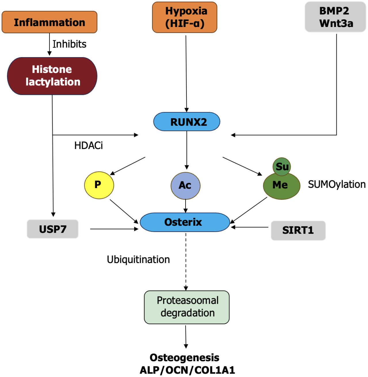
Figure 1 Schematic illustration of major post-translational modifications regulating osteogenic differentiation in oral-derived stem cells.
This diagram illustrates how key post-translational modifications (PTMs) - such as phosphorylation, acetylation, methylation, SUMOylation, histone lactylation, and ubiquitination - work together to regulate the osteogenic differentiation of oral-derived stem cells. External factors like inflammation, hypoxia (hypoxia-inducible factor-alpha), and osteoinductive signals (bone morphogenetic protein 2, Wnt3a) activate runt-related transcription factor 2 (RUNX2), the master transcription factor for osteogenesis. The function and stability of RUNX2 are finely tuned by multiple PTMs. Histone lactylation promotes RUNX2 activation, while inflammation suppresses this modification. Further downstream, osterix drives the expression of osteogenic genes (alkaline phosphatase, osteocalcin, collagen type I) and is regulated by ubiquitination-mediated degradation. Positive regulators such as histone deacetylases inhibitors, USP7, and sirtuin 1 enhance the activity of RUNX2 and osterix by modifying their post-translational states. Together, these mechanisms highlight the crucial role of PTMs in integrating external signals and internal pathways during osteogenic commitment. HIF-α: Hypoxia-inducible factor-alpha; BMP2: Bone morphogenetic protein 2; RUNX2: Runt-related transcription factor 2; HDACi: Histone deacetylases inhibitor; P: Phosphorylation; Me: Methylation; Ac: Acetylation; Su: SUMOylation; SIRT1: Sirtuin 1; ALP: Alkaline phosphatase; OCN: Osteocalcin; COL1A1: Collagen type I.
- Citation: Shi ZJ, Liu W. Post-translational modifications in osteogenic differentiation of oral-derived stem cells: Mechanisms and clinical implications. World J Stem Cells 2025; 17(9): 109662
- URL: https://www.wjgnet.com/1948-0210/full/v17/i9/109662.htm
- DOI: https://dx.doi.org/10.4252/wjsc.v17.i9.109662


