©The Author(s) 2025.
World J Clin Cases. Nov 6, 2025; 13(31): 110734
Published online Nov 6, 2025. doi: 10.12998/wjcc.v13.i31.110734
Published online Nov 6, 2025. doi: 10.12998/wjcc.v13.i31.110734
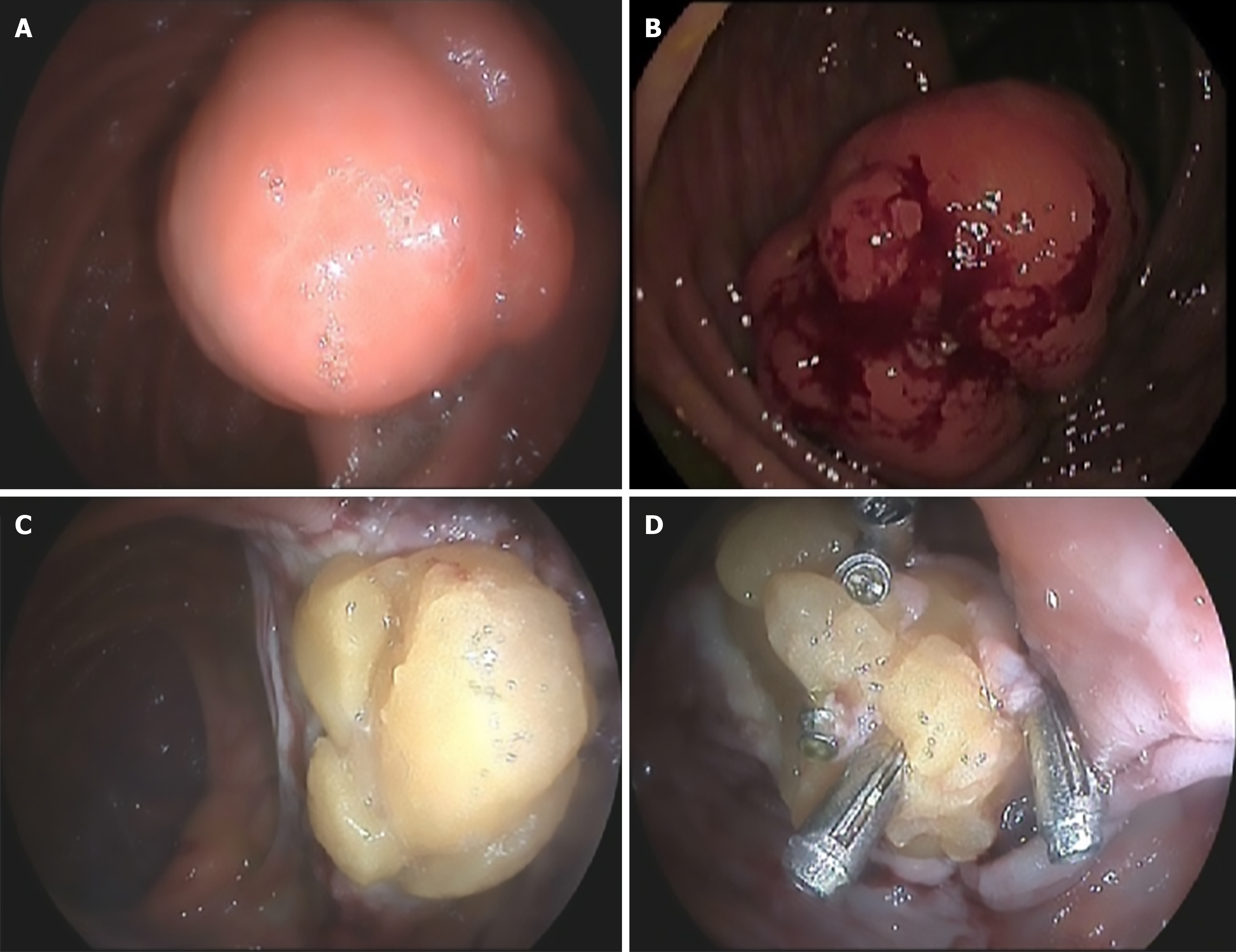
Figure 1 Images of the colonic lesion during therapeutic colonoscopy.
A: Initial appearance of the lesion prior to treatment. Central ulceration is observed; B: Pre-treatment image showing spontaneous bleeding; C: Post-polypectomy scar. Adipose tissue can be seen protruding from the defect; D: Attempt to close the scar using through-the-scope clips.
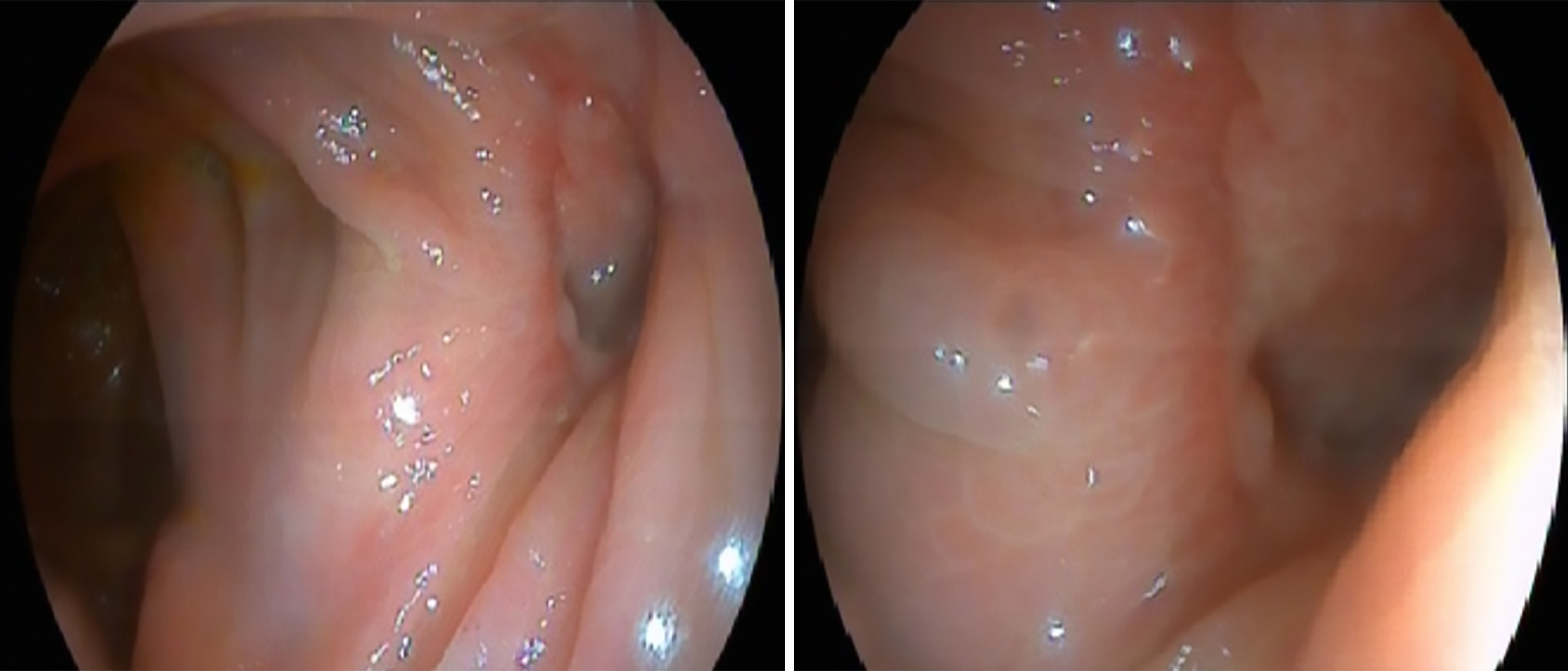
Figure 2 Post-polypectomy orifice observed during the follow-up colonoscopy performed during hospital admission.
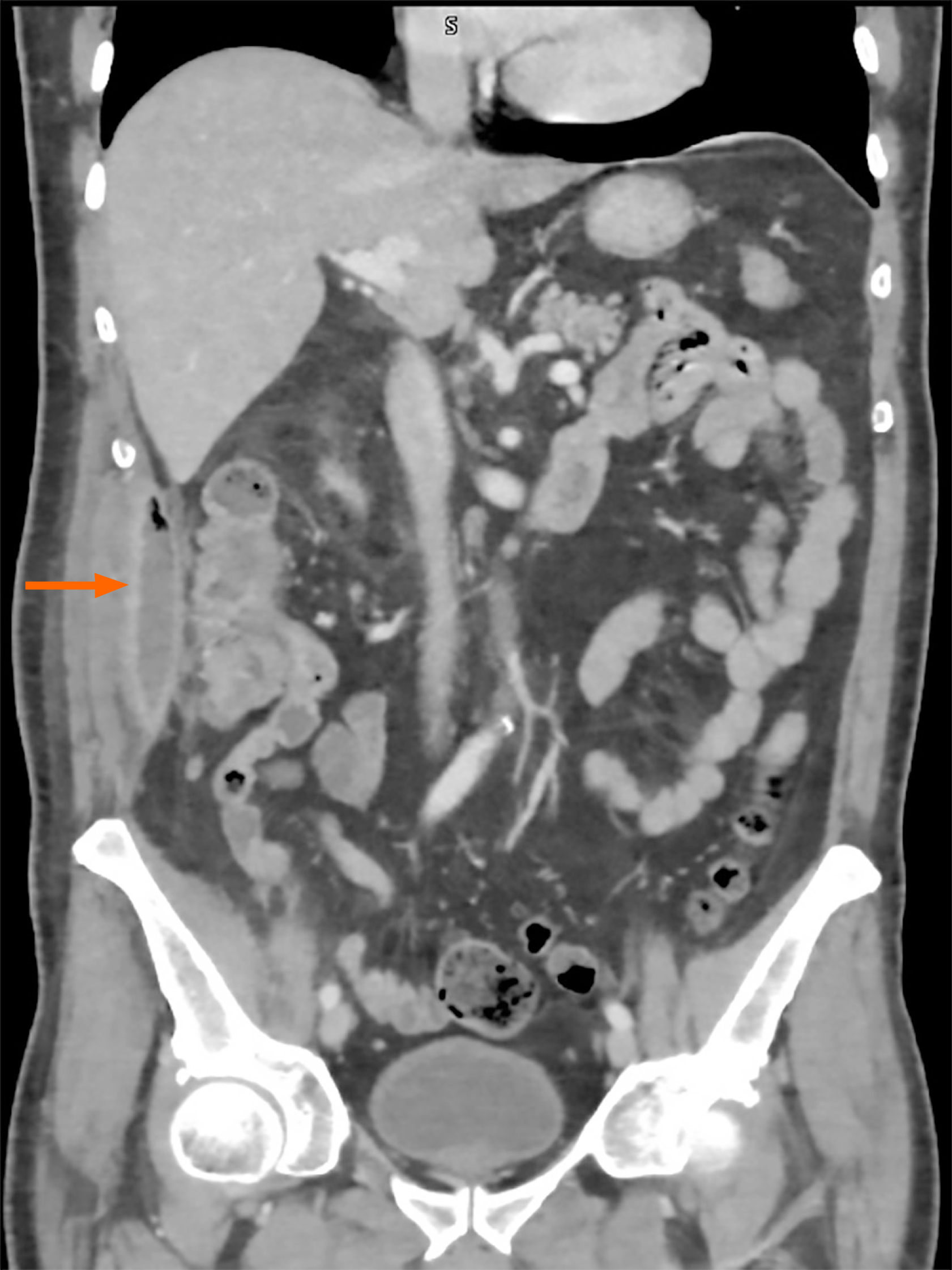
Figure 3 Abdominal computed tomography scan image.
The abscess collection adjacent to the right colon is indicated by the arrow.
- Citation: Suárez M, Martínez R, Santiago-Ramos PC. Not losing sight of the bigger picture of complications associated with post-polypectomy syndrome: A case report and review of literature. World J Clin Cases 2025; 13(31): 110734
- URL: https://www.wjgnet.com/2307-8960/full/v13/i31/110734.htm
- DOI: https://dx.doi.org/10.12998/wjcc.v13.i31.110734


