©The Author(s) 2025.
World J Gastrointest Oncol. Sep 15, 2025; 17(9): 110376
Published online Sep 15, 2025. doi: 10.4251/wjgo.v17.i9.110376
Published online Sep 15, 2025. doi: 10.4251/wjgo.v17.i9.110376
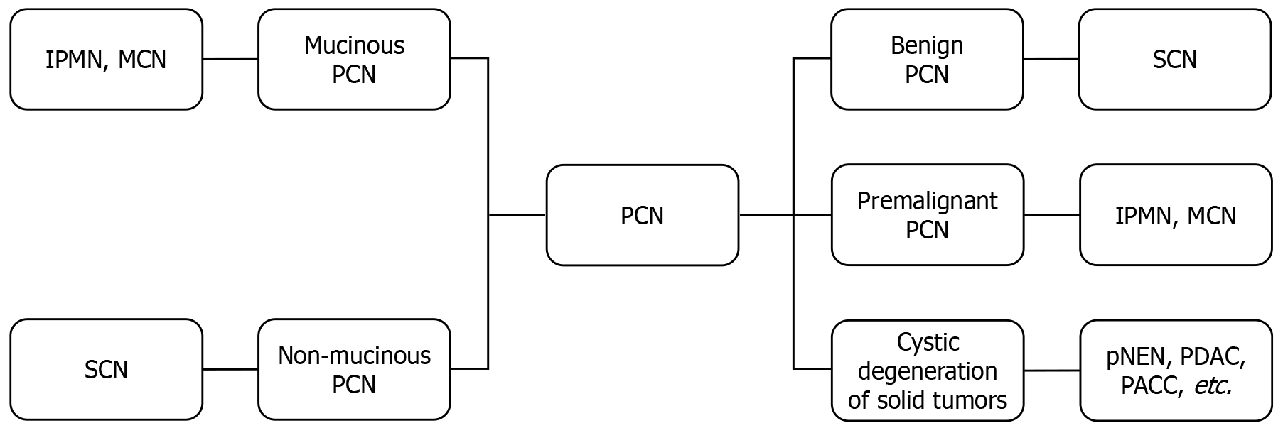
Figure 1 Classification of pancreatic cystic neoplasms.
IPMN: Intraductal papillary mucinous neoplasm; MCN: Mucinous cystic neoplasm; PCN: Pancreatic cystic neoplasm; SCN: Serous cystic neoplasm; pNEN: Pancreatic neuroendocrine neoplasm; PDAC: Pancreatic ductal adenocarcinoma; PACC: Pancreatic acinar cell carcinoma.
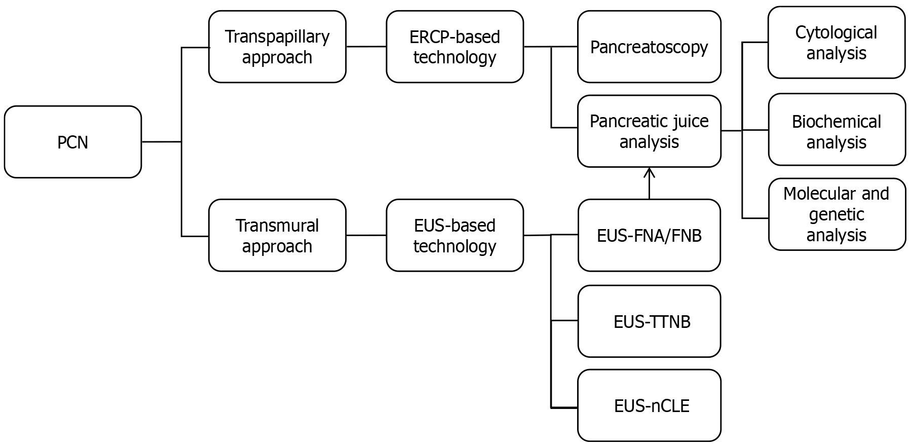
Figure 2 Endoscopic diagnosis of pancreatic cystic neoplasms.
PCN: Pancreatic cystic neoplasm; ERCP: Endoscopic retrograde cholan
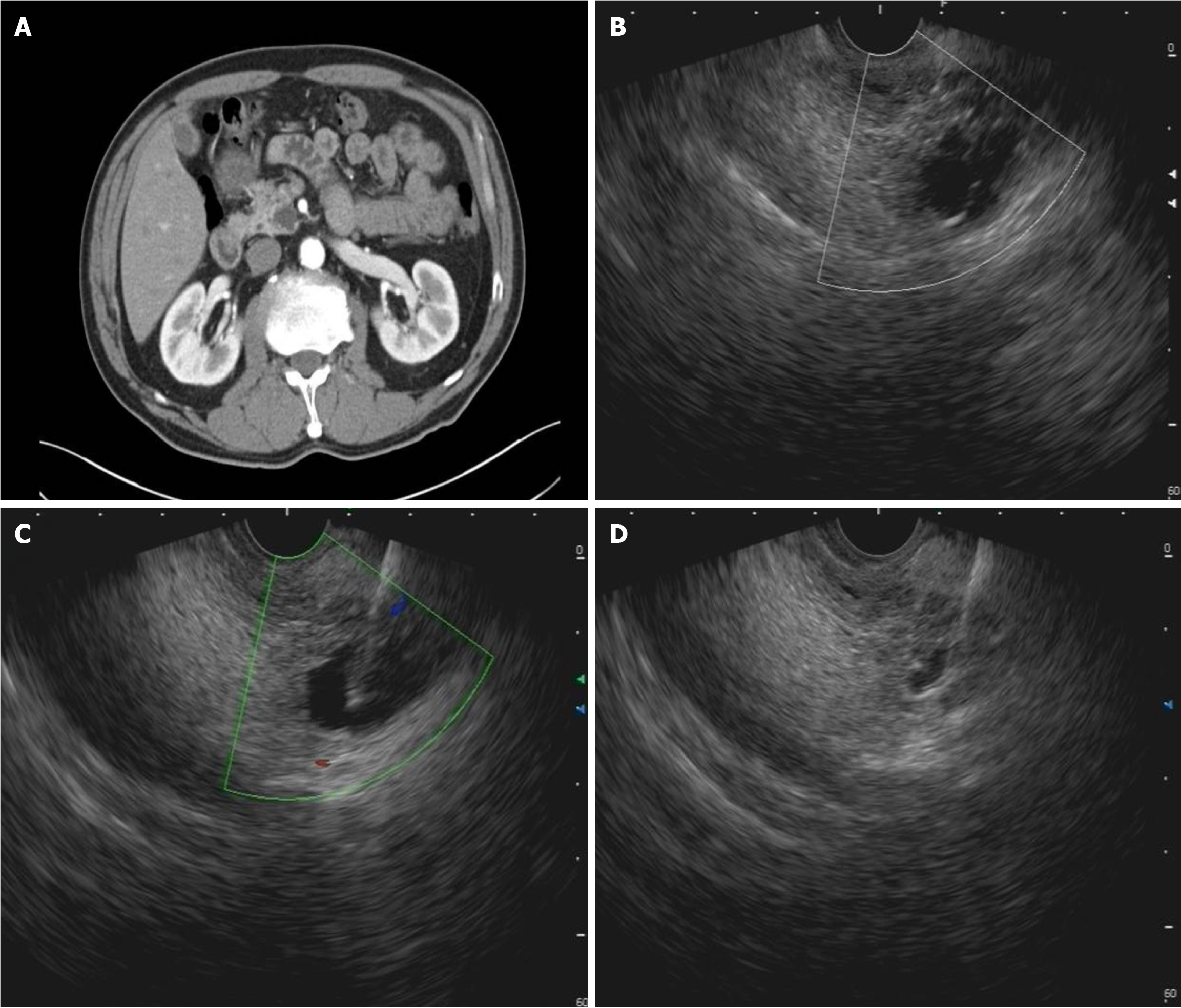
Figure 3 Endoscopic ultrasound-guided fine-needle aspiration.
A: Cross-sectional view of a pancreatic cystic neoplasm; B: Endoscopic ultrasound (EUS)-confirmed an anechoic pancreatic lesion; C: EUS-guided fine-needle aspiration; D: EUS image of the same lesion after cyst fluid aspiration.
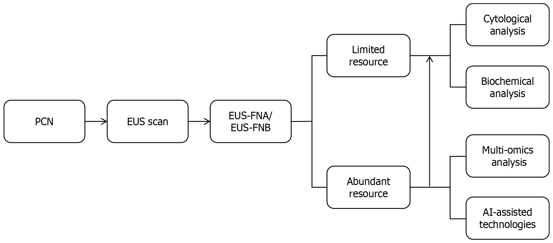
Figure 4 Endoscopic ultrasound differential diagnosis of pancreatic cystic neoplasms.
PCN: Pancreatic cystic neoplasm; EUS: Endoscopic ultrasound; FNA: Fine-needle aspiration; FNB: Fine-needle biopsy; AI: Artificial intelligence.
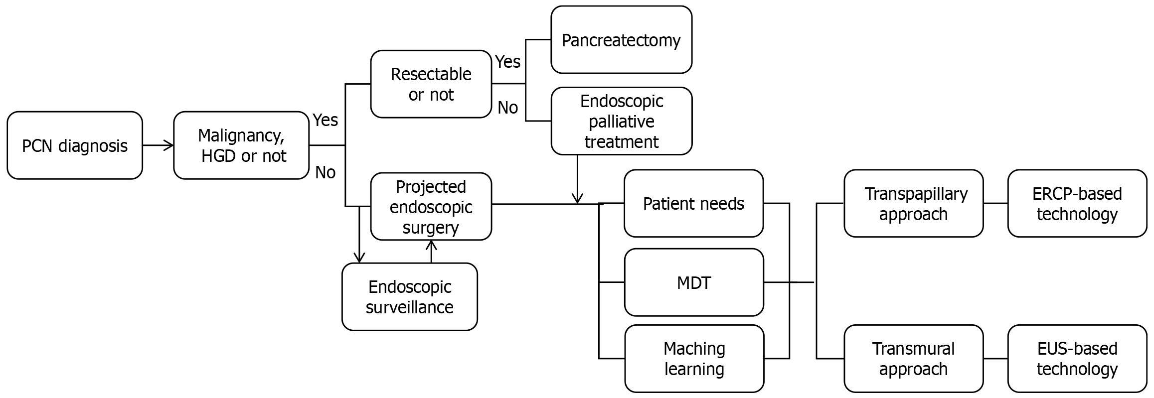
Figure 5 Endoscopic treatment flowchart of pancreatic cystic neoplasms.
PCN: Pancreatic cystic neoplasm; HGD: High-grade dysplasia; MDT: Multidisciplinary team; EUS: Endoscopic ultrasound; ERCP: Endoscopic retrograde cholangiopancreatography.
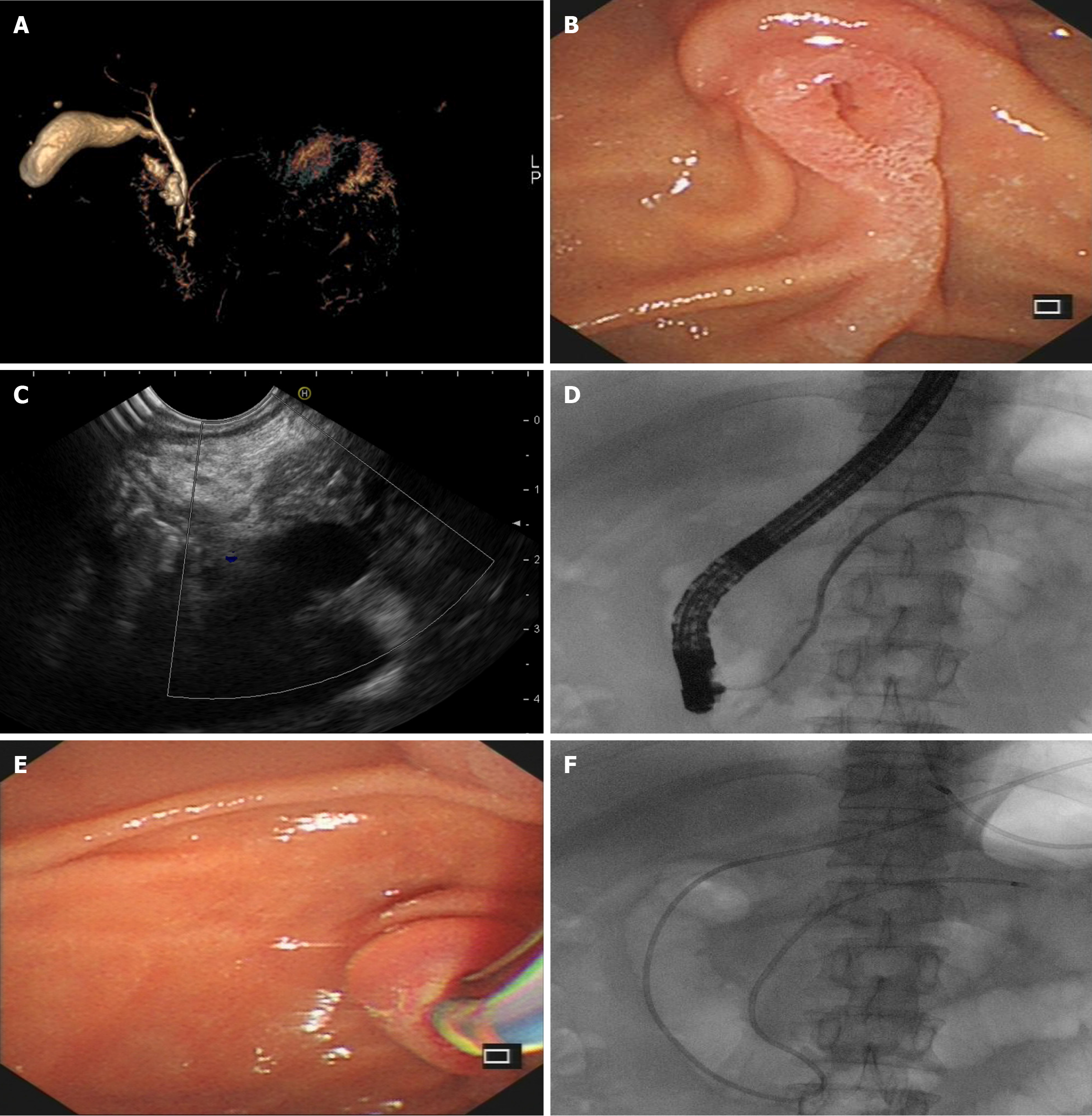
Figure 6 Endoscopic retrograde cholangiopancreatography management of pancreatic cystic neoplasm.
A: Magnetic resonance cholan
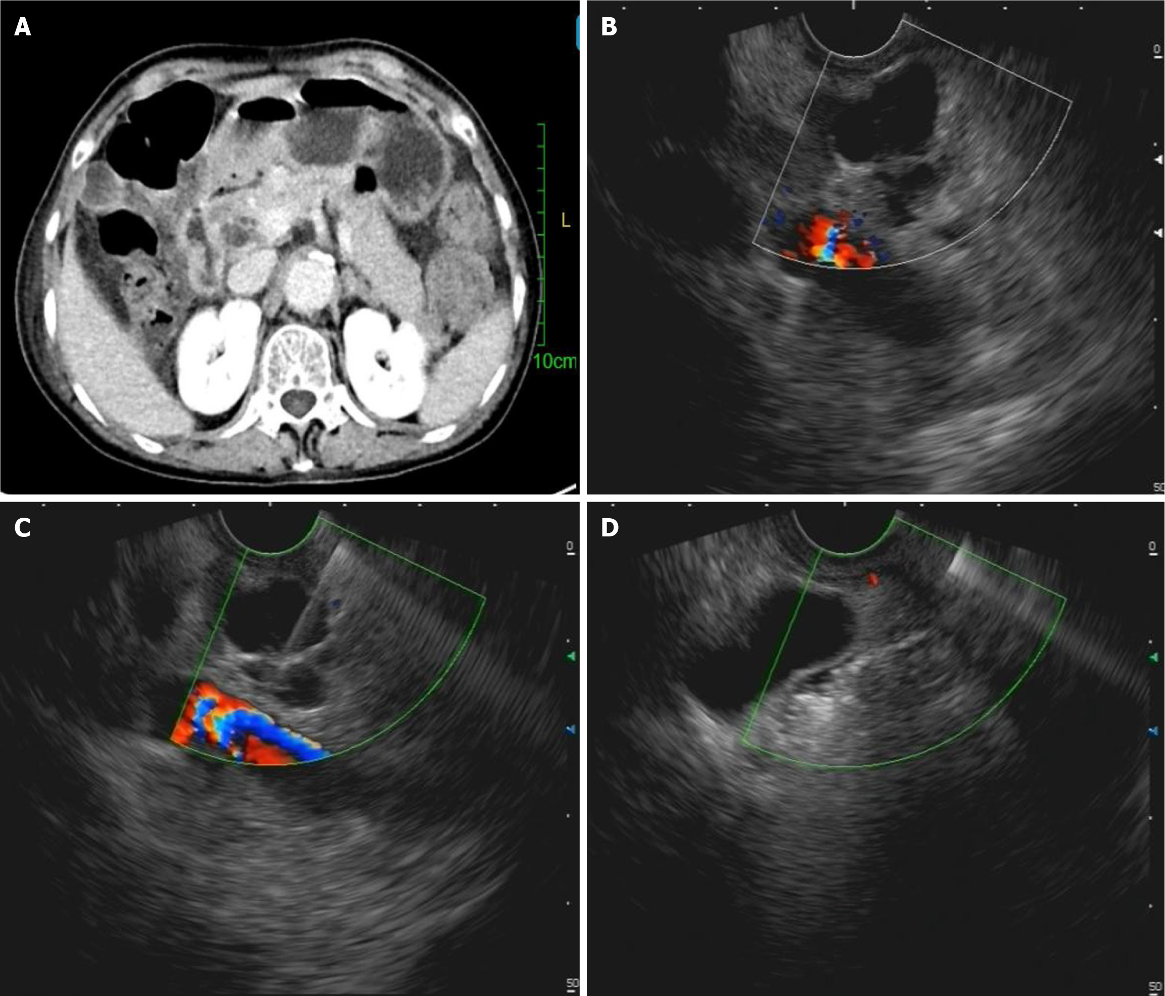
Figure 7 Endoscopic ultrasound management of pancreatic cystic neoplasm.
A: Cross-sectional view of a pancreatic cystic neoplasm; B: Endoscopic ultrasound (EUS)-confirmed anechoic pancreatic lesion; C: EUS-guided fine-needle aspiration; D: EUS-guided pancreatic cyst ablation.
- Citation: Zeng Y, Zhang JW, Yang J. Endoscopic management of pancreatic cystic neoplasms. World J Gastrointest Oncol 2025; 17(9): 110376
- URL: https://www.wjgnet.com/1948-5204/full/v17/i9/110376.htm
- DOI: https://dx.doi.org/10.4251/wjgo.v17.i9.110376


