Published online Oct 16, 2021. doi: 10.12998/wjcc.v9.i29.8871
Peer-review started: June 3, 2021
First decision: July 15, 2021
Revised: July 16, 2021
Accepted: August 5, 2021
Article in press: August 5, 2021
Published online: October 16, 2021
Processing time: 129 Days and 1.6 Hours
Synovial sarcoma (SS) is a highly malignant tumor of unknown histological origin. This tumor can occur in various parts of the body, including those without synovial structures, but mainly in and around the joints, mostly in the lower extremities. Primary intracranial SSs are remarkably rare. This paper aims to report a case of primary intracranial SS with hemorrhage.
A 35-year-old male patient suffered a headache and slurred speech during manual labor and was sent to the emergency department. Through imaging examination, the patient was considered to have high-grade glioma complicated with hemorrhage and was treated with craniotomy. Postoperative pathology revealed SS. positron emission tomography/computed tomography was performed, which ruled out the possibility of metastasis to the intracranial from other parts of the body. Postoperative radiotherapy was given to the patient, during which radiation necrosis occurred. Sixteen months after craniotomy, cranial magnetic resonance imaging revealed recurrence of the tumor.
Primary intracranial SS is a rare malignant tumor. Primary intracranial SS with hemorrhage and radiation necrosis should be carefully monitored during postoperative radiotherapy. Surgical resection of the tumor combined with postoperative radiotherapy and chemotherapy is currently used, but the prognosis is poor.
Core Tip: This paper presents a rare case of primary intracranial synovial sarcoma (SS) with hemorrhage. Through imaging examination, the patient was considered to have high-grade glioma complicated with hemorrhage and was treated with craniotomy. Postoperative pathology revealed SS. Postoperative radiotherapy was given, during which radiation necrosis occurred. By reviewing the diagnostic and therapeutic history and analyzing the clinical and radiological manifestations, a better understanding of the characteristics of primary intracranial SS can be achieved, helping to improve diagnosis and treatment.
- Citation: Wang YY, Li ML, Zhang ZY, Ding JW, Xiao LF, Li WC, Wang L, Sun T. Primary intracranial synovial sarcoma with hemorrhage: A case report. World J Clin Cases 2021; 9(29): 8871-8878
- URL: https://www.wjgnet.com/2307-8960/full/v9/i29/8871.htm
- DOI: https://dx.doi.org/10.12998/wjcc.v9.i29.8871
Synovial sarcoma (SS) is a rare malignant soft-tissue tumor that occurs at any age and in any part of the body, but mainly in and around the joints, mostly in the lower extremities of middle-aged patients. The tumor constitutes 5%-10% of soft-tissue sarcomas[1].
Intracranial SS, especially primary intracranial SS, is rare. This article reports a case of SS originating from intracranial SS. The authors believe that this is the second case of an SS patient with a tumor complicated with hemorrhage[2]. This article aims to understand the characteristics of primary intracranial SS and help improve its diagnosis and treatment by reviewing the diagnostic and treatment history and analyzing the clinical and imaging findings.
A 35-year-old man was sent to the emergency department due to headache and slurred speech.
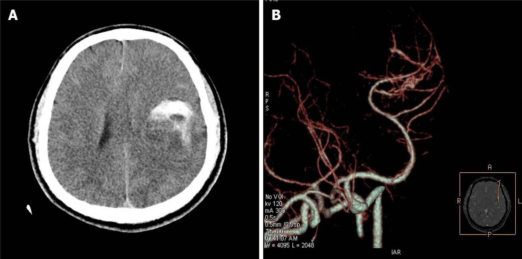
This patient was sent to the emergency department (The First Affiliated Hospital of Xinxiang Medical University, Xinxiang, Henan Province, China) for treatment due to a sudden headache and slurred speech during manual labor 11 h previously. Head computed tomography angiography (CTA) revealed left middle cerebral artery malformation with hemorrhage. The patient was hospitalized.
The patient was physically healthy and had no abnormal medical history.
The patient has smoked approximately 20 cigarettes a day for 10 years and has not quit smoking. He and his younger brother are twins. His younger brother, who has a history of epilepsy since childhood, has been treated with oral carbamazepine.
The patient was mildly lethargic and slurred. His right upper, right lower, and left limb muscle strength was Grade 3, 4 and 5, respectively. Physiological reflex was present, the Babinski sign on the right was positive, and other pathological signs were not elicited.
White blood cell count was increased (12.2 × 10⁹/L), platelet count was decreased (109 × 109/L), and phosphorus was increased in the electrolyte test (1.39 mmol/L). The results of other routine laboratory biochemical tests showed no abnormalities.
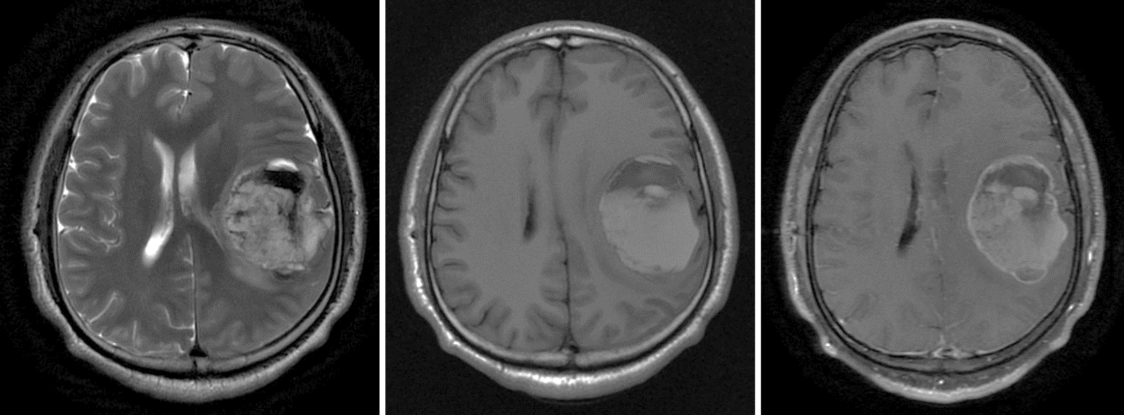
CT and CTA indicated left intracerebral lesions with hemorrhage and left middle cerebral artery arteriovenous malformation, respectively (Figure 1). Preoperative magnetic resonance imaging (MRI) revealed a mass of mixed-signal shadows in the left frontotemporal parietal lobe, approximately 51.3 mm × 54.2 mm × 60.0 mm, and the wall and substantial part of the lesion were enhanced (Figure 2). Radiological diagnosis of the left frontotemporal parietal lobe was solid cystic mass, which was possibly high-grade glioma with hemorrhage.
The patient was diagnosed with primary intracranial SS.
Whole cerebral arteriography was performed because of arteriovenous malformation of the left middle cerebral artery, as suggested by CTA, and no vascular malformation was found. Preoperative examinations were conducted to exclude surgical contraindications. Craniotomy was performed. The dura mater had high tension during the operation, and the brain tissue was expanded. The tumor was gray–red, soft in texture, with a rich blood supply, accompanied by hemorrhage. The M2 segment of the middle cerebral artery and its branches were located inside the tumor. The tumor was resected in pieces, and all the resected tumors were finally removed. Antibiotics, dehydration to lower intracranial pressure, and other related treatments were given after the operation.
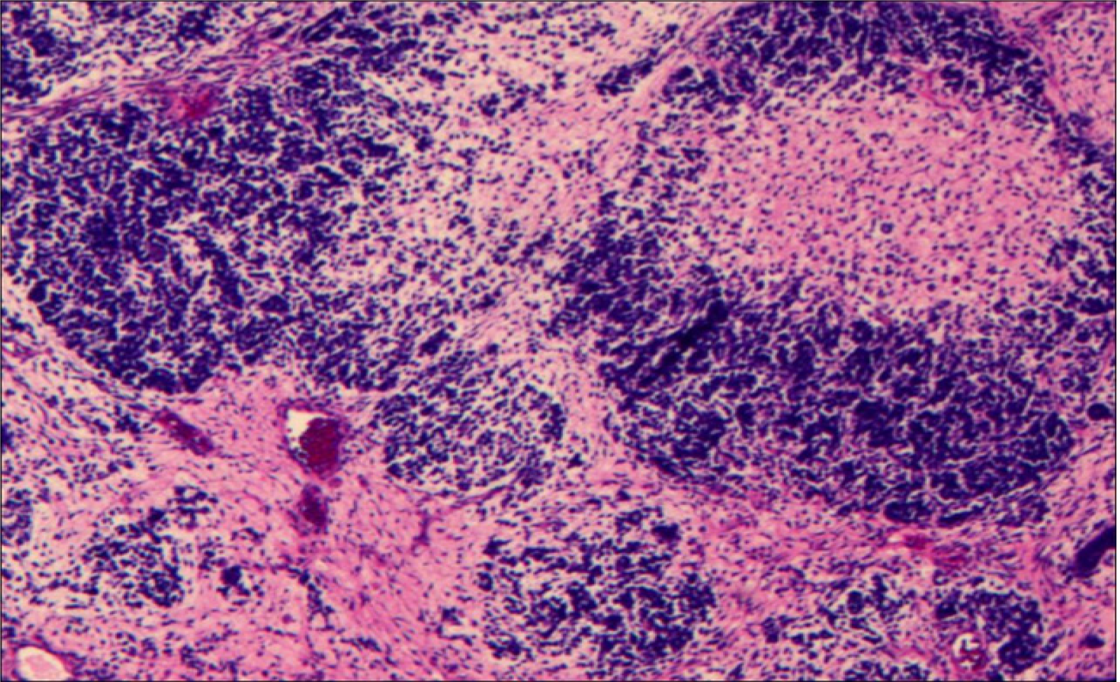
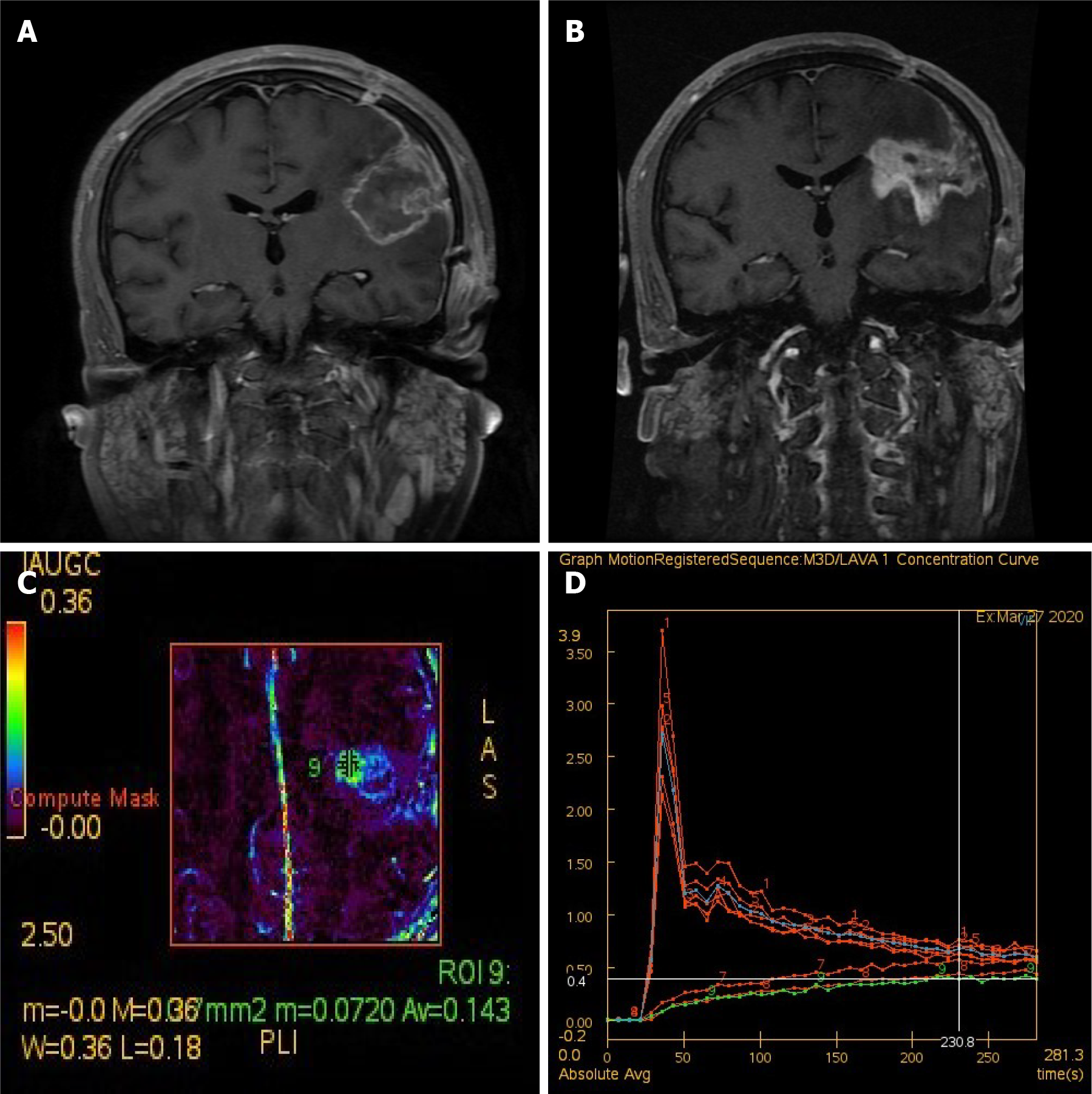
Pathological examination of the tumor tissue and brain tissue in the surrounding edematous area was performed. Dark red–gray–white tissue was observed, approximately 9.5 cm × 9 cm × 2 cm in size. The section was purplish brown and partly gray–white. Microscopically, the tumor cells showed diffused growth with cellular atypia, and mitotic figures (Figure 3). The immunohistochemical results showed the following: GFAP(-), IDH1(R132H)(-), oligo-2(-), S-100(-), TARX(+), CK(-), EMA(-), Ki67(+70%), vimentin(+), CD56(+), NAPSIN-A(-), TTF-1(-), INI1(+), CD34(-), CD99(-), FLi-1(+), NSE(-), PLAP(-), PR(+/-), Sall-4(-), SATB-2(+), SMA(-), Syn(-), Bcl-2(+), and TLE1(+). Further genetic testing of SYT (SS18) was negative for gene breaks, suggesting that SS18 had no fusion with other genes. SS usually occurs in the extremities. Thus, a whole-body positron emission tomography/CT were performed to search for a potential primary tumor. However, no abnormality was found. Postoperative head MRI was also re-examined (Figure 4A).
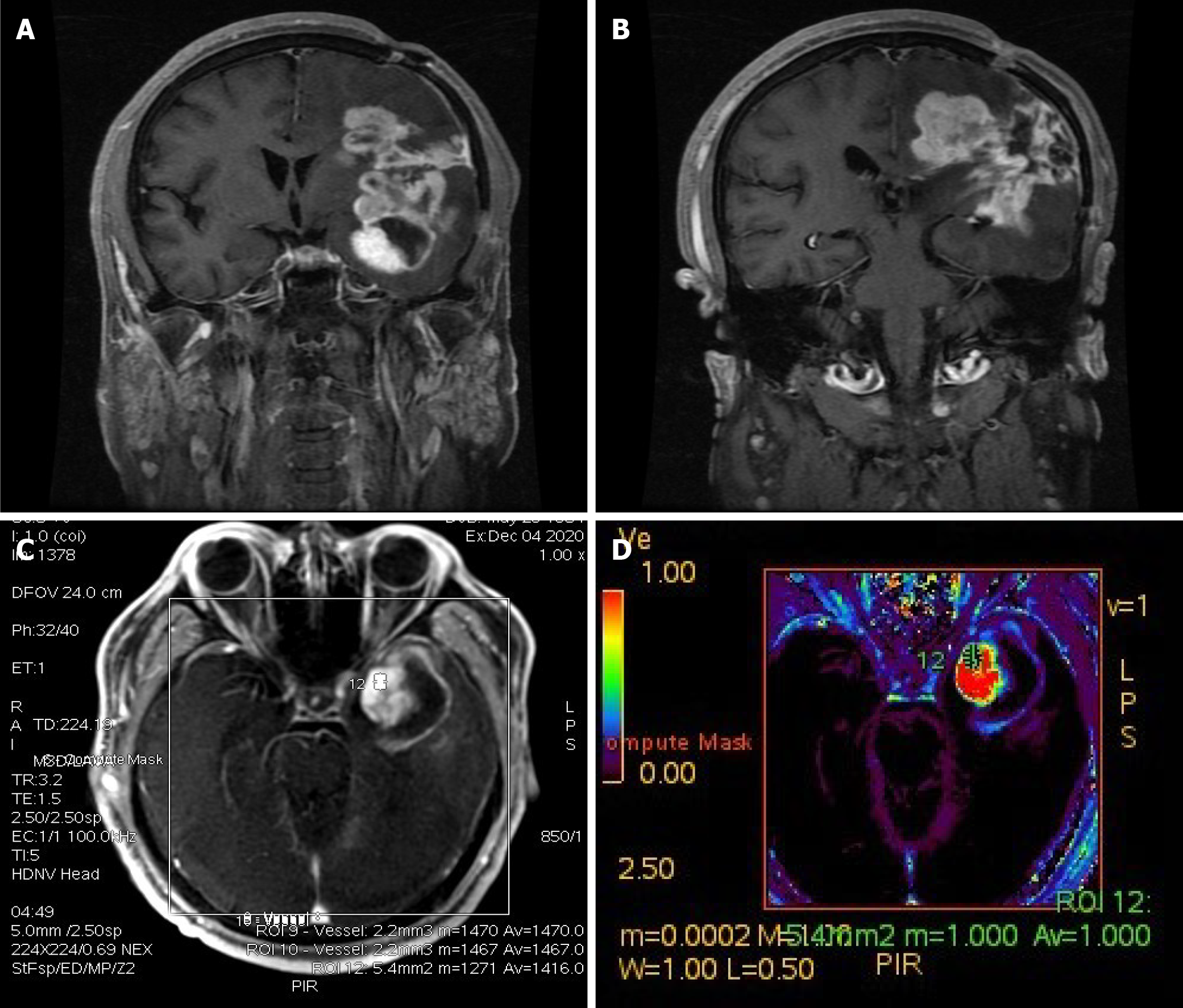
Radiotherapy was administered 2 wk after the operation with 95% PGTV 2.2 Gy × 30 F. Six months after craniotomy (3 mo after radiotherapy), perfusion-weighted imaging (PWI) was performed in the outpatient department (Figure 4B-D): The lesion was markedly enlarged, but this enhancement showed hypoperfusion, which was considered to be radiation necrosis, and bevacizumab was given. Sixteen months after craniotomy, the patient developed headaches, and PWI was repeated for detection of tumor recurrence (Figure 5).
SS is a clinically rare malignant tumor. SS is generally believed to be a soft tissue malignant tumor originating from the joints, synovia, and tendon sheath synovia, which usually occurs in the extremities of younger adults. However, SS may occur in any part of the body, such as the heart, kidney, throat and tongue, with an increase in case reports[3-6]. Therefore, SS is considered to be a malignant tumor with an unknown histological origin. The literature review revealed that cases of intracranial primary SS are remarkably rare, and diagnosis by surgery and pathology is uncommon. This article reports a case of SS originating from intracranial SS. The authors believe that this is the second case of SS complicated with hemorrhage. This tumor was initially misdiagnosed as glioma. The final diagnosis was SS after surgical and pathological examination.
The clinical manifestations of primary intracranial SS lack specificity, and diagnosis depends on the location, size and complications of the tumor. The clinical manifestations of primary intracranial SS include headache, nausea, vomiting, limb hemiplegia, and slurred speech. This patient was sent to the emergency department for treatment due to SS with hemorrhage. The clinical manifestations were headache, slurred speech, and hemiplegia. The literature on primary intracranial SSs with hemorrhage is summarized in Table 1. Similarly, imaging examination of intracranial primary SS also lacks specificity[2]. CT mainly manifests as dense or low-density shadows, such as nodular, lobulated masses, and most of the boundaries are clear. CT of this patient showed a round tumor with clear borders, and the part of the tumor with hemorrhage showed high-density shadows. MRI mostly reveals the shadow of soft-tissue masses with clear boundaries, with high- or low-signal intensity on T1-weighted imaging (WI) and slightly high signal intensity on T2WI. Mixed signals of low, equal, and high may appear with the occurrence of cystic degeneration, necrosis, and hemorrhage in the mass. MRI of this patient showed long/short T1WI signals, and T2WI demonstrated mixed signals. The wall and substantial part of the lesion were enhanced, which was consistent with the MRI findings of previous cases reported in the literature[7].
| Characteristics | Present case | Patel et al[2] |
| Sex | Male | Male |
| Age (yr) | 35 | 21 |
| Clinical presentation | Headaches and slurred speech | Headaches, ataxia, left hemianopsia, left arm weakness |
| Intracranial findings | Solid and cystic mass with hemorrhage in the left frontotemporal parietal lobe | Right parietal mass with hemorrhage |
| Imaging examinations | CT (left intracerebral lesions with hemorrhage); MRI revealed a mass of mixed-signal shadows in the left frontotemporal parietal lobe, approximately 51.3 mm × 54.2 mm × 60.0 mm, and the wall and substantial part of the lesion were enhanced | CT (right parietal heterogeneous, hyperdense mass with a large medial hematoma) |
| Treatment | (1) Craniotomy, GTR; and (2) RT | (1) Decompression and clot evacuation; (2) RT; (3) Craniotomy, GTR; and (4) Chemotherapy |
| Outcome and Follow-up | The tumor recurred 16 mo after the craniotomy | Survived for at least two years |
Diagnosis of primary intracranial SS requires pathological examination and immunohistochemistry due to the lack of specificity in clinical manifestations and imaging findings. Primary intracranial SS is generally large in volume and usually lobulated and nodular. The section of the tumor is fish-like, gray white, or gray red. Gross observation of the present case showed that the tumor had dark red and gray and white tissues, approximately 9.5 cm × 9 cm × 2 cm in size, purple–brown in section, and partly gray–white, which is consistent with previous reports[7]. SS can be classified microscopically according to the composition of epithelial and/or spindle cells. The classification is as follows: (1) Biphasic type: Contains spindle and epithelioid cells; (2) Monophasic type: Comprises only spindle or epithelioid cells; and (3) Poorly differentiated SS type[7,8]. Vimentin and Bcl-2 are consistently expressed in SS tumor cells, as seen by immunohistochemistry. CD99 and CD56 are frequently expressed but not SMA, S-100, CD34 and GFAP[9,10]. The immunohistochemical results of tumor cells in this patient were consistent with those reported in the literature, including vimentin(+), Bcl-2(+), CD56(+), SMA(-), GFAP(-), S-100(-) and CD34(-), as well as CD99(-). In addition, Ki-67 proliferation index of 70% was observed in the most proliferative zone of the tumor.
The cytogenetic marker of SS is currently believed to be the mutual translocation of the SYT gene on chromosome 18 and SSX1 or SSX2 gene on chromosome X and the formation of a new chimeric gene SYT–SSX1 or SYT–SSX2. More than 90% of SSs possess this molecular characteristic[11]. Unfortunately, the genetic test of our patient for SYT (SS18) was negative for gene breaks, suggesting the absence of fusion of other SS18 genes. However, the disease was finally concluded to be a primary intracranial SS after comprehensive consideration of patient history, clinical manifestations, imaging data, and pathological examination, as well as multidisciplinary discussion among neurosurgery, imaging and pathology departments.
The current recommendation for the treatment of primary intracranial SS is to remove the tumor as far as possible while ensuring the patient’s safety, followed by postoperative adjuvant radiotherapy and chemotherapy. Reports indicate that the degree of tumor resection is closely related to prognosis[10]. The recommended dose of postoperative adjuvant radiotherapy is 60-70 Gy[12,13]. A 95% PGTV 2.2 Gy × 30 F radiotherapy plan for our patient was performed, but he developed radiation necrosis 3 mo after radiotherapy. In addition, SS is remarkably prone to metastasis, particularly to the lungs, because it is a highly malignant tumor, and related monitoring approaches, such as chest radiography, should be performed during follow-up[10].
Primary intracranial SS is a remarkably rare malignant tumor with atypical clinical and imaging manifestations. Diagnosis requires pathological and cytogenetic examination. The current treatment methods are surgical resection combined with postoperative radiotherapy and chemotherapy. Numerous experiences and lessons can be learned from the treatment of our patient. He and his brother are twins, and his brother has had a history of epilepsy since childhood. The possible relation of this kind of primary intracranial SS to the history of the patient should be further studied. In addition, primary intracranial SS with hemorrhage and radiation necrosis should be carefully monitored during postoperative radiotherapy. Overall, there are few cases of primary intracranial SS, and the lack of long-term follow-up data complicates the diagnosis and identification. Therefore, the diagnosis, treatment and prognosis of primary intracranial SS need to be studied in further cases.
Primary intracranial synovial sarcoma is a remarkably rare malignant tumor. Primary intracranial SS with hemorrhage and radiation necrosis should be carefully monitored during postoperative radiotherapy. Surgical resection of the tumor combined with postoperative radiotherapy and chemotherapy is currently used, but the prognosis is poor.
| 1. | Stacchiotti S, Van Tine BA. Synovial Sarcoma: Current Concepts and Future Perspectives. J Clin Oncol. 2018;36:180-187. [RCA] [PubMed] [DOI] [Full Text] [Cited by in Crossref: 84] [Cited by in RCA: 90] [Article Influence: 11.3] [Reference Citation Analysis (0)] |
| 2. | Patel M, Li L, Nguyen HS, Doan N, Sinson G, Mueller W. Primary Intracranial Synovial Sarcoma. Case Rep Neurol Med. 2016;2016:5608315. [RCA] [PubMed] [DOI] [Full Text] [Full Text (PDF)] [Cited by in Crossref: 2] [Cited by in RCA: 7] [Article Influence: 0.7] [Reference Citation Analysis (0)] |
| 3. | Veshti A, Prifti EM, Ikonomi M. Primary cardiac synovial sarcoma originating from the mitral valve causing left ventricular outflow tract obstruction. Heart Surg Forum. 2015;18:E112-E113. [RCA] [PubMed] [DOI] [Full Text] [Cited by in Crossref: 2] [Cited by in RCA: 2] [Article Influence: 0.2] [Reference Citation Analysis (0)] |
| 4. | El Chediak A, Mukherji D, Temraz S, Nassif S, Sinno S, Mahfouz R, Shamseddine A. Primary synovial sarcoma of the kidney: a case report of complete pathological response at a Lebanese tertiary care center. BMC Urol. 2018;18:40. [RCA] [PubMed] [DOI] [Full Text] [Full Text (PDF)] [Cited by in Crossref: 11] [Cited by in RCA: 16] [Article Influence: 2.0] [Reference Citation Analysis (0)] |
| 5. | Madabhavi I, Bhardawa V, Modi M, Patel A, Sarkar M. Primary synovial sarcoma (SS) of larynx: An unusual site. Oral Oncol. 2018;79:80-82. [RCA] [PubMed] [DOI] [Full Text] [Cited by in Crossref: 5] [Cited by in RCA: 11] [Article Influence: 1.4] [Reference Citation Analysis (0)] |
| 6. | Su Z, Zhang J, Gao P, Shi J, Qi M, Chen L, Wang X. Synovial sarcoma of the tongue: report of a case and review of the literature. Ann R Coll Surg Engl. 2018;100:e118-e122. [RCA] [PubMed] [DOI] [Full Text] [Cited by in Crossref: 5] [Cited by in RCA: 7] [Article Influence: 0.9] [Reference Citation Analysis (0)] |
| 7. | Nuwal P, Dixit R, Shah NS, Samaria A. Primary monophasic synovial sarcoma lung with brain metastasis diagnosed on transthoracic FNAC: Report of a case with literature review. Lung India. 2012;29:384-387. [RCA] [PubMed] [DOI] [Full Text] [Full Text (PDF)] [Cited by in Crossref: 13] [Cited by in RCA: 13] [Article Influence: 0.9] [Reference Citation Analysis (0)] |
| 8. | Lin YJ, Yang QX, Tian XY, Li B, Li Z. Unusual primary intracranial dural-based poorly differentiated synovial sarcoma with t(X; 18)(p11; q11). Neuropathology. 2013;33:75-82. [RCA] [PubMed] [DOI] [Full Text] [Cited by in Crossref: 11] [Cited by in RCA: 12] [Article Influence: 0.9] [Reference Citation Analysis (0)] |
| 9. | Xiao GY, Pan BC, Tian XY, Li Y, Li B, Li Z. Synovial sarcoma in cerebellum: a case report and literature review. Brain Tumor Pathol. 2014;31:68-75. [RCA] [PubMed] [DOI] [Full Text] [Cited by in Crossref: 13] [Cited by in RCA: 18] [Article Influence: 1.3] [Reference Citation Analysis (0)] |
| 10. | Zhang G, Xiao B, Huang H, Zhang Y, Zhang X, Zhang J, Wang Y. Intracranial synovial sarcoma: A clinical, radiological and pathological study of 16 cases. Eur J Surg Oncol. 2019;45:2379-2385. [RCA] [PubMed] [DOI] [Full Text] [Cited by in Crossref: 8] [Cited by in RCA: 18] [Article Influence: 2.6] [Reference Citation Analysis (0)] |
| 11. | Fletcher CD, Unni K, Mertens F. World Health Organization classification of tumours. Pathology and genetics of tumours of soft tissue and bone. Oxford, Oxford University Press, 2002: 430. |
| 12. | ten Heuvel SE, Hoekstra HJ, Bastiaannet E, Suurmeijer AJ. The classic prognostic factors tumor stage, tumor size, and tumor grade are the strongest predictors of outcome in synovial sarcoma: no role for SSX fusion type or ezrin expression. Appl Immunohistochem Mol Morphol. 2009;17:189-195. [RCA] [PubMed] [DOI] [Full Text] [Cited by in Crossref: 39] [Cited by in RCA: 38] [Article Influence: 2.2] [Reference Citation Analysis (0)] |
| 13. | Andrä C, Rauch J, Li M, Ganswindt U, Belka C, Saleh-Ebrahimi L, Ballhausen H, Nachbichler SB, Roeder F. Excellent local control and survival after postoperative or definitive radiation therapy for sarcomas of the head and neck. Radiat Oncol. 2015;10:140. [RCA] [PubMed] [DOI] [Full Text] [Full Text (PDF)] [Cited by in Crossref: 19] [Cited by in RCA: 30] [Article Influence: 2.7] [Reference Citation Analysis (0)] |
Open-Access: This article is an open-access article that was selected by an in-house editor and fully peer-reviewed by external reviewers. It is distributed in accordance with the Creative Commons Attribution NonCommercial (CC BY-NC 4.0) license, which permits others to distribute, remix, adapt, build upon this work non-commercially, and license their derivative works on different terms, provided the original work is properly cited and the use is non-commercial. See: http://creativecommons.org/Licenses/by-nc/4.0/
Manuscript source: Unsolicited manuscript
Specialty type: Clinical neurology
Country/Territory of origin: China
Peer-review report’s scientific quality classification
Grade A (Excellent): 0
Grade B (Very good): B
Grade C (Good): 0
Grade D (Fair): 0
Grade E (Poor): 0
P-Reviewer: Macciò A S-Editor: Yan JP L-Editor: A P-Editor: Wang LYT


