©The Author(s) 2021.
World J Clin Cases. Nov 16, 2021; 9(32): 9889-9895
Published online Nov 16, 2021. doi: 10.12998/wjcc.v9.i32.9889
Published online Nov 16, 2021. doi: 10.12998/wjcc.v9.i32.9889
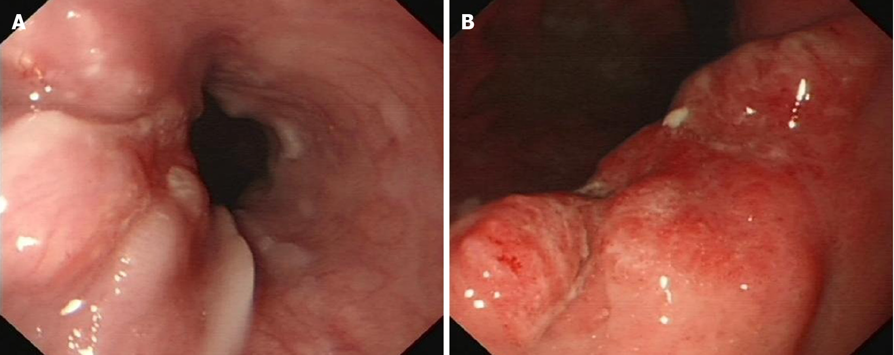
Figure 1 Preoperative endoscopy.
A: Preoperative endoscopy showed one soft nodular neoplasm with superficial erosion along with the lower esophagus; B: Preoperative endoscopy showed a nodular neoplasm with superficial erosion and irregular boundary in the stomach angular notch.
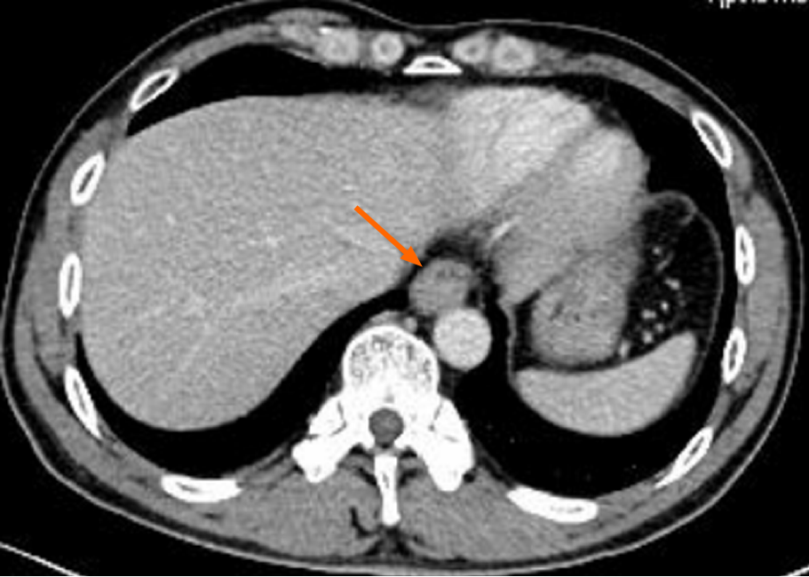
Figure 2 Preoperative computed tomography.
Preoperative computed tomography showed that the wall of the lower esophagus was eccentrically thickening and enhanced, and the esophageal lumen became narrowed obviously (orange arrow).
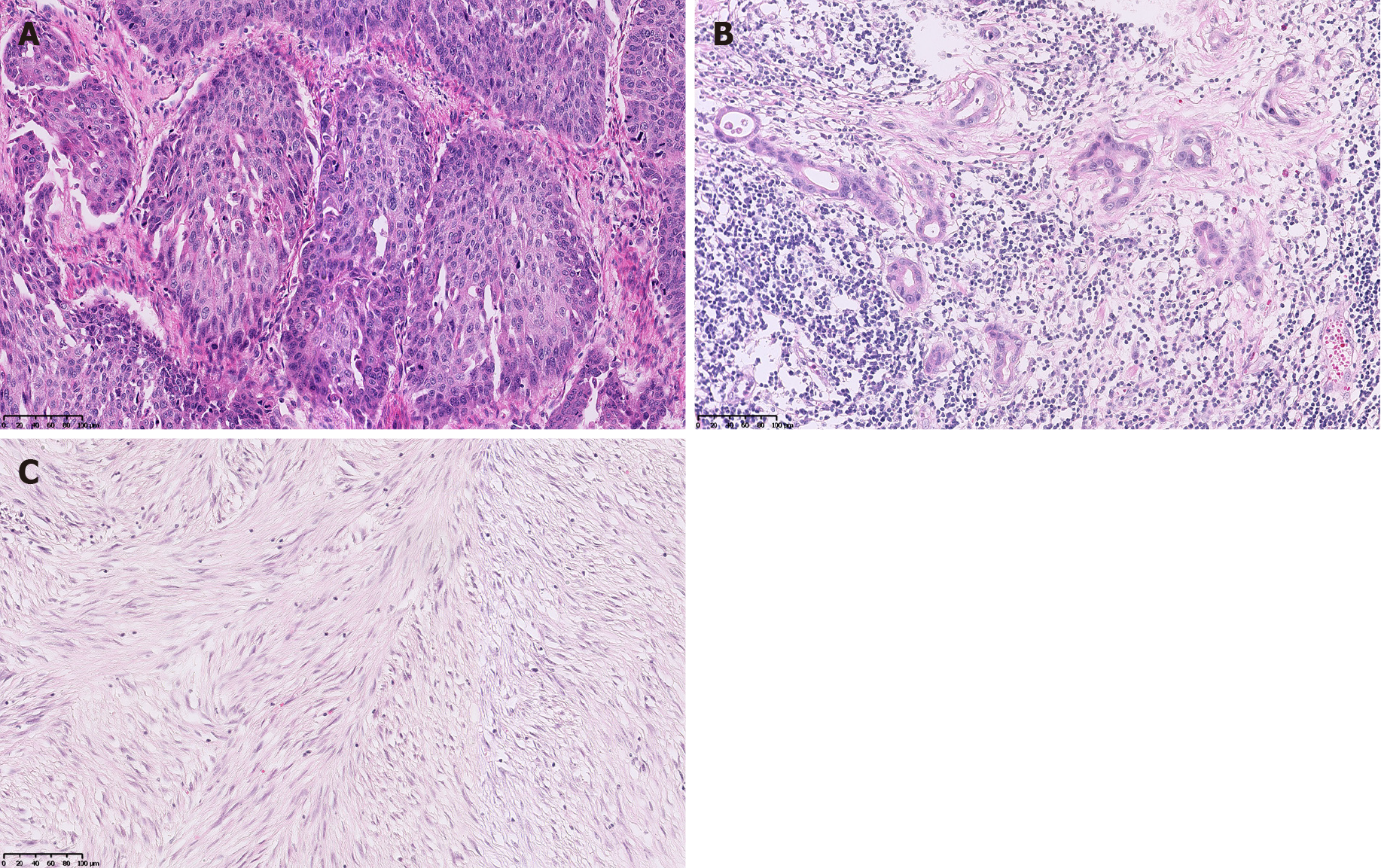
Figure 3 Postoperative pathology.
A: Postoperative pathology showed that the esophageal tumor was a moderately differentiated squamous cell carcinoma with lymph nodes metastases (pT3N2M0, G2, stage IIIB); B: Postoperative pathology showed that the gastric tumor was a moderately to poorly differentiated adenocarcinoma (tubular adenocarcinoma and signet-ring cell carcinomaa, Laurén mixed type) with lymph node metastases (pT3N2M0, G2-G3, stage IIIA); C: Postoperative pathology showed that the jejunal tumor was a gastrointestinal stromal tumor of spindle cell type (high-risk).
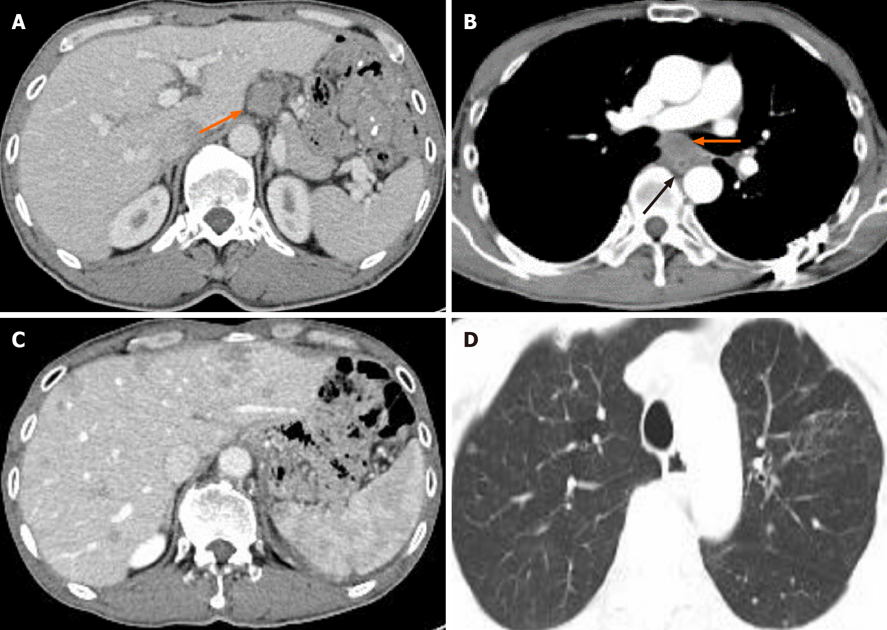
Figure 4 Computed tomography re-examination.
A: Computed tomography (CT) re-examination showed that the hepatogastric ligamentous lymph node enlarged 21 mo after surgery (orange arrow); B: CT re-examination showed that the local thickening of the esophageal anastomotic site (black arrow) and enlarged subcarinal lymph nodes (orange arrow); C: CT re-examination showed multiple nodules in the liver; D: CT re-examination showed multiple nodules scattered in both lungs emerging 28 mo after surgery.
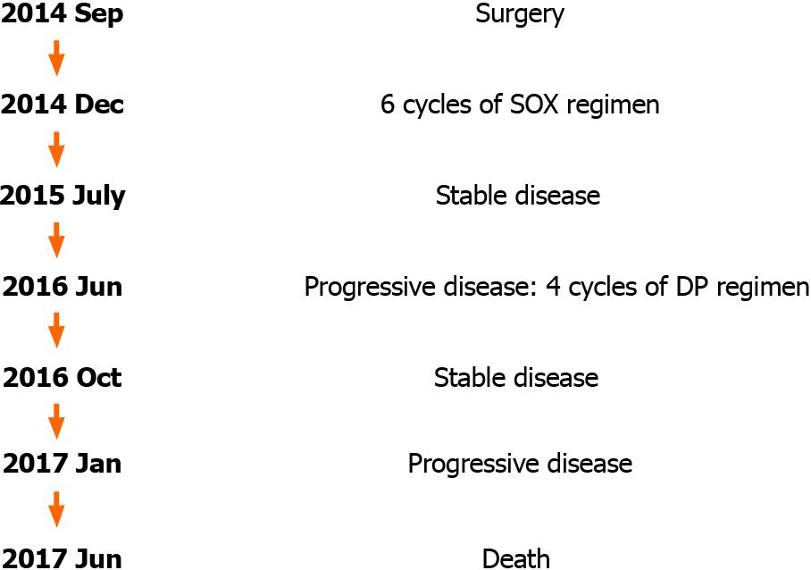
Figure 5 Timeline.
The patient was diagnosed with multiple primary malignancies in September 2014 and was treated by radical surgery and adjuvant chemotherapy. Abdominal lymph node metastasis occurred in June 2016 (21 mo after surgery). Additional chemotherapy was carried out but was not well tolerated, and he refused further therapy. Widespread metastases occurred soon, and the patient eventually died in June 2017 (33 mo after surgery). SOX: Oxaliplatin 200 mg d1 combined with Tegafur Gimeracil Oteracil Potassium capsules 50 mg bid d1-14; DP: Docetaxel 110 mg d1 combined with Cisplatin 35 mg d1-d3.
- Citation: Li Y, Ye LS, Hu B. Synchronous multiple primary malignancies of the esophagus, stomach, and jejunum: A case report. World J Clin Cases 2021; 9(32): 9889-9895
- URL: https://www.wjgnet.com/2307-8960/full/v9/i32/9889.htm
- DOI: https://dx.doi.org/10.12998/wjcc.v9.i32.9889


