©The Author(s) 2021.
World J Clin Cases. Oct 26, 2021; 9(30): 9228-9235
Published online Oct 26, 2021. doi: 10.12998/wjcc.v9.i30.9228
Published online Oct 26, 2021. doi: 10.12998/wjcc.v9.i30.9228
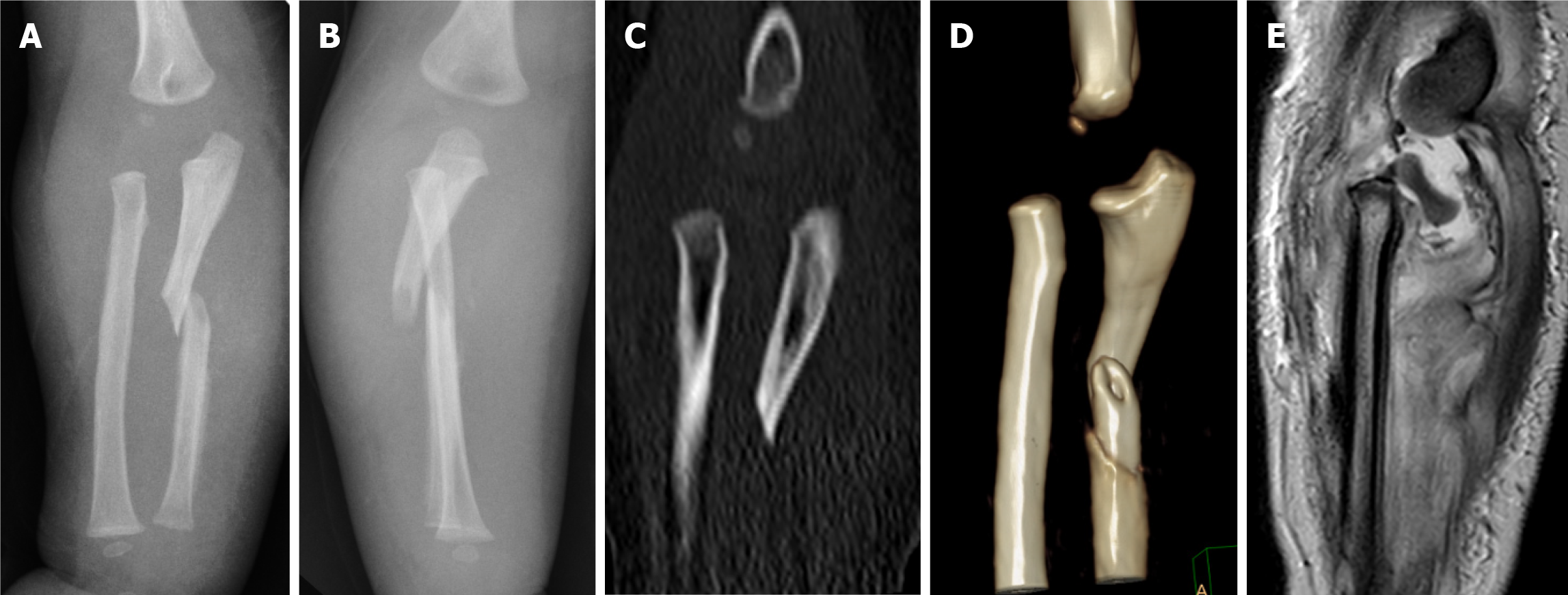
Figure 1 Images before surgery.
A and B: Plain radiographs in the anteroposterior (A) and lateral (B) views of the affected forearm. There was a short oblique fracture of the ulna. The congruency of the radiocapitellar joint misalignment led to a suspicion that a Monteggia fracture dislocation existed; C and D: Multiplanar computed tomography reconstruction (C) and three-dimensional reconstruction scans (D) of the right elbow, indicating that the proximal metaphysic of the radius was anteriorly displaced; E: Magnetic resonance imaging showing a Salter-Harris type I fracture of the radial head epiphysis. The cartilaginous radial head was tilted by 60° but remained intra-articular.
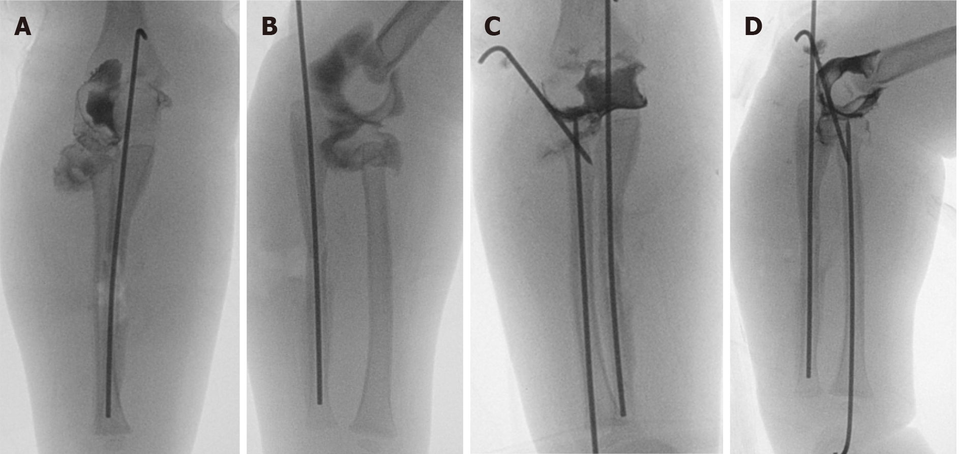
Figure 2 Intraoperative images.
A and B: Intraoperative anteroposterior (AP) (A) and lateral (B) arthrogram showing a Salter-Harris type I fracture of the radial head epiphysis. After the ulnar fracture was reduced, the radial metaphysis and capitellar alignment seemed restored. However, the cartilaginous radial head was still tilted; C and D: AP (C) and lateral (D) arthrograms showing almost anatomical reduction of the fracture of the ulna and fracture of the radial neck.
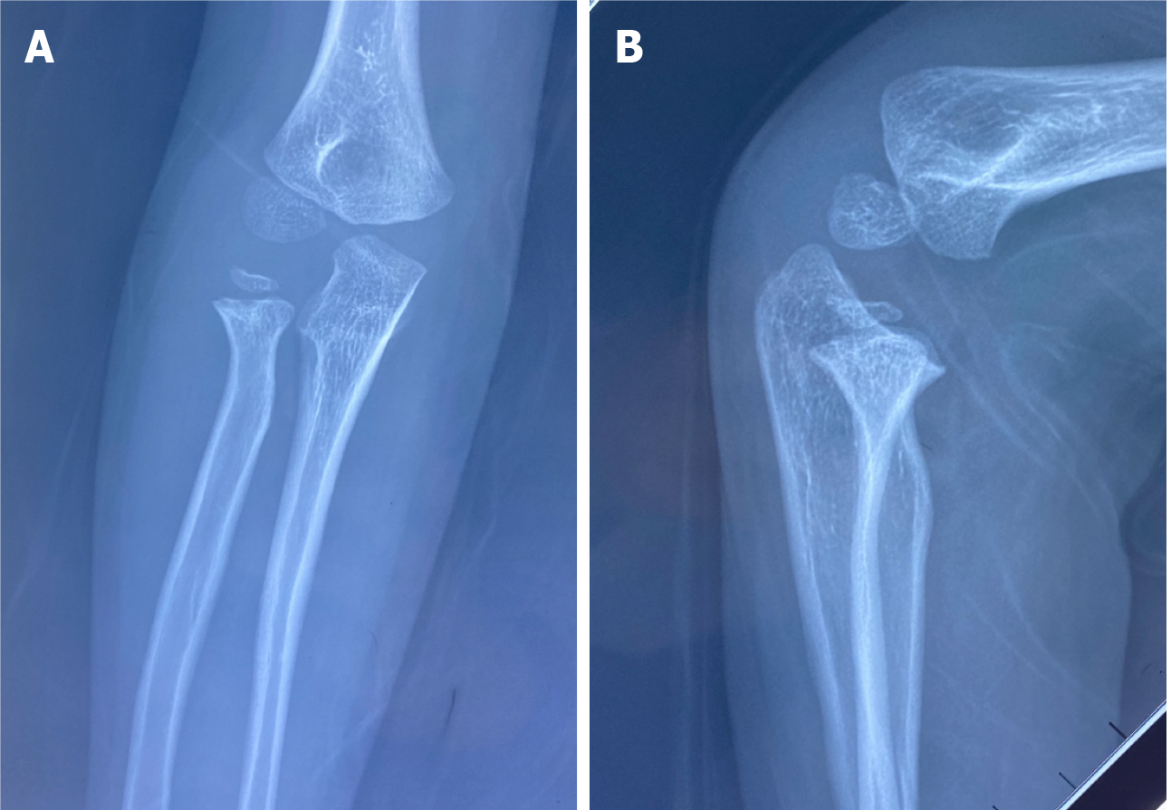
Figure 3 Follow-up images.
Radiographs at the 5-year follow-up revealed successful union of both bones. A: AP radiograph view; B: lateral radiograph view.
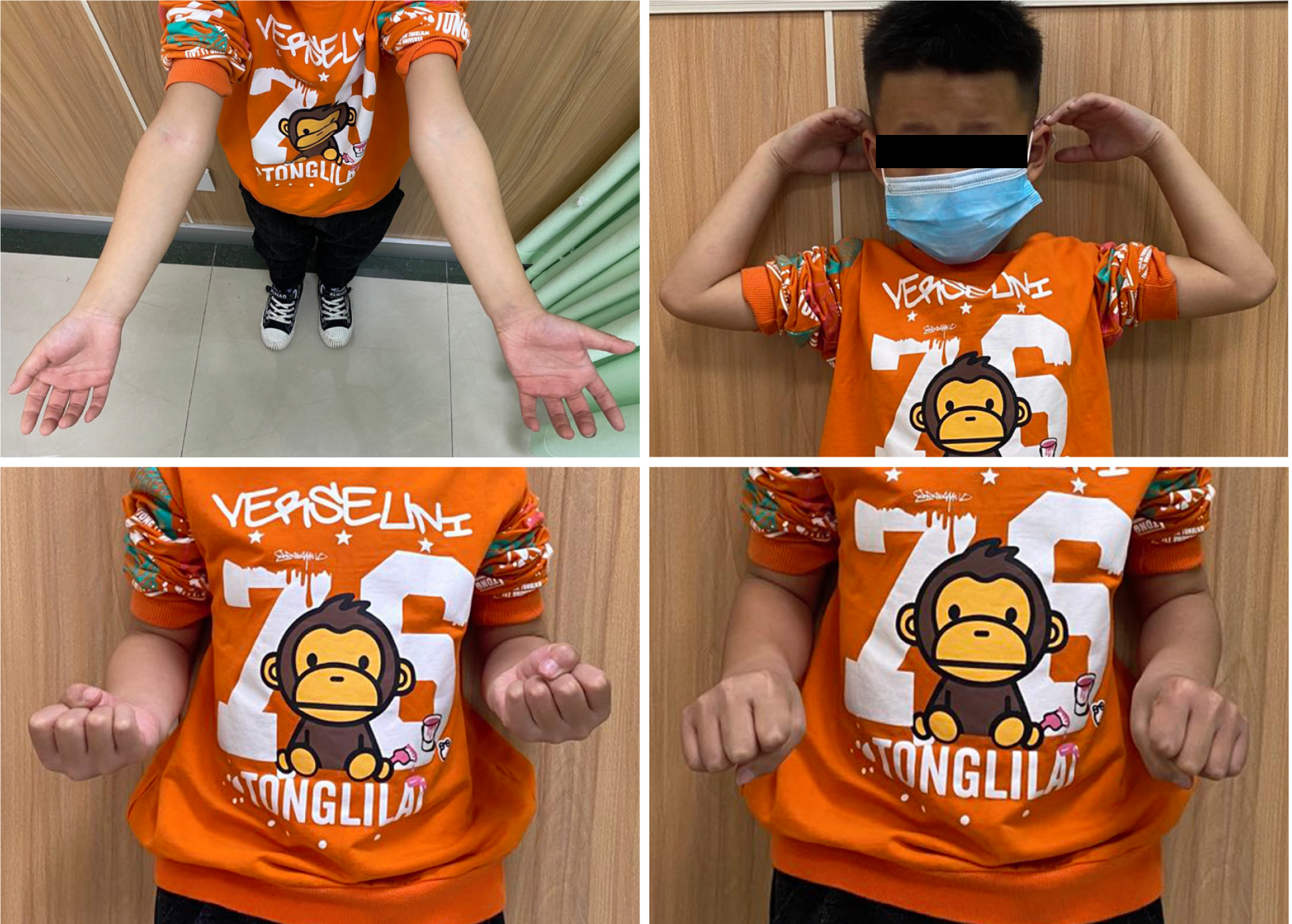
Figure 4 Functional results at the 5-year follow-up.
- Citation: Li ML, Zhou WZ, Li LY, Li QW. Monteggia type-I equivalent fracture in a fourteen-month-old child: A case report. World J Clin Cases 2021; 9(30): 9228-9235
- URL: https://www.wjgnet.com/2307-8960/full/v9/i30/9228.htm
- DOI: https://dx.doi.org/10.12998/wjcc.v9.i30.9228


