©The Author(s) 2021.
World J Clin Cases. Oct 26, 2021; 9(30): 9211-9217
Published online Oct 26, 2021. doi: 10.12998/wjcc.v9.i30.9211
Published online Oct 26, 2021. doi: 10.12998/wjcc.v9.i30.9211
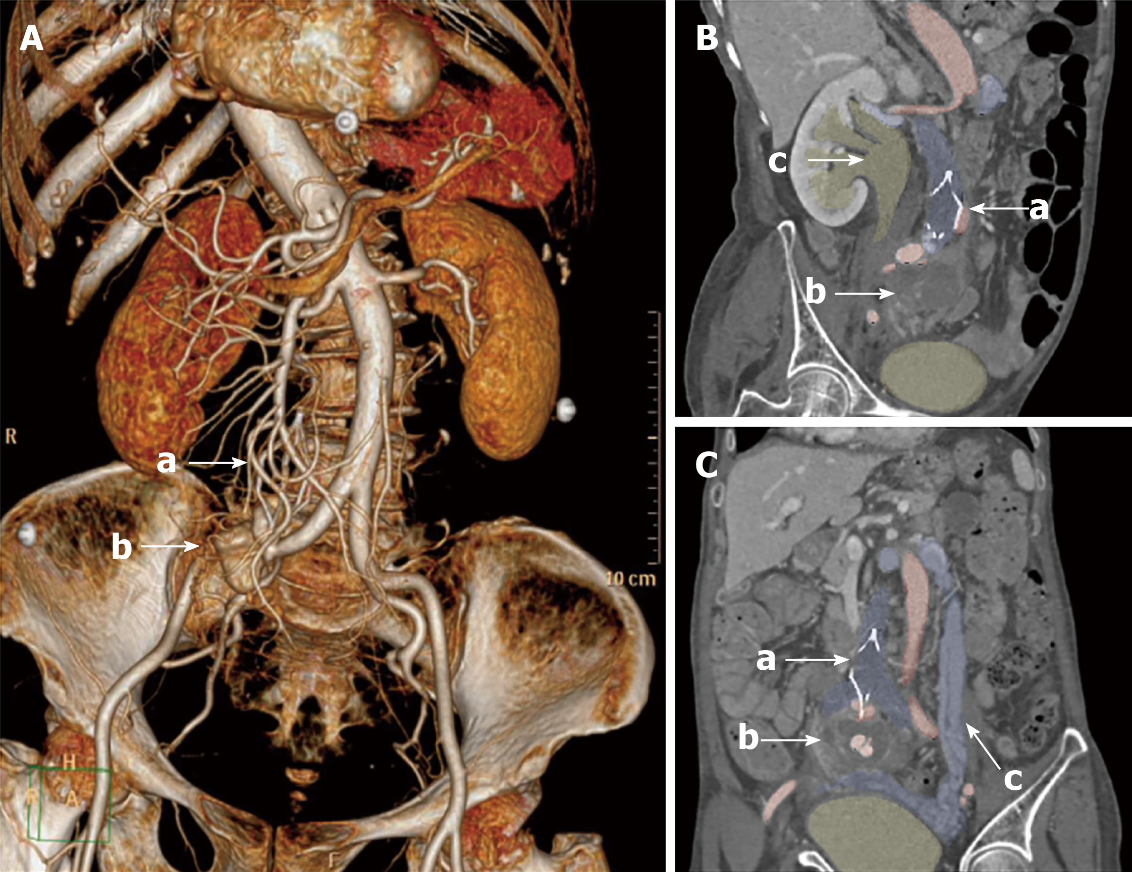
Figure 1 Preoperative computed tomography of patient.
A: Preoperative computed tomography indicated the retrieval hook of inferior vena cava filter (arrow a) penetrated into the right common iliac vein and artery causing pseudoaneurysm (arrow b). Scoliosis was also found in the computed tomography scan; B: The right ureteral was compressed by the pseudoaneurysm (arrow b) with ipsilateral hydronephrosis (arrow c); C: The infrarenal inferior vena cava and bilateral common iliac veins were occluded with significant pelvic varicosities (arrow d).
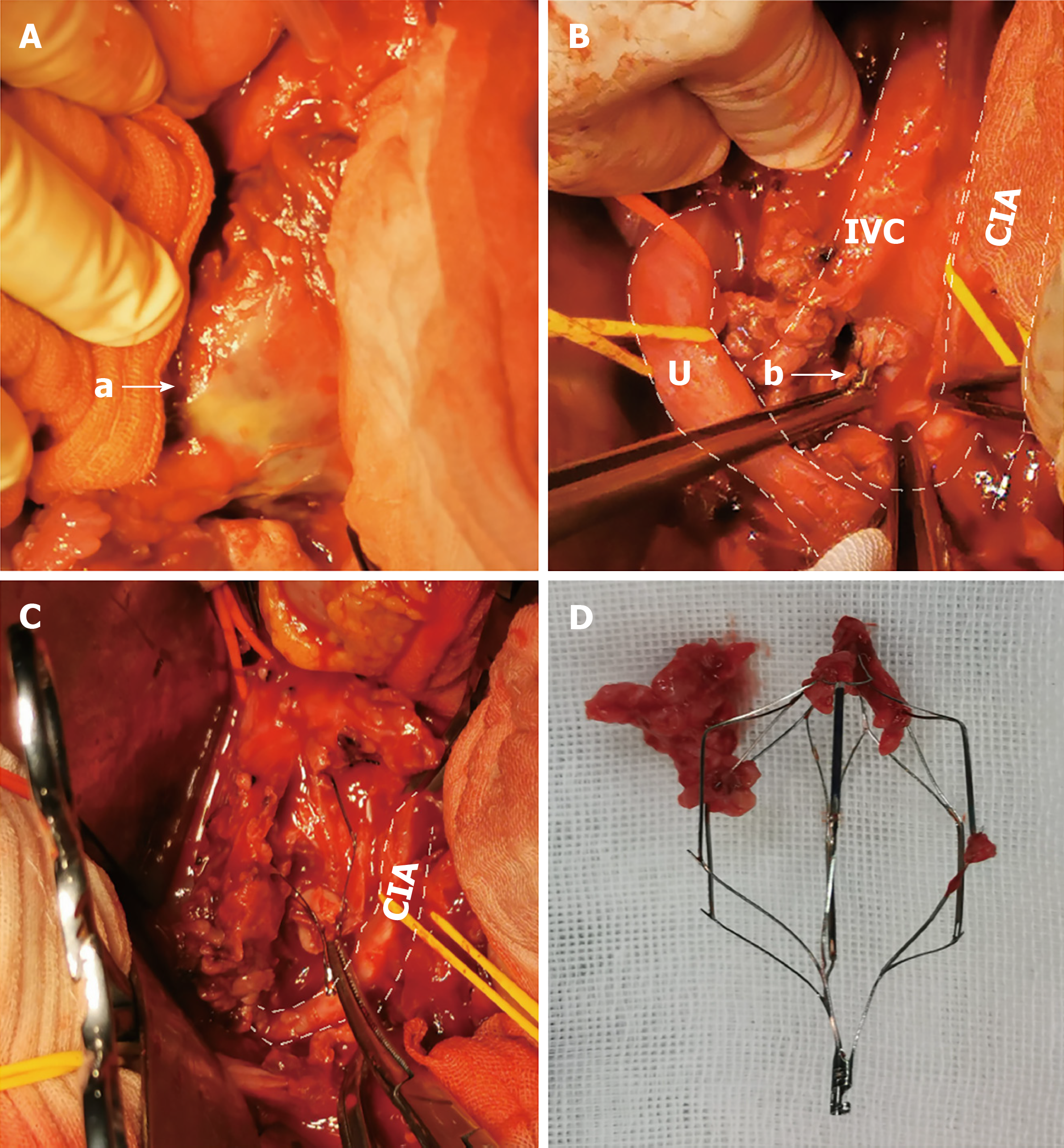
Figure 2 Intraoperative images of patients.
A: Right iliac pseudoaneurysm with abscess (arrow a); B: The indwelling inferior vena cava filter (arrow b) penetrated the right common iliac vein and artery, and the ureter was dilated; C: The inferior vena cava filter was embedded in the organized thrombus. The inferior vena cava was open, while no back-bleeding of lumbar veins were found; D: The double-basket Visee WXF-32 IVC filter. Image designations: U: Ureter; IVC: Inferior vena cava; CIA: Common iliac artery.
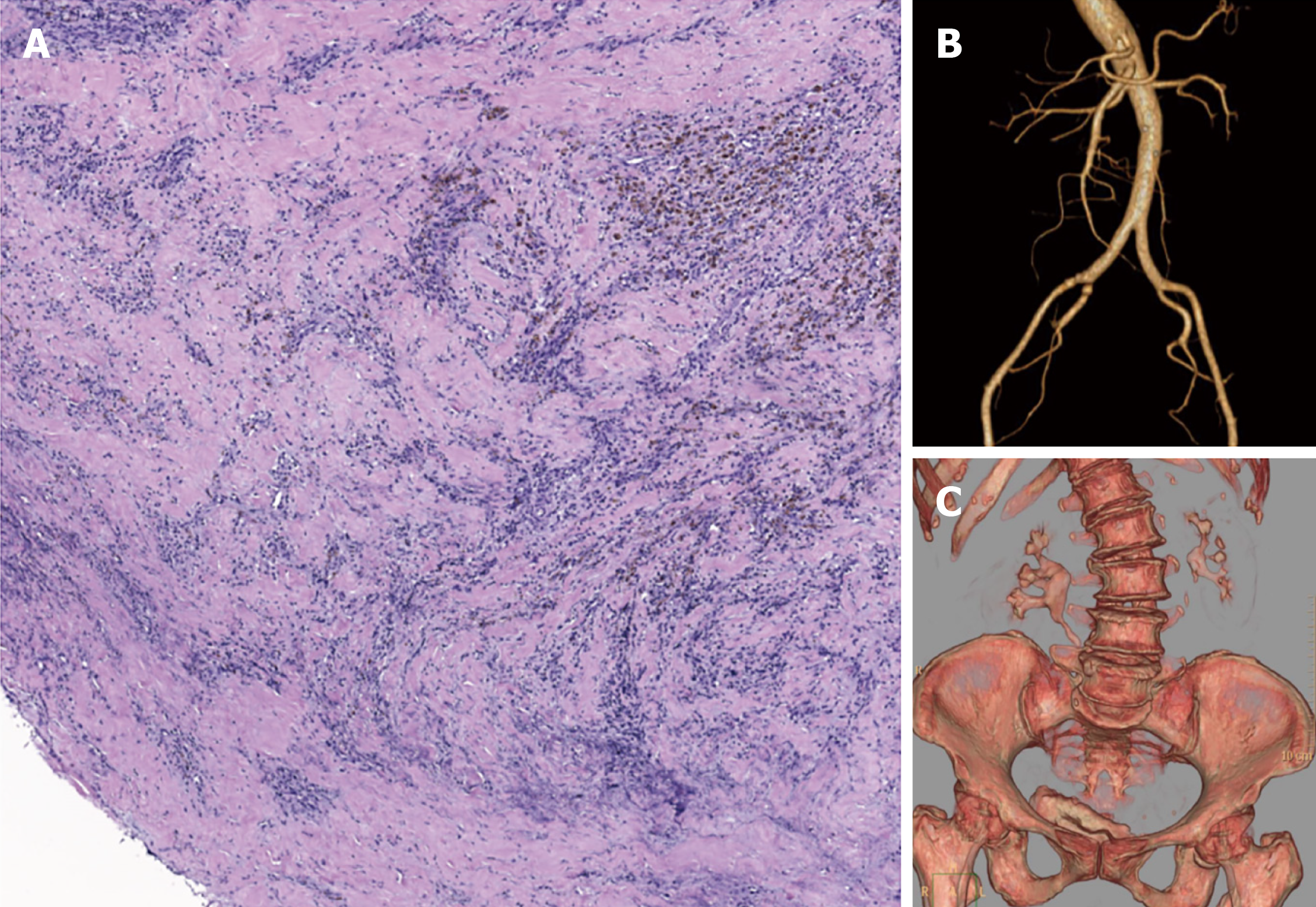
Figure 3 Surgical pathology and 6-mo follow-up computed tomography.
A: Organized thrombus of inferior vena cava with massive inflammatory cell infiltration; B: Computed tomography urography indicated mild right hydronephrosis; C: Computed tomography angiography indicated patency of bilateral iliac arteries.
- Citation: Weng CX, Wang SM, Wang TH, Zhao JC, Yuan D. Successful management of infected right iliac pseudoaneurysm caused by penetration of migrated inferior vena cava filter: A case report. World J Clin Cases 2021; 9(30): 9211-9217
- URL: https://www.wjgnet.com/2307-8960/full/v9/i30/9211.htm
- DOI: https://dx.doi.org/10.12998/wjcc.v9.i30.9211


