©The Author(s) 2019.
World J Clin Cases. Sep 6, 2019; 7(17): 2536-2541
Published online Sep 6, 2019. doi: 10.12998/wjcc.v7.i17.2536
Published online Sep 6, 2019. doi: 10.12998/wjcc.v7.i17.2536
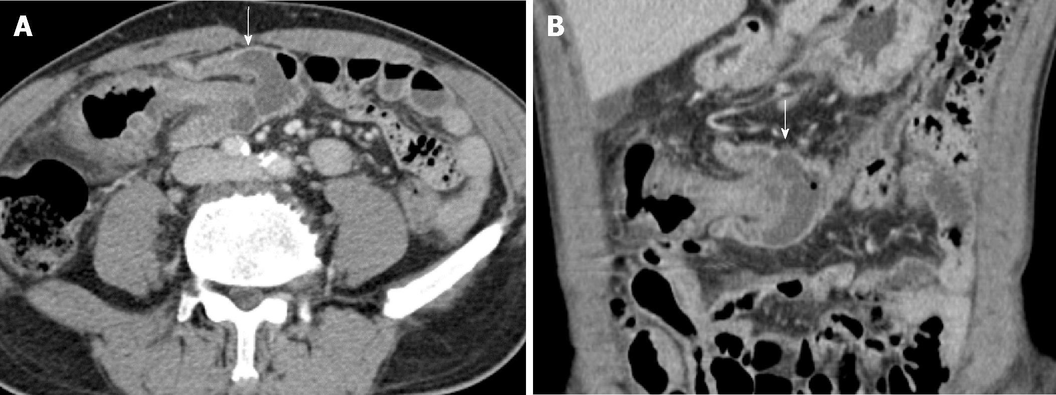
Figure 1 Pre-treatment abdominopelvic computed tomography.
A and B: Axial and coronal reformatted images of contrast-enhanced computed tomography show a colo-colonic intussusception with subepithelial edema in mid transverse colon (white arrows). There is no evidence of visible tumor at intussusception in computed tomography.
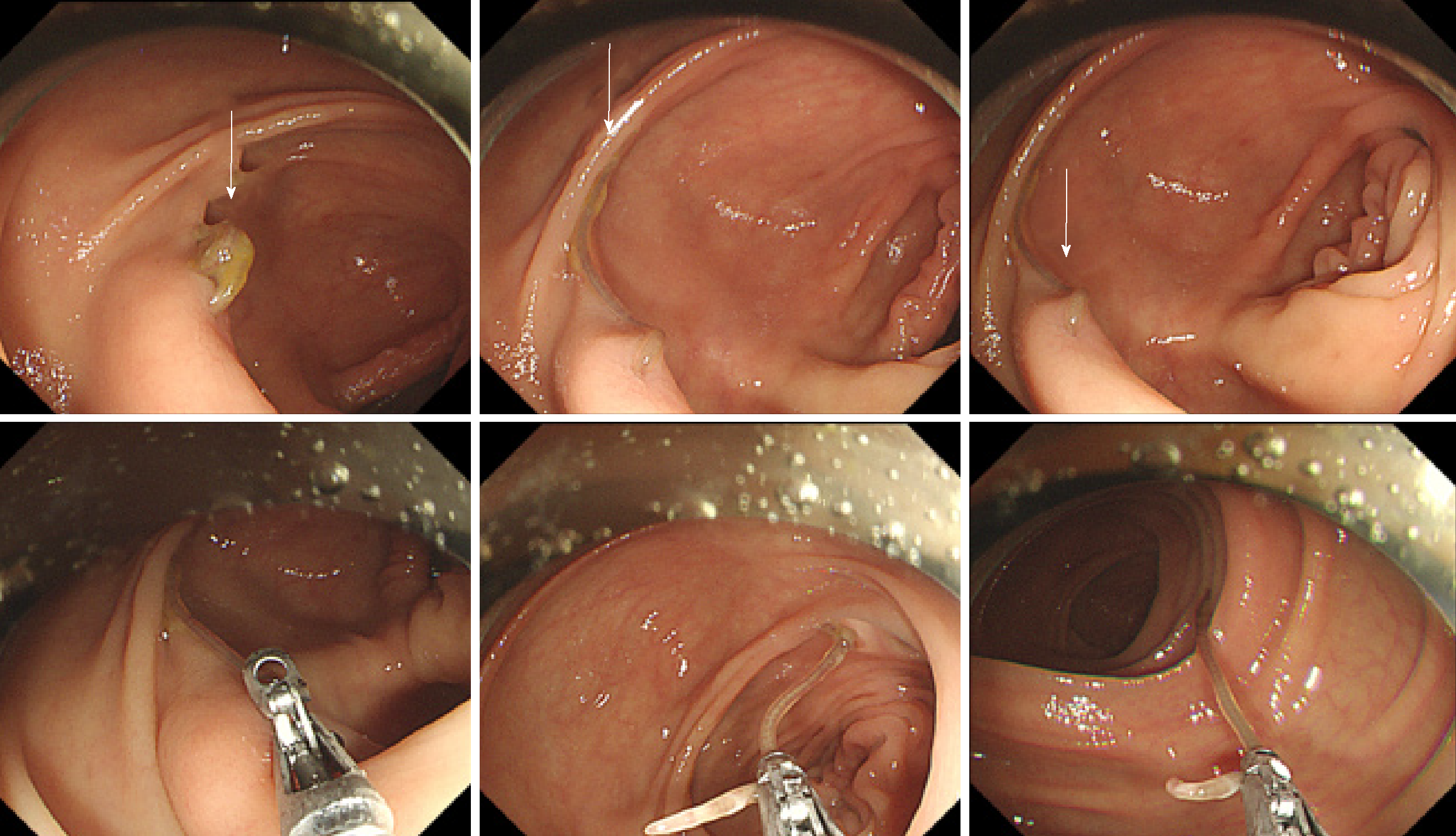
Figure 2 Series of endoscopic image of descending colon demonstrating biopsy forceps removing Anisakis simplex (arrow).
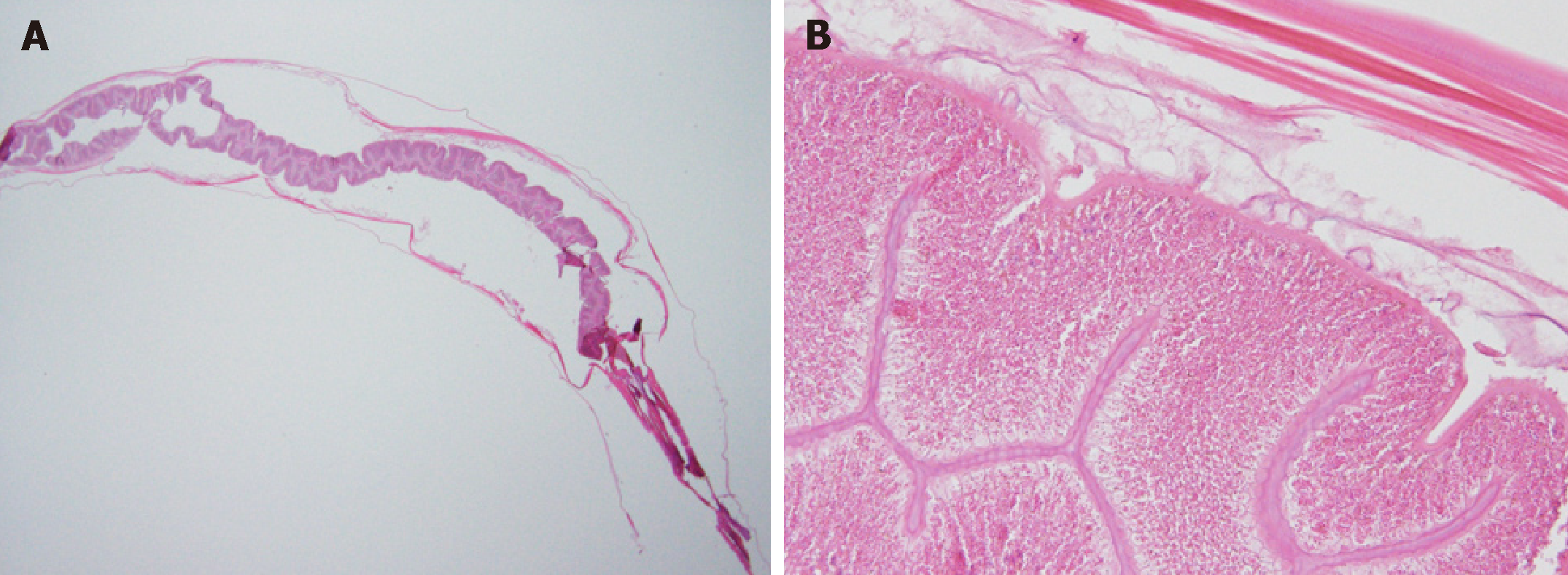
Figure 3 Histopathologic images of the Anisakis body.
Histopathological findings of the specimen. A: Longitudinal section of Anisakis larva. The nematode is 2.9 cm in length and 0.1 cm in width (hematoxylin and eosin stain; × 12.5); B: High magnification showing cuticle and intestine (× 200).
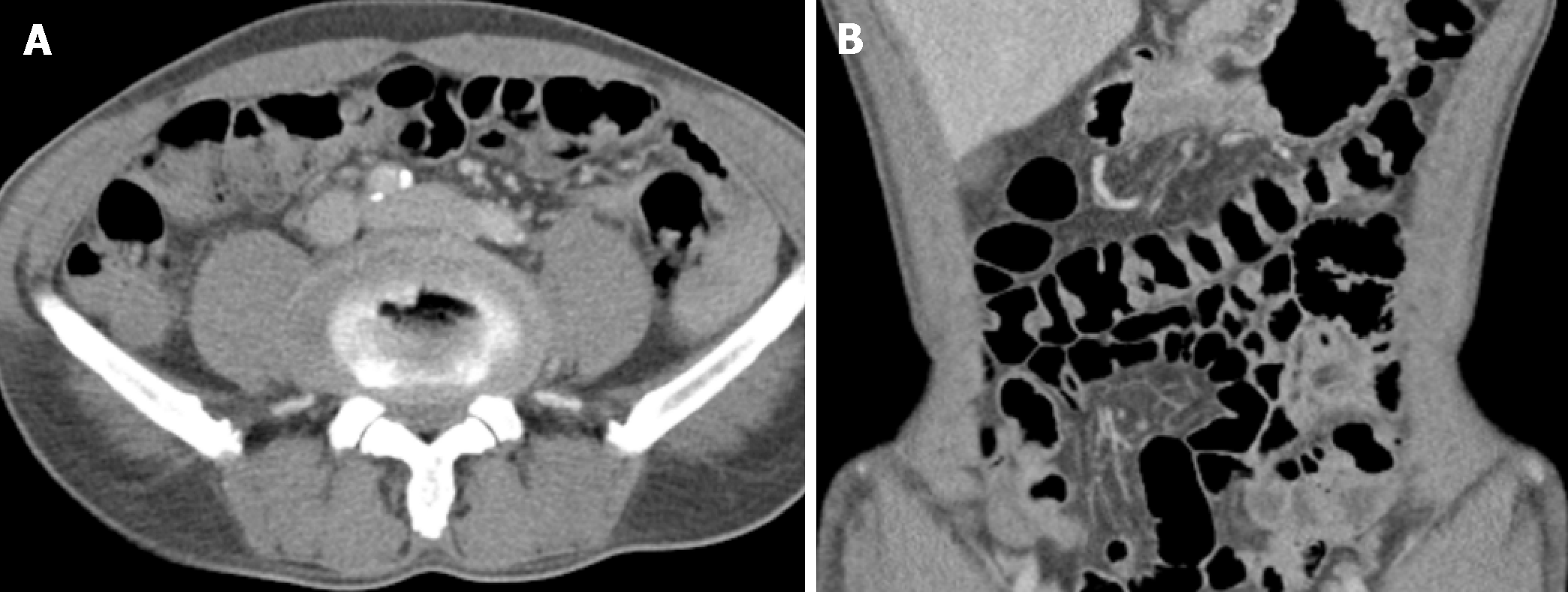
Figure 4 Post treatment abdominopelvic computed tomography.
A and B: After removal state of parasite, repeated computed tomography images demonstrate the disappearance of colo-colonic intussusception.
- Citation: Choi YI, Park DK, Cho HY, Choi SJ, Chung JW, Kim KO, Kwon KA, Kim YJ. Adult intussusception caused by colonic anisakis: A case report. World J Clin Cases 2019; 7(17): 2536-2541
- URL: https://www.wjgnet.com/2307-8960/full/v7/i17/2536.htm
- DOI: https://dx.doi.org/10.12998/wjcc.v7.i17.2536


