©The Author(s) 2025.
World J Clin Cases. Nov 16, 2025; 13(32): 111551
Published online Nov 16, 2025. doi: 10.12998/wjcc.v13.i32.111551
Published online Nov 16, 2025. doi: 10.12998/wjcc.v13.i32.111551
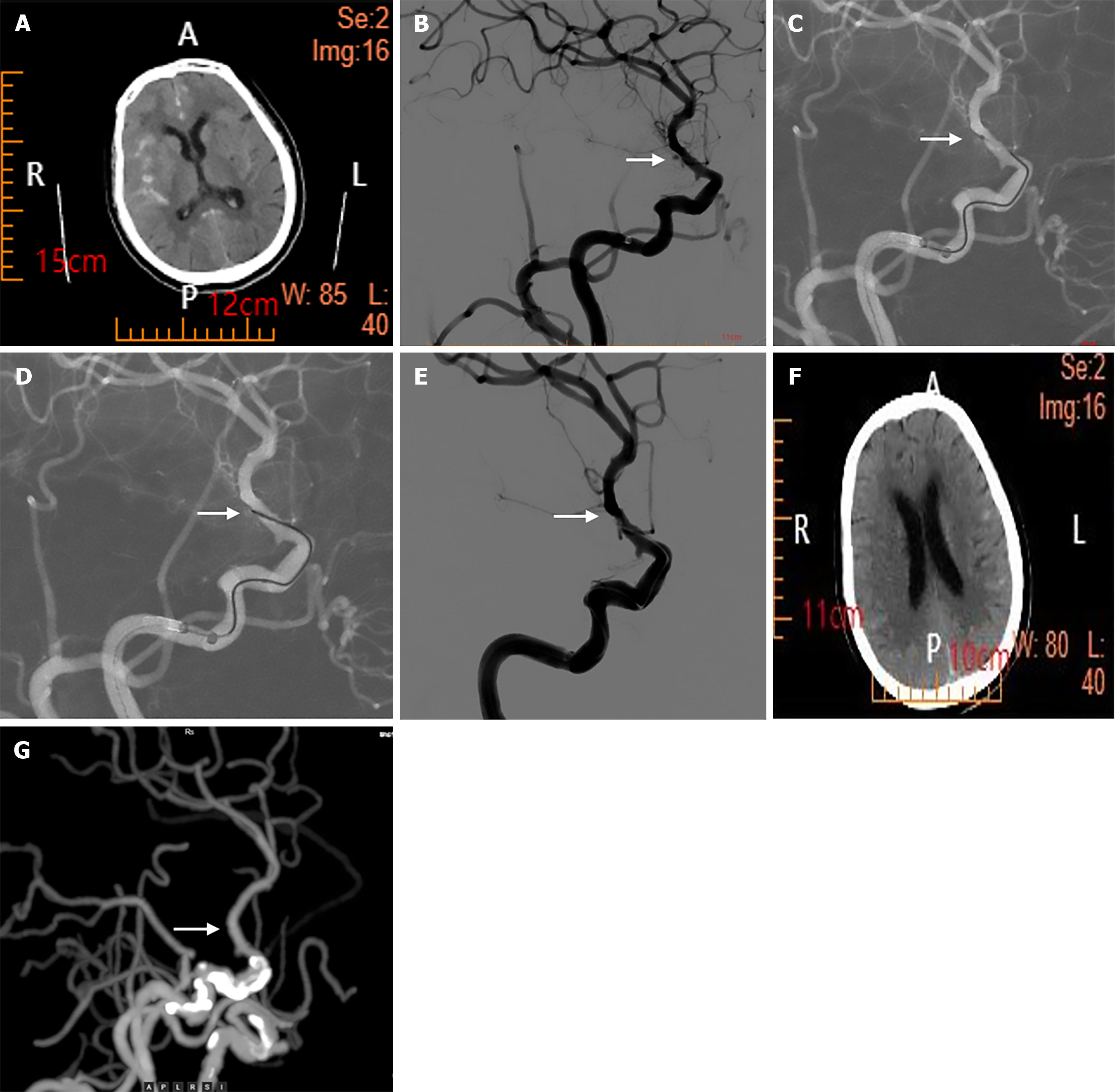
Figure 1 Computed tomography and angiography.
A: Computed tomography(CT) scan showing increased density in the posterior horns of the bilateral ventricles and partial sulci, diagnosed as a subarachnoid hemorrhage; B: Cerebral angiography image showing a saccular bulge in the choroidal segment of the internal carotid artery without contrast agent extravasation, which was speculated to be an arterial ampulla. In addition, a blister-like aneurysm ruptured and bled (arrow); C and D: Show that we used microguidewire electrocoagulation surgery to treat the aneurysm (arrow); E: Shows that after the third continuous electrocoagulation, revisualization of the aneurysm was absent (arrow); F: CT scan reexamination 3 days after surgery shows no obvious subarachnoid hemorrhage; G: CT angiography reexamination 3 months after surgery revealed no contrast agent extravasation in the original lesion, with a good prognosis (arrow).
- Citation: Zhang ZY, Zhang XY, Lu GZ, Wang SL, Hao JH, Zhang LY. Endovascular electrocoagulation for treating a blister-like microaneurysm with an extremely narrow neck: A case report. World J Clin Cases 2025; 13(32): 111551
- URL: https://www.wjgnet.com/2307-8960/full/v13/i32/111551.htm
- DOI: https://dx.doi.org/10.12998/wjcc.v13.i32.111551


