©The Author(s) 2025.
World J Clin Cases. Nov 6, 2025; 13(31): 112023
Published online Nov 6, 2025. doi: 10.12998/wjcc.v13.i31.112023
Published online Nov 6, 2025. doi: 10.12998/wjcc.v13.i31.112023
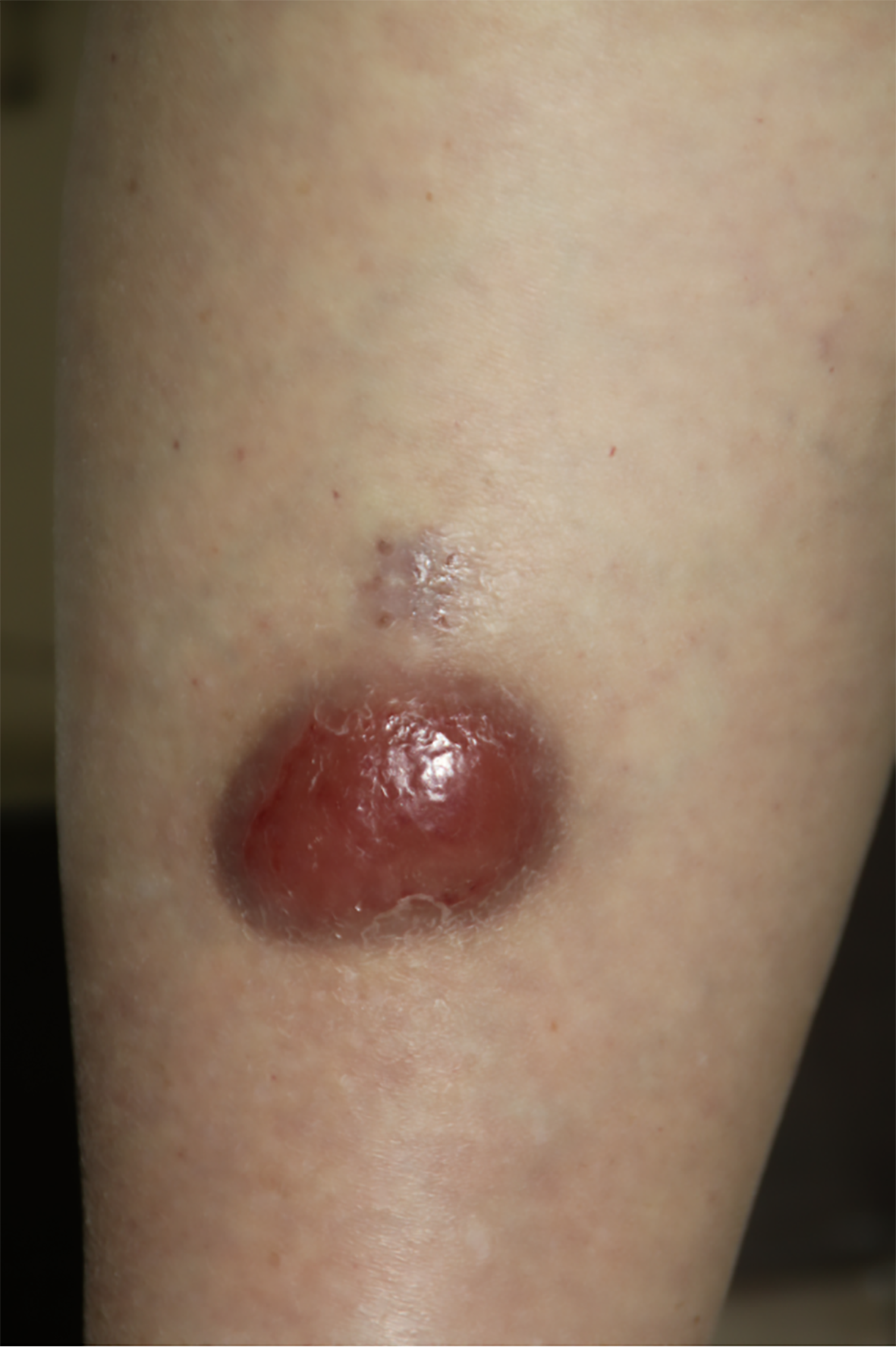
Figure 1 Clinical presentation.
A solitary, erythematous nodule measuring 6 cm × 4 cm on the anterior aspect of the right lower leg, demonstrating smooth surface morphology and firm consistency.
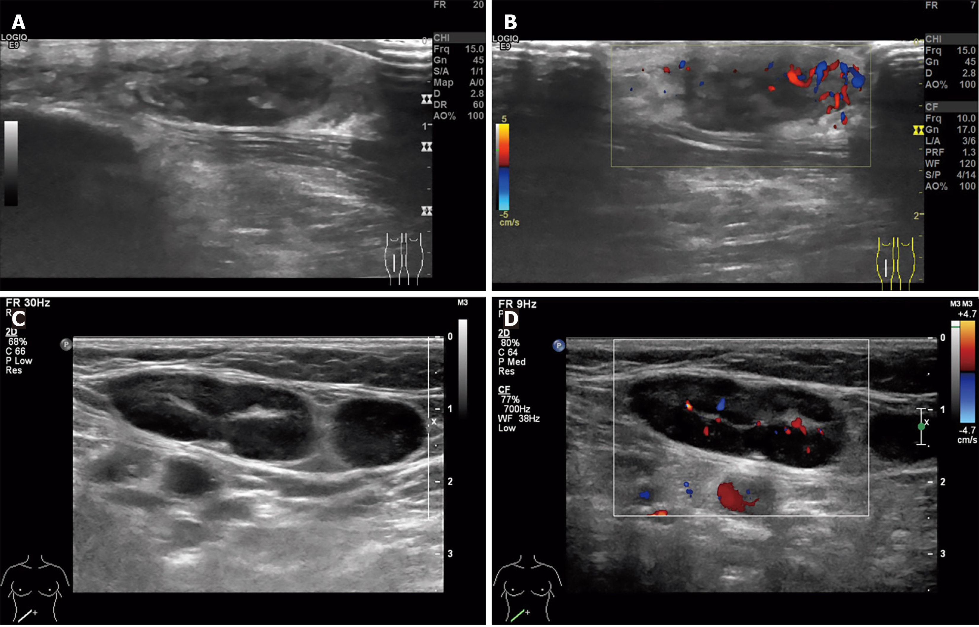
Figure 2 Ultrasound imaging of the right lower leg mass and right inguinal lymph nodes.
A: A hypoechoic subcutaneous mass (25 mm × 9 mm × 25 mm) in the mid-right lower leg, exhibiting irregular margins and an ill-defined shape; B: The adjacent tissue demonstrates increased echogenicity, accompanied by prominent peripheral vascularity; C: Multiple enlarged lymph nodes are observed in the right inguinal region, with the largest measuring 30 mm × 13 mm and showing asymmetrical cortical thickening; D: Intranodal blood flow appears as punctate/Linear vascular signals.
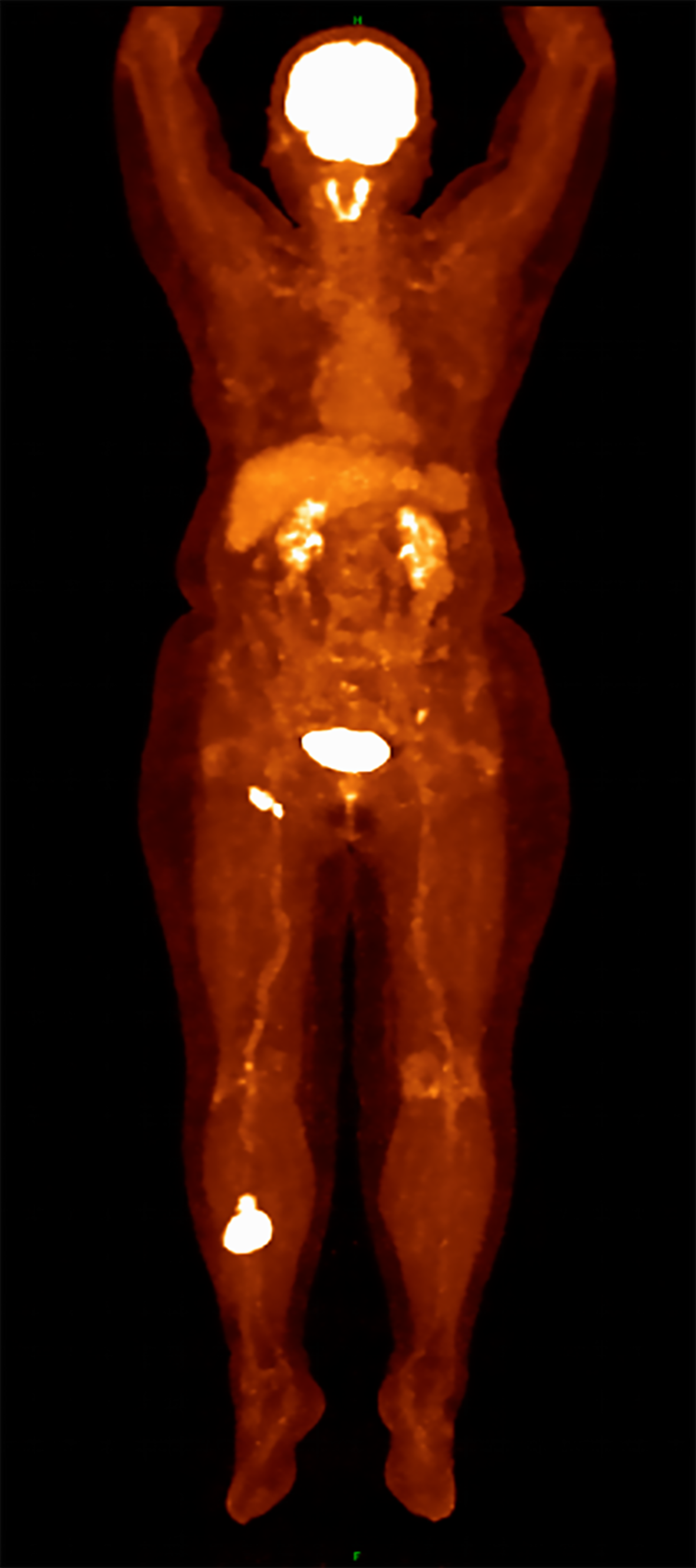
Figure 3 Positron emission tomography-computed tomography imaging findings.
Coronal view demonstrating fluorodeoxyglucose-avid soft tissue mass in the right lower extremity (SUVmax 15.95). Axial view showing multiple hypermetabolic right inguinal lymph nodes (SUVmax 10.54). Both are consistent with lymphomatous involvement.
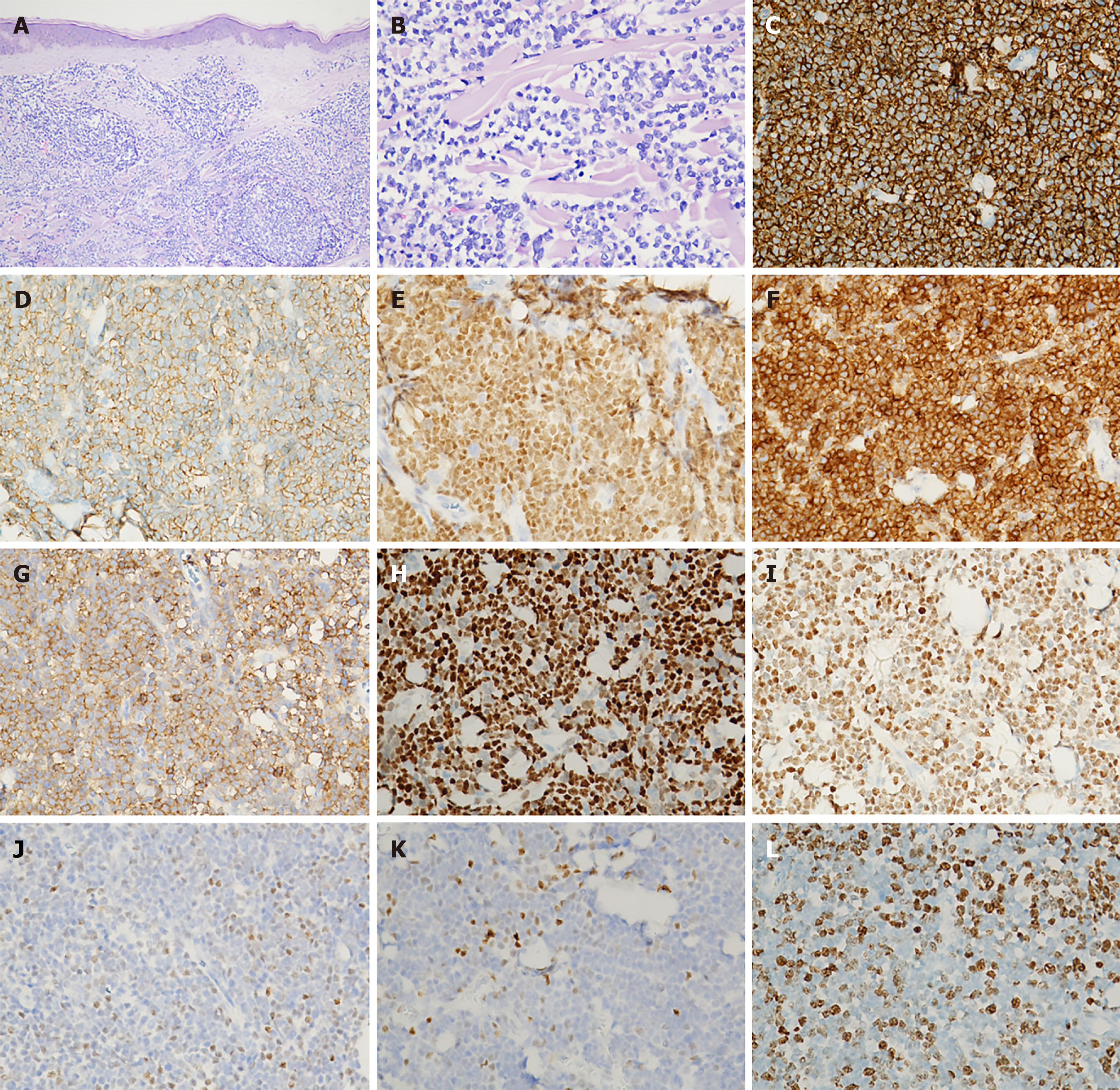
Figure 4 Histopathological and immunohistochemical results of the nodule.
A: Mild thickening of the epidermal stratum spinosum, with a clear separation between the epidermis and the dermis [hematoxylin & eosin (H&E), × 400]; B: The dermis and subcutaneous fat were densely infiltrated by lymphoid cells. These cells exhibited large, irregular nuclei with abnormal features and occasionally underwent cell division (H&E, × 400); C-I: Tumor cells were diffuse positive for (C) CD20 (× 400), (D) CD79a (× 400), (E) PAX5 (× 400), (F) Bcl-2 (× 400), (G) CD5 (× 400), (H) Cyclin D1 (× 400), and (I) SOX11 (× 400); J and K: C-MYC shows scattered small focal areas of positivity (× 400); L: Ki-67 hotspots exhibit approximately 60% positivity (× 400).
- Citation: Zhou PY, Chen W, Wang L. Mantle cell lymphoma presenting primarily as cutaneous lesions: A case report. World J Clin Cases 2025; 13(31): 112023
- URL: https://www.wjgnet.com/2307-8960/full/v13/i31/112023.htm
- DOI: https://dx.doi.org/10.12998/wjcc.v13.i31.112023


