©The Author(s) 2025.
World J Clin Cases. Oct 26, 2025; 13(30): 110330
Published online Oct 26, 2025. doi: 10.12998/wjcc.v13.i30.110330
Published online Oct 26, 2025. doi: 10.12998/wjcc.v13.i30.110330
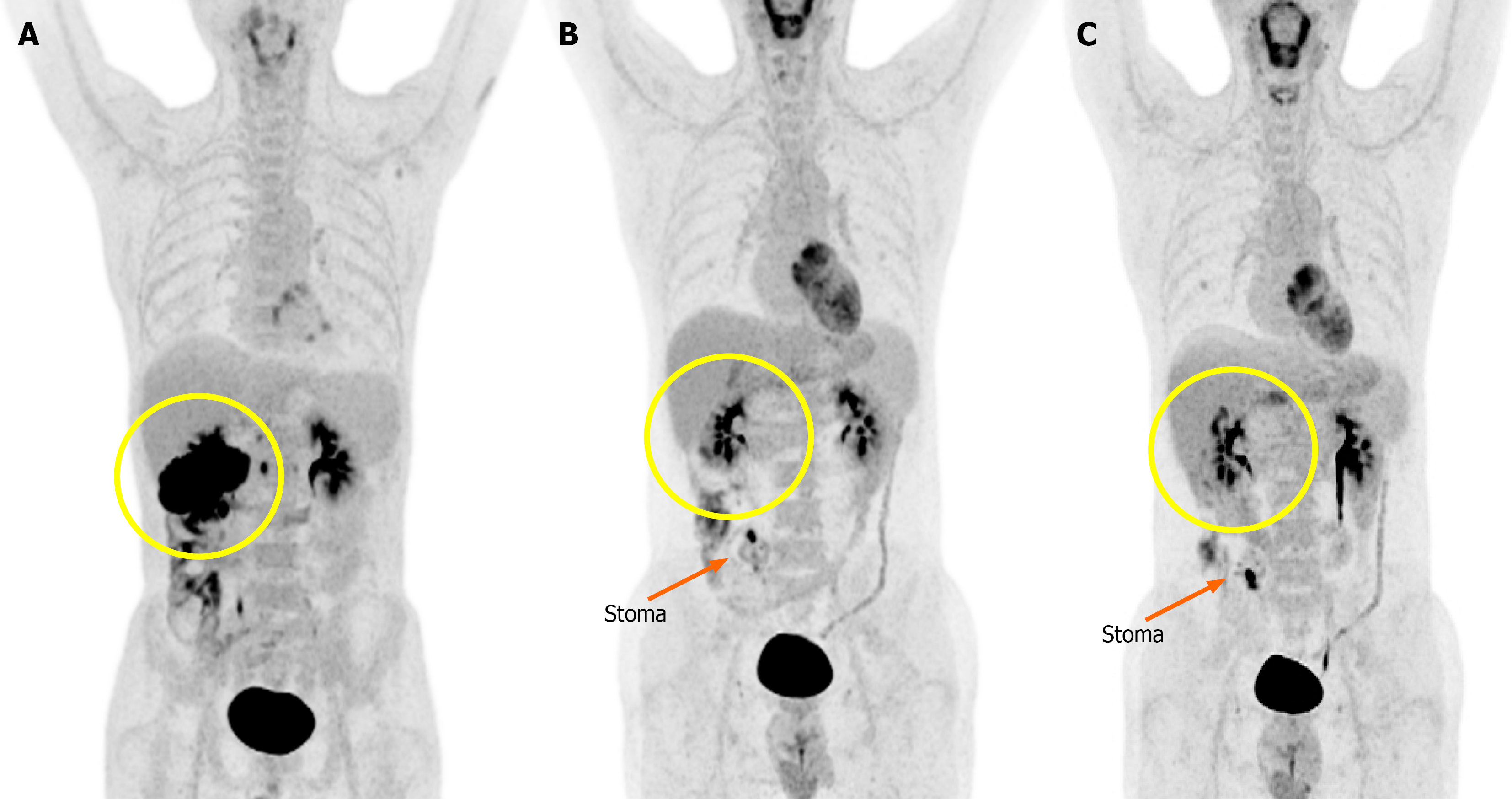
Figure 1 Serial fluorodeoxyglucose positron emission tomography-computed tomography.
A: Baseline imaging showed intense fluorodeoxyglucose uptake in the hepatic flexure of the colon, multiple hypermetabolic liver lesions, and suspected peritoneal involvement; B: After 7 months of pembrolizumab treatment, a marked reduction in fluorodeoxyglucose uptake at the primary site and metastatic lesions was observed, indicating a significant treatment response; C: After completion of pembrolizumab treatment, complete resolution of prior hypermetabolic activity at the primary tumor site was observed. A new focal uptake was seen near the stoma and gallbladder bed, suggestive of either post-treatment inflammation or possible recurrence. The primary tumor site is marked with a yellow circle, and the stoma location is indicated by a orange arrow.
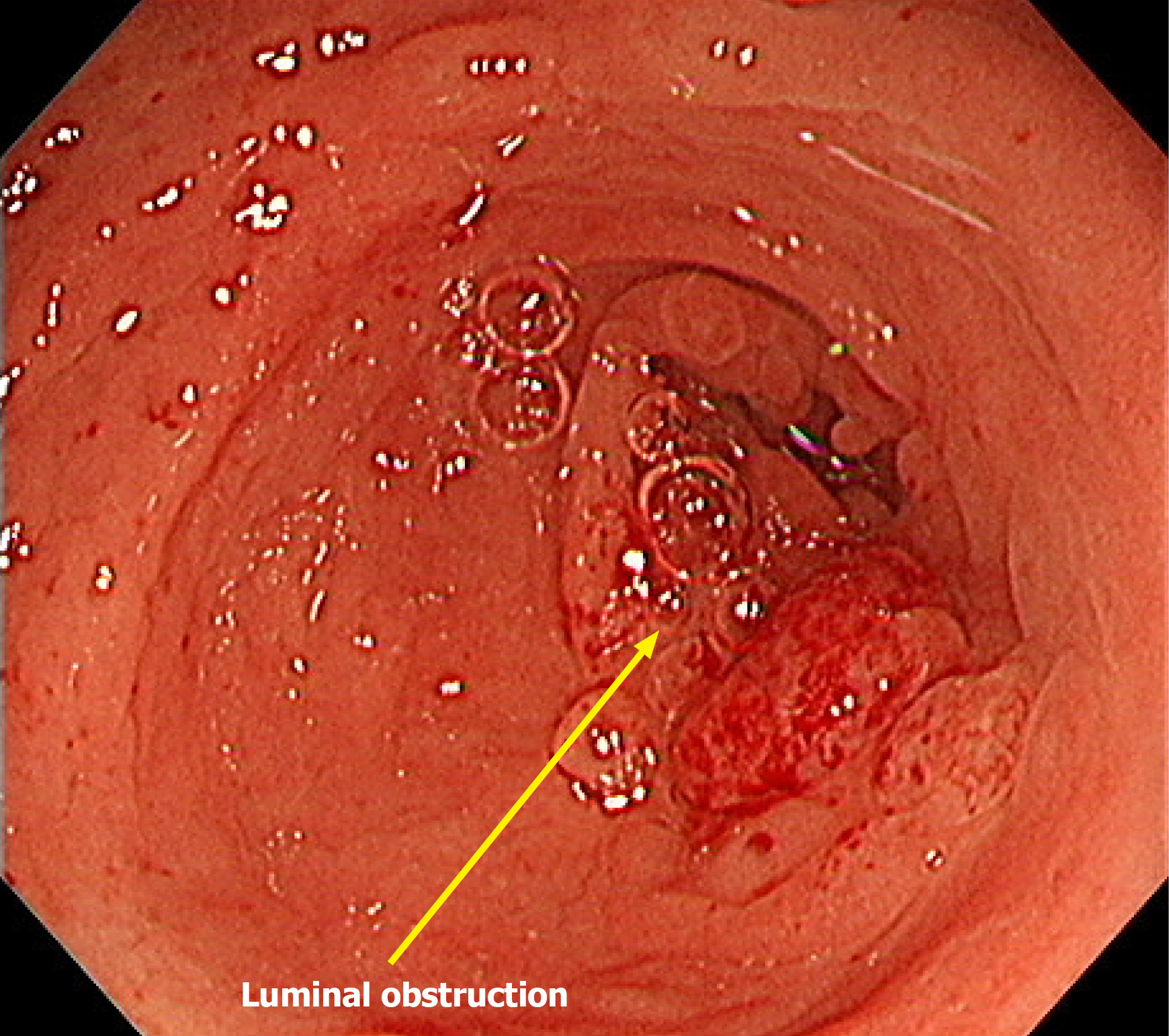
Figure 2 Colonoscopy performed after receiving immunotherapy for 17 months.
The scope was inserted through the anus and advanced to the transverse colon, but further passage was not possible due to luminal obstruction at the tumor site.
Figure 3 Axial Abdominopelvic computed tomography during the treatment course.
A: Baseline imaging revealed a large soft tissue mass in the proximal transverse colon with invasion into adjacent structures; B: After 6 months of pembrolizumab treatment, a marked reduction in tumor size was revealed; C: After 20 months of pembrolizumab treatment, radiologic complete response of the primary lesion was observed. The yellow circles indicate the location of the primary tumor across the treatment timeline. The orange arrow indicates the primary tumor in the colon with suspected invasion into the pancreas.
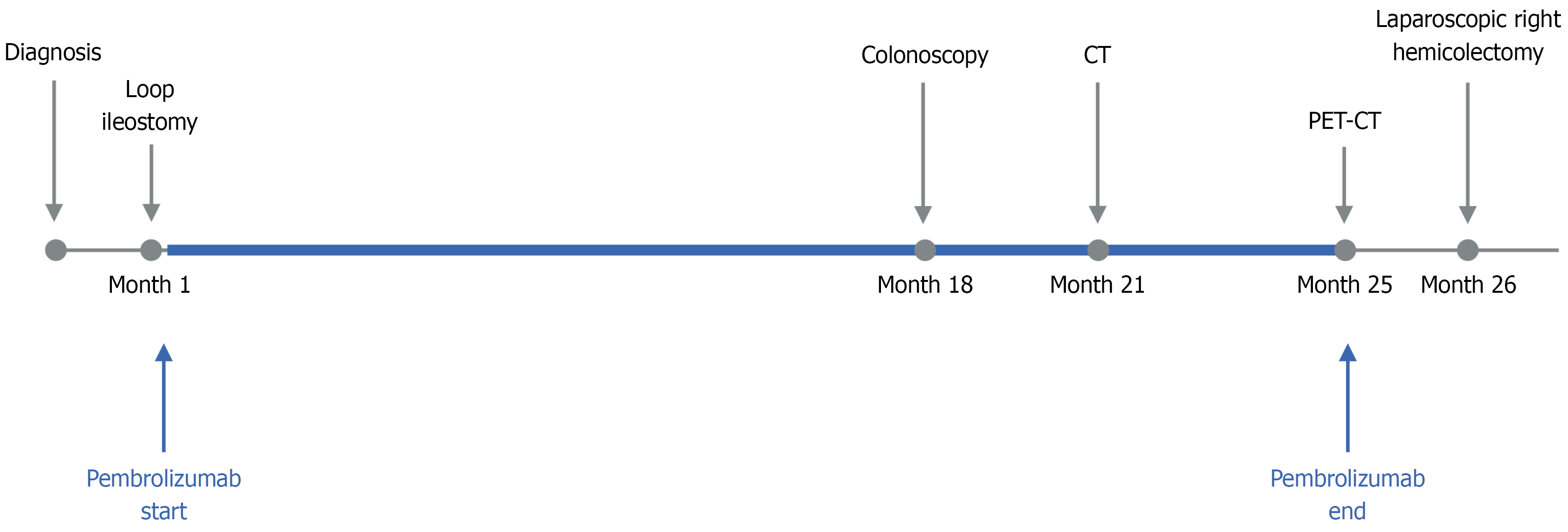
Figure 4 Clinical timeline of diagnosis, treatment, and surgery in a patient.
Clinical timeline showing key events from diagnosis to surgery. Pembrolizumab treatment began after loop ileostomy and continued for 36 cycles over approximately 2 years. Colonoscopy (Month 17), computed tomography (Month 21), and positron emission tomography/computed tomography (Month 25) were performed during follow-up, culminating in a laparoscopic right hemicolectomy at Month 26.
- Citation: Lee C, Kim MH, Choi ET, Park IJ, Lim SB, Yoon YS, Kim CW, Lee JL, Park EJ. Spontaneous colonic transection following pathologic complete response to pembrolizumab in high microsatellite instability colorectal cancer: A case report and review of literature. World J Clin Cases 2025; 13(30): 110330
- URL: https://www.wjgnet.com/2307-8960/full/v13/i30/110330.htm
- DOI: https://dx.doi.org/10.12998/wjcc.v13.i30.110330


