©The Author(s) 2025.
World J Clin Cases. Oct 26, 2025; 13(30): 110314
Published online Oct 26, 2025. doi: 10.12998/wjcc.v13.i30.110314
Published online Oct 26, 2025. doi: 10.12998/wjcc.v13.i30.110314
Figure 1 Preoperative evaluation.
A: Diffuse distension of the small and large intestines on axial-view abdominal computed tomography; B: Long segmental thickening of the rectosigmoid colonic wall and perirectal fat stranding with a clear vesicorectal fat plane (white arrow) on sagittal-view abdominal computed tomography; C: Obstructed lumen with mucosal swelling (white circle) located up to 5 cm from the anal verge on colonoscopy.
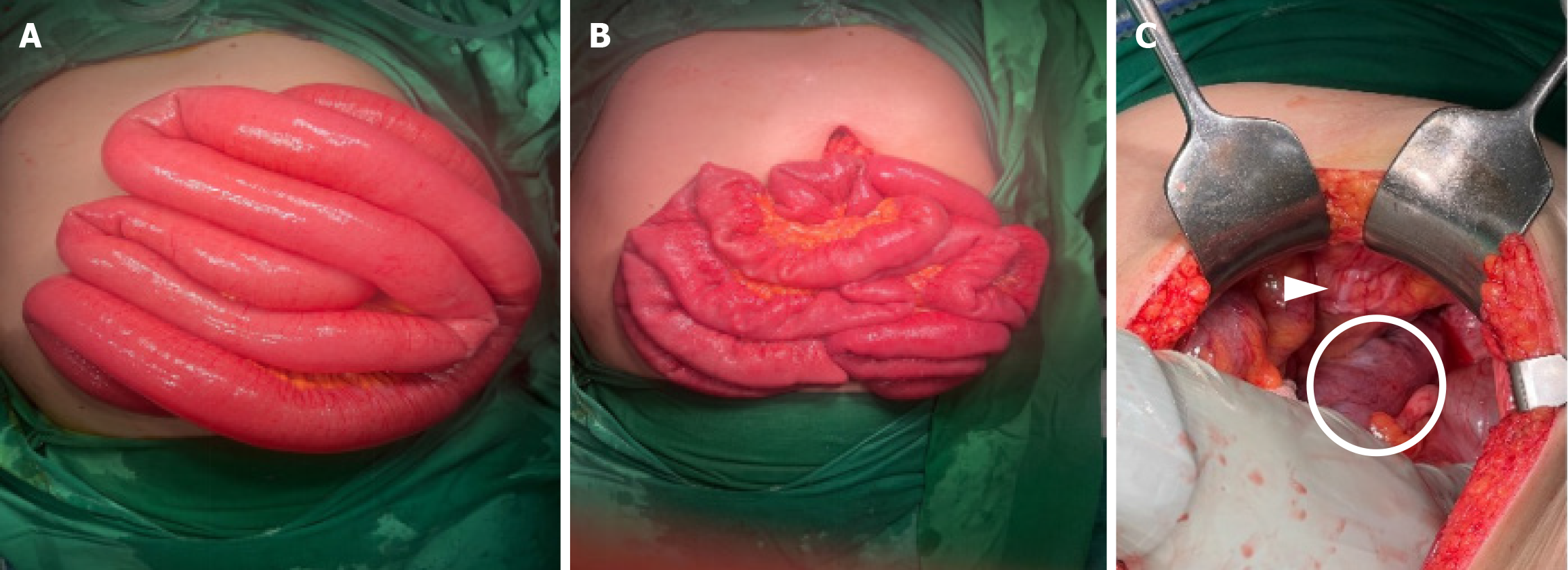
Figure 2 Intraoperative images.
A: Distension of the intestine from the ligament of Treitz to the ileocecal valve and ascending colon; B: No apparent adhesion or obstructive lesion observed after manual decompression; C: Distended sigmoid colon with marked inflammatory changes in the colonic wall extending from the sigmoid colon to the rectum (white circle), without any evidence of urinary bladder invasion (white arrowhead).
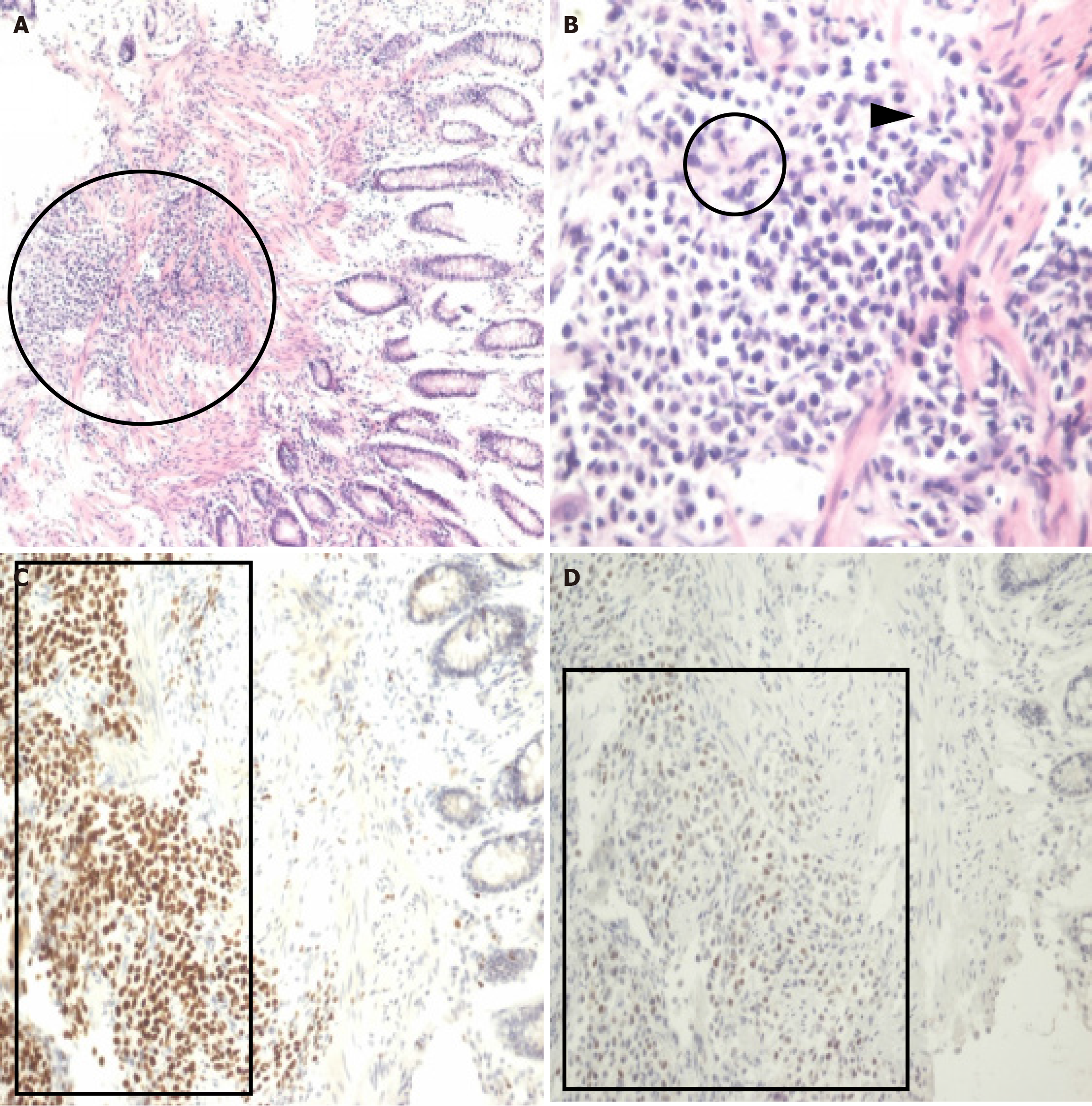
Figure 3 Histopathologic findings.
A: Discohesive tumor cells infiltrating the muscularis mucosa of the colon (black circle; hematoxylin–eosin staining, × 100); B: Plasmacytoid cells with abundant eosinophilic cytoplasm (black circle) and eccentric nuclei (black arrowhead; hematoxylin–eosin staining, × 400); C: Immunohistochemical staining for GATA-3 indicated nuclear positivity (black box; immunoperoxidase method, × 200); D: P63 staining revealed nuclear positivity (black box; immunoperoxidase method, × 200).
- Citation: Chen GY, Tsai HL, Chen PJ, Chen YC, Wang JY, Lin CY, Yin HL. Plasmacytoid urothelial carcinoma of the urinary bladder with colorectal metastasis and mimicking colitis: A case report. World J Clin Cases 2025; 13(30): 110314
- URL: https://www.wjgnet.com/2307-8960/full/v13/i30/110314.htm
- DOI: https://dx.doi.org/10.12998/wjcc.v13.i30.110314


