©The Author(s) 2024.
World J Clin Cases. Mar 6, 2024; 12(7): 1333-1338
Published online Mar 6, 2024. doi: 10.12998/wjcc.v12.i7.1333
Published online Mar 6, 2024. doi: 10.12998/wjcc.v12.i7.1333
Figure 1 Computed tomography images.
A: Occupational lesion of the posterior segment of the upper lobe of the left lung; B: Prostate enlarged, surface not smooth and indistinctly demarcated from bladder on the anterior surface; wall of rectum markedly thickened.
Figure 2 Rectal biopsy: Heterogeneous cells with extrusion injury are seen in the mucosal interstitium.
A: Original magnification: × 100; B: Original magnification: × 100.
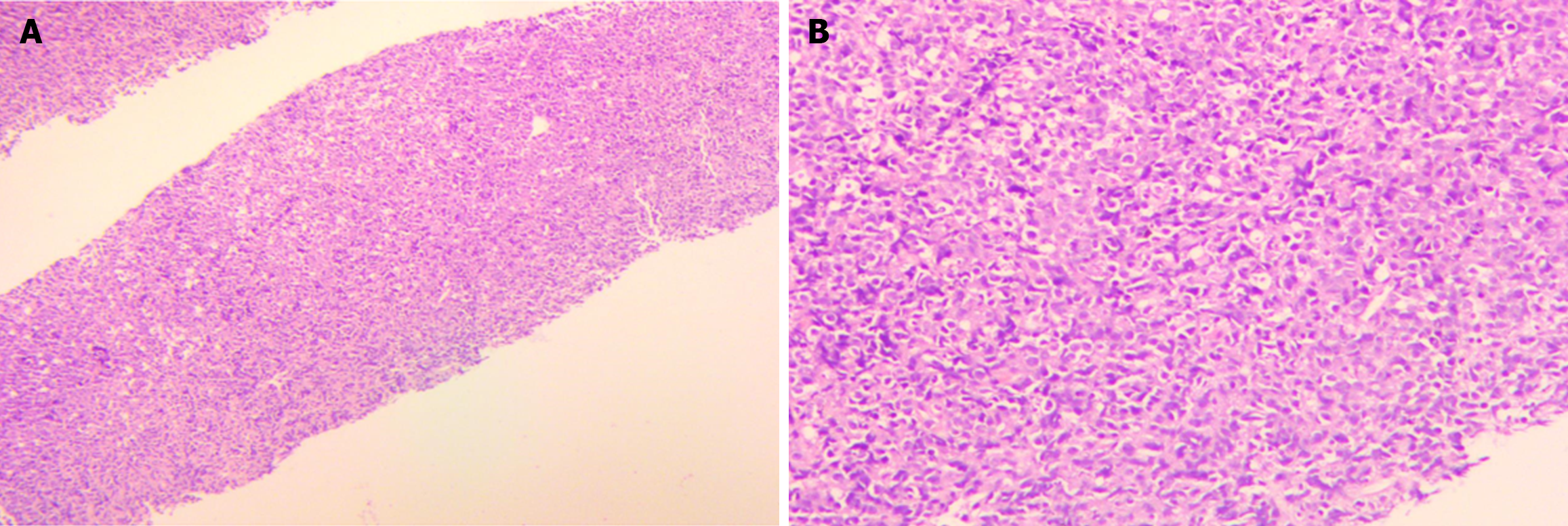
Figure 3 Lung biopsy: Malignant tumour, consider non-Hodgkin's lymphoma.
A: Original magnification: × 40; B: Original magnification: × 100.
Figure 4 Tumor changes after 2 cycles of chemotherapy with the CHOP regimen.
A: Changes of chest lesions after chemotherapy; B: Changes of rectal lesions after chemotherapy.
- Citation: Liang JL, Bu YQ, Peng LL, Zhang HZ. Heterochronous multiple primary prostate cancer and lymphoma: A case report. World J Clin Cases 2024; 12(7): 1333-1338
- URL: https://www.wjgnet.com/2307-8960/full/v12/i7/1333.htm
- DOI: https://dx.doi.org/10.12998/wjcc.v12.i7.1333


