Copyright
©The Author(s) 2024.
World J Clin Cases. Dec 16, 2024; 12(35): 6834-6839
Published online Dec 16, 2024. doi: 10.12998/wjcc.v12.i35.6834
Published online Dec 16, 2024. doi: 10.12998/wjcc.v12.i35.6834
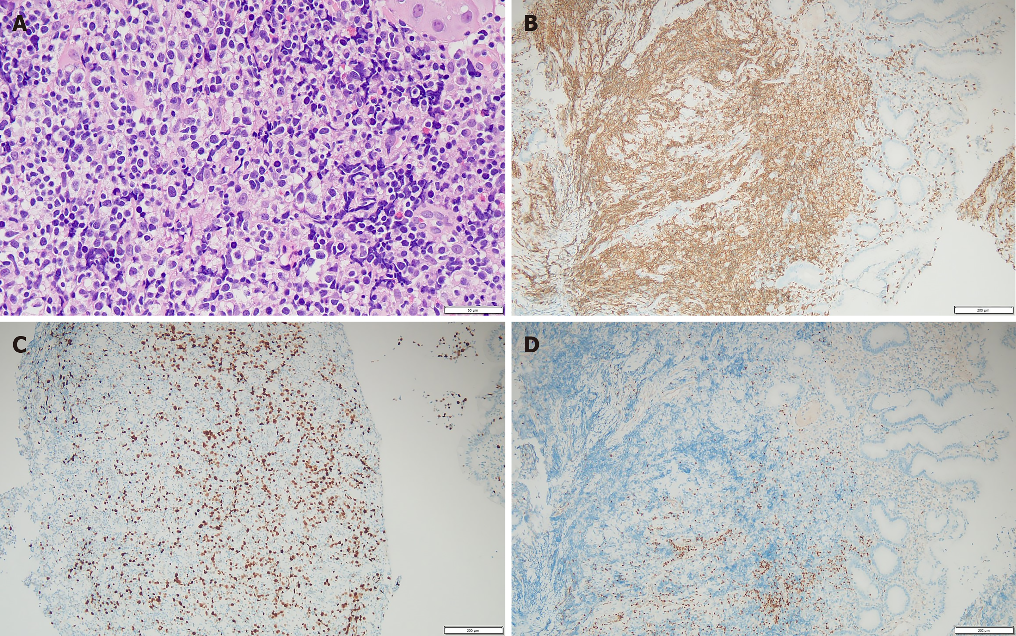
Figure 1 Histopathological findings.
A: Hematoxylin and eosin staining of stomach of magnification of 400 ×; B-D: Results of immunohistochemistry revealing the tumor was CD3 positive, Ki-67 positive, and PAX positive (magnification 100 ×).
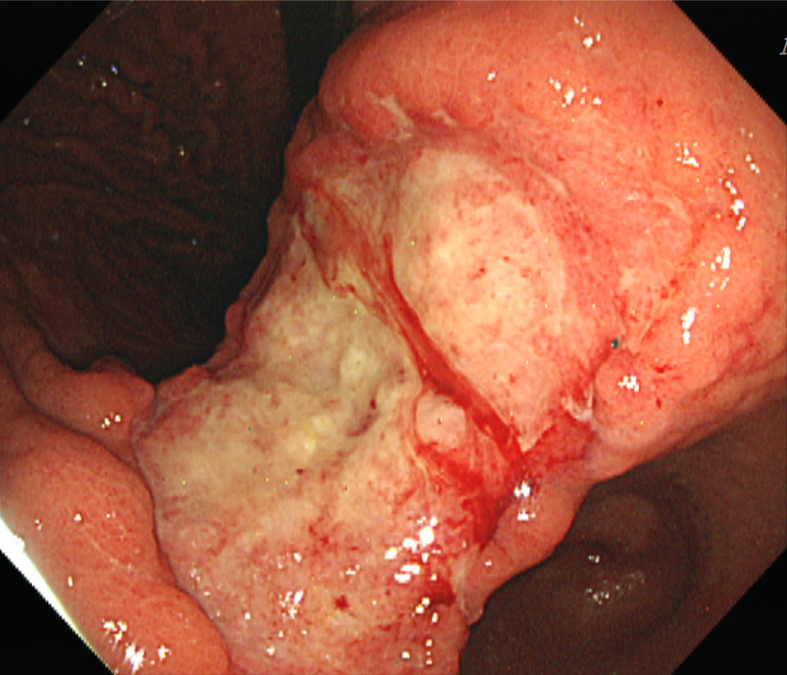
Figure 2 Endoscopic finding.
A huge irregular ulcerative lesion in the gastric cardia.
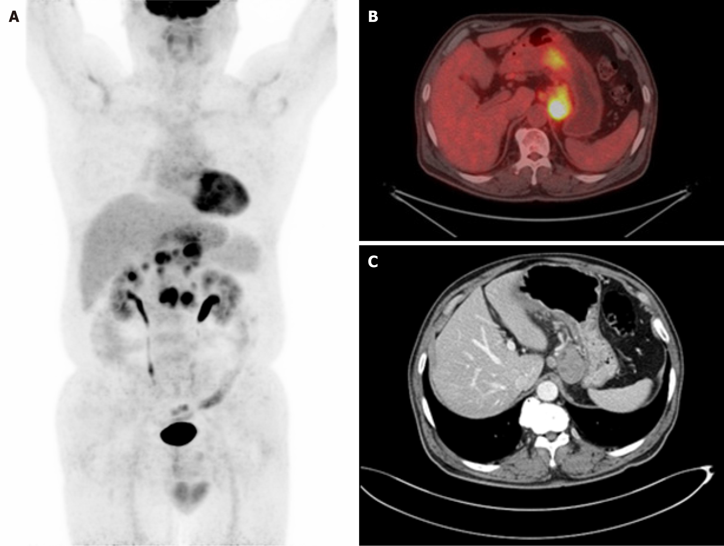
Figure 3 Underwent imaging.
A and B: Positron emission tomography showed hypermetabolic mass located in the lower body; C: Computed tomography showed diffuse wall thickening of the posterior lesser curvature of the gastric body with metastatic nodes.
- Citation: Jang HR, Lee K, Lim KH. Rare primary gastric peripheral T-cell lymphoma not otherwise specified: A case report. World J Clin Cases 2024; 12(35): 6834-6839
- URL: https://www.wjgnet.com/2307-8960/full/v12/i35/6834.htm
- DOI: https://dx.doi.org/10.12998/wjcc.v12.i35.6834


