©The Author(s) 2024.
World J Clin Cases. Sep 6, 2024; 12(25): 5805-5813
Published online Sep 6, 2024. doi: 10.12998/wjcc.v12.i25.5805
Published online Sep 6, 2024. doi: 10.12998/wjcc.v12.i25.5805
Figure 1 Pathology of puncture.
A: Left lung; B: Pelvic mass.
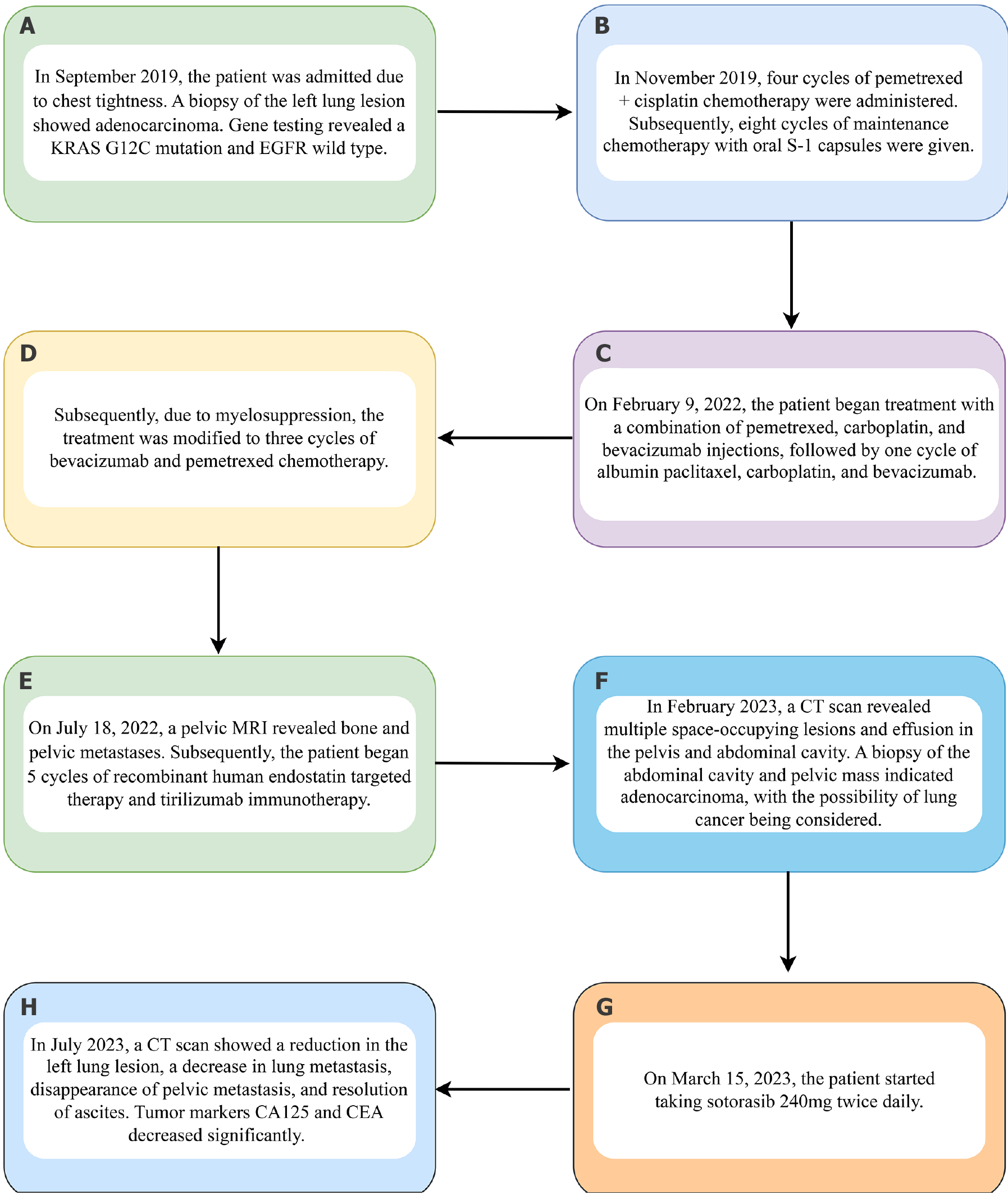
Figure 2 Treatment flow chart.
A: September 2019: Diagnosis with lung cancer and type of gene mutation; B: November 2019: Four cycles of pemetrexed + cisplatin, followed by eight oral tegafur maintenance cycles; C: Adjustment of medication due to myelosuppression; D: Chemotherapy and targeted therapy on 9 February 2022; E: Received immunotherapy on 18 July 2022; F: Metastasis occurred again in February 2023; G: In July 2023, the tumor regressed and the patient improved; H: Sotorasib started on 15 March 2023.
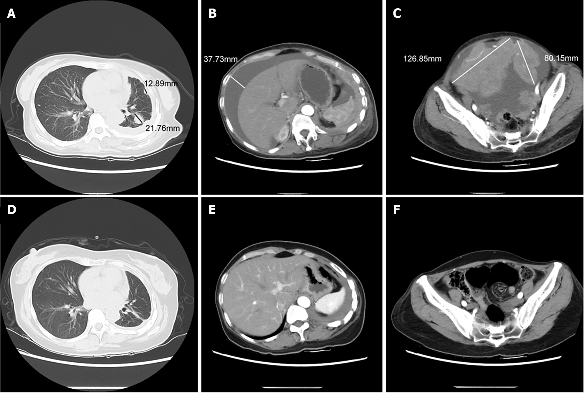
Figure 3 Computed tomography tracking of changes in lung, peritoneal fluid, and pelvic mass before and after treatment with sotorasib.
A: Lungs before treatment in the sotorasib group; B: Abdomen before sotorasib treatment; C: Pelvis before sotorasib treatment; D: Lungs after sotorasib treatment; E: Abdomen after sotorasib treatment; F: Pelvis after sotorasib treatment.
Figure 4 Changes in tumor markers.
A: Carcinoembryonic antigen (CEA) values; B: Carbohydrate antigen 125 (CA125) values.
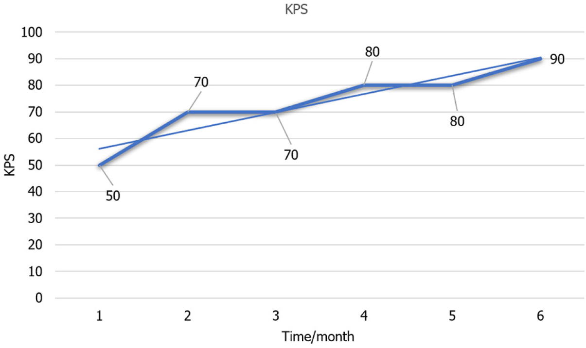
Figure 5 Changes in Karnofsky Performance Status score.
KPS: Karnofsky Performance Status.
- Citation: Wang MX, Zhu P, Shi Y, Sun QM, Dong WH. Returning from the afterlife? Sotorasib treatment for advanced KRAS mutant lung cancer: A case report. World J Clin Cases 2024; 12(25): 5805-5813
- URL: https://www.wjgnet.com/2307-8960/full/v12/i25/5805.htm
- DOI: https://dx.doi.org/10.12998/wjcc.v12.i25.5805


