Copyright
©The Author(s) 2023.
World J Clin Cases. Mar 6, 2023; 11(7): 1506-1512
Published online Mar 6, 2023. doi: 10.12998/wjcc.v11.i7.1506
Published online Mar 6, 2023. doi: 10.12998/wjcc.v11.i7.1506
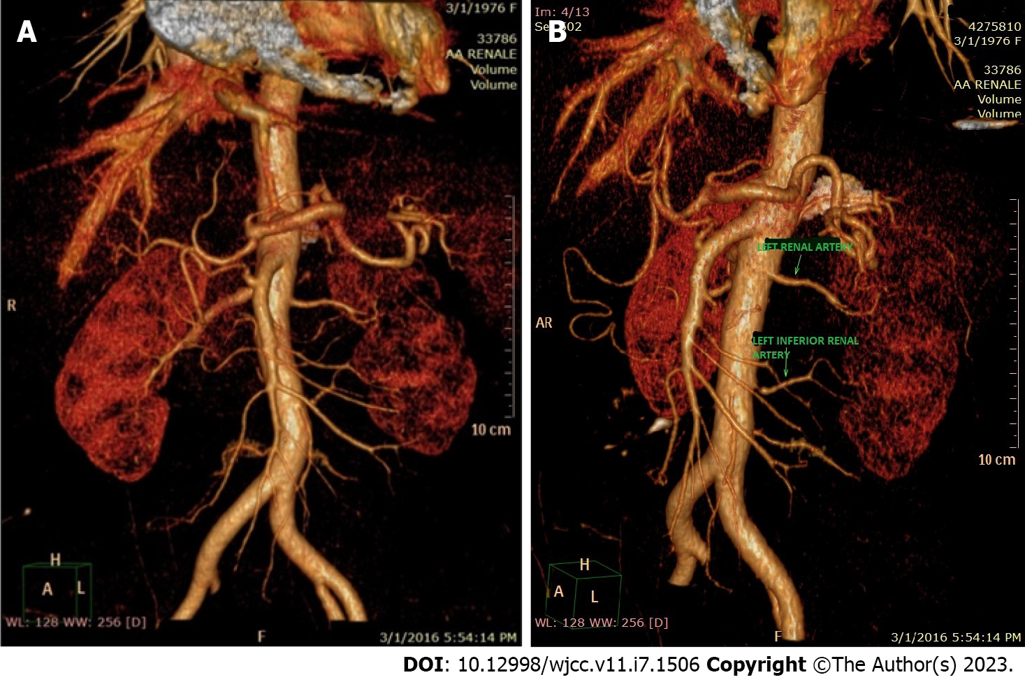
Figure 1 3D Renal Computed Tomography image showing two left renal arteries.
A: 3D reconstruction of the descending aorta; B: Two left renal arteries supplying the left kidney.
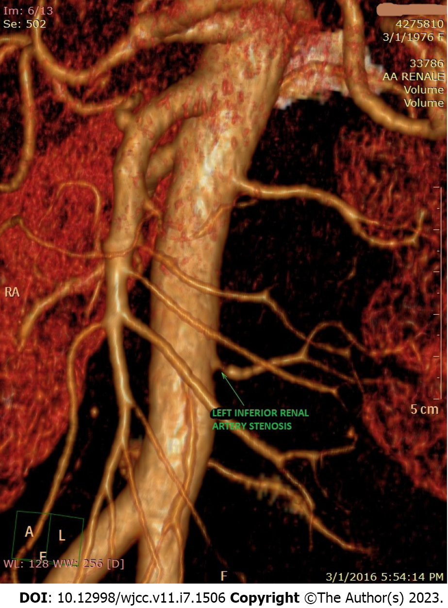
Figure 2 3D image of renal computed tomography with contrast showing the area of ostial stenosis of the left accessory renal artery.
- Citation: Calinoiu A, Guluta EC, Rusu A, Minca A, Minca D, Tomescu L, Gheorghita V, Minca DG, Negreanu L. Accessory renal arteries - a source of hypertension: A case report. World J Clin Cases 2023; 11(7): 1506-1512
- URL: https://www.wjgnet.com/2307-8960/full/v11/i7/1506.htm
- DOI: https://dx.doi.org/10.12998/wjcc.v11.i7.1506


