©The Author(s) 2023.
World J Clin Cases. Dec 16, 2023; 11(35): 8372-8378
Published online Dec 16, 2023. doi: 10.12998/wjcc.v11.i35.8372
Published online Dec 16, 2023. doi: 10.12998/wjcc.v11.i35.8372
Figure 1 Initial chest images suggesting an impression of thoracic empyema.
A: At the initial visit, chest radiography revealed a large pleural effusion in the right thorax (orange arrows) with almost complete opacification of the right lung field; B: Coronal view of chest contrast-enhanced computed tomography confirmed multiple right-side lobulated pleural effusions (orange arrows); C: Axial view of chest contrast-enhanced computed tomography confirmed multiple right-side lobulated pleural effusions (orange arrows).
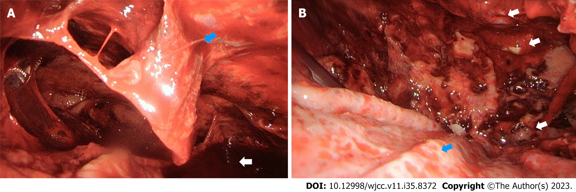
Figure 2 Operative findings during video-assisted thoracoscopic surgery.
A: Video-assisted thoracoscopic surgery indicated massive bloody pleura effusion (white arrow). Adhesion bands and peels are shown (blue arrow); B: Multiple pleural nodules were found after removing the bloody pleural effusion and pneumolysis (white arrows). Peel-coating over the visceral pleura of the right lung was observed (blue arrow). Then pulmonary docortication was performed.
Figure 3 Histological and immunohistochemistry staining of pleural tumors yielded a diagnosis of epithelioid subtype of malignant mesothelioma.
A: Hematoxylin & eosin staining showed poorly differentiated carcinoma characterized by nested tumor growth pattern composed of tumor cells with epithelioid cytoplasm, high nuclear/cytoplasmic ratio, enlarged nuclei, and marked nucleoli infiltrating in the stromal of the pleural peel tissue; B: Immunohistochemistry staining of pleural tumors showed negative immunoreactivity to calretinin; C: Immunohistochemistry staining of pleural tumors showed immunoreactivity to the WT1, determining a diagnosis of malignant pleural mesothelioma of epithelioid subtype.
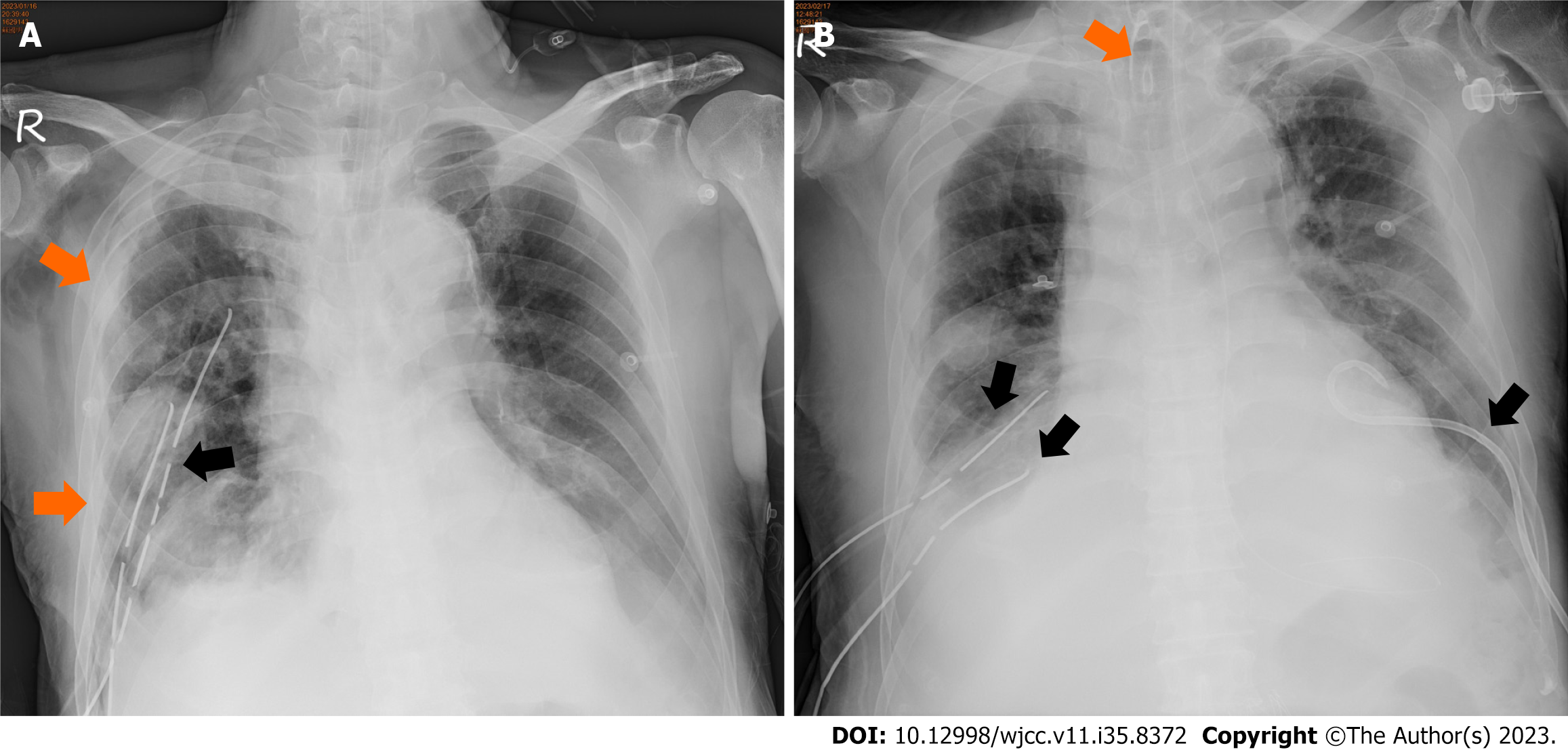
Figure 4 Postoperative chest plain films showing refractory bilateral pleural effusion and disease severity.
A: Postoperative chest plain film exhibiting improved expansion of the right lung. Minimal bilateral pleural effusions (orange arrows) and two chest tubes in the right pleural cavity (black arrow) were observed; B: Chest plain film on the 14th POD revealing bilateral pleural effusion after bilateral pleural catheter drainage (black arrows). A tracheostomy was performed due to respiratory failure (orange arrow).
- Citation: Yao YH, Kuo YS. Malignant pleural mesothelioma mimics thoracic empyema: A case report. World J Clin Cases 2023; 11(35): 8372-8378
- URL: https://www.wjgnet.com/2307-8960/full/v11/i35/8372.htm
- DOI: https://dx.doi.org/10.12998/wjcc.v11.i35.8372


