©The Author(s) 2023.
World J Clin Cases. Aug 6, 2023; 11(22): 5391-5397
Published online Aug 6, 2023. doi: 10.12998/wjcc.v11.i22.5391
Published online Aug 6, 2023. doi: 10.12998/wjcc.v11.i22.5391
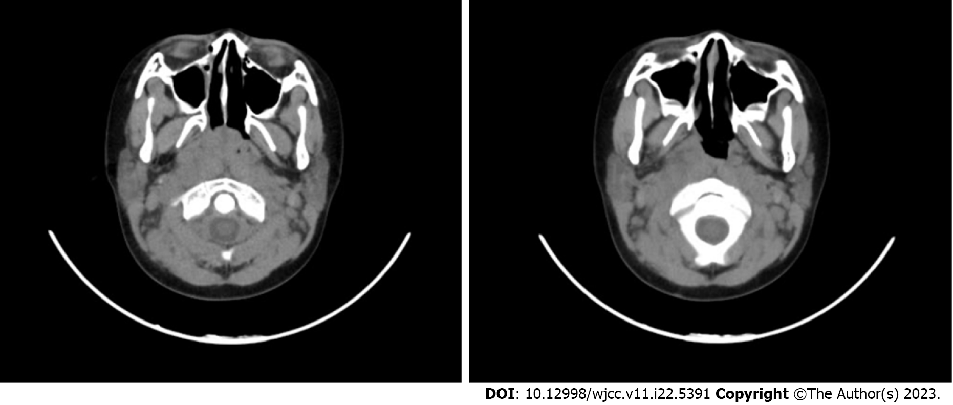
Figure 1 Neck computed tomography of the patient on the first day of hospitalisation.
Computed tomography scan of the neck showed widening of the retropharyngeal space with liquid hypodensity, thickening of the pharyngeal lymphatic ring, and multiple slightly large lymph nodes in the neck space.
Figure 2 Neck computed tomography of the patient on the fifteenth day of hospitalisation.
Computed tomography scan suggested the presence of multiple small lymph nodes in the neck, which were reduced in size compared to the film scanned on the first day. Additionally, oropharyngeal and retropharyngeal space effusion were disappeared.
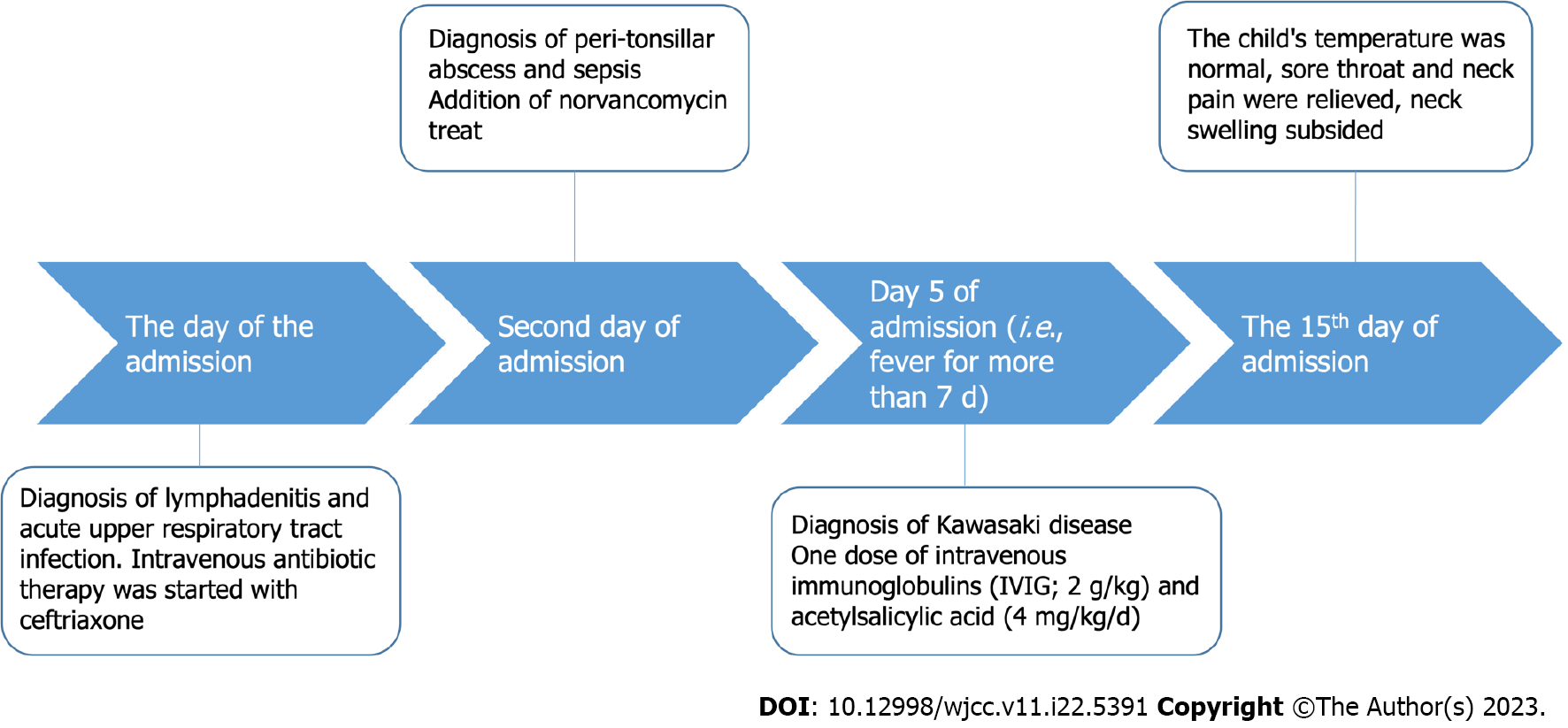
Figure 3 The timeline of diagnosis and treatment of Kawasaki disease in this child.
- Citation: Huo LM, Li LM, Peng HY, Wang LJ, Feng ZY. Kawasaki disease with peritonsillar abscess as the first symptom: A case report. World J Clin Cases 2023; 11(22): 5391-5397
- URL: https://www.wjgnet.com/2307-8960/full/v11/i22/5391.htm
- DOI: https://dx.doi.org/10.12998/wjcc.v11.i22.5391


