Copyright
©The Author(s) 2023.
World J Clin Cases. Apr 16, 2023; 11(11): 2549-2558
Published online Apr 16, 2023. doi: 10.12998/wjcc.v11.i11.2549
Published online Apr 16, 2023. doi: 10.12998/wjcc.v11.i11.2549
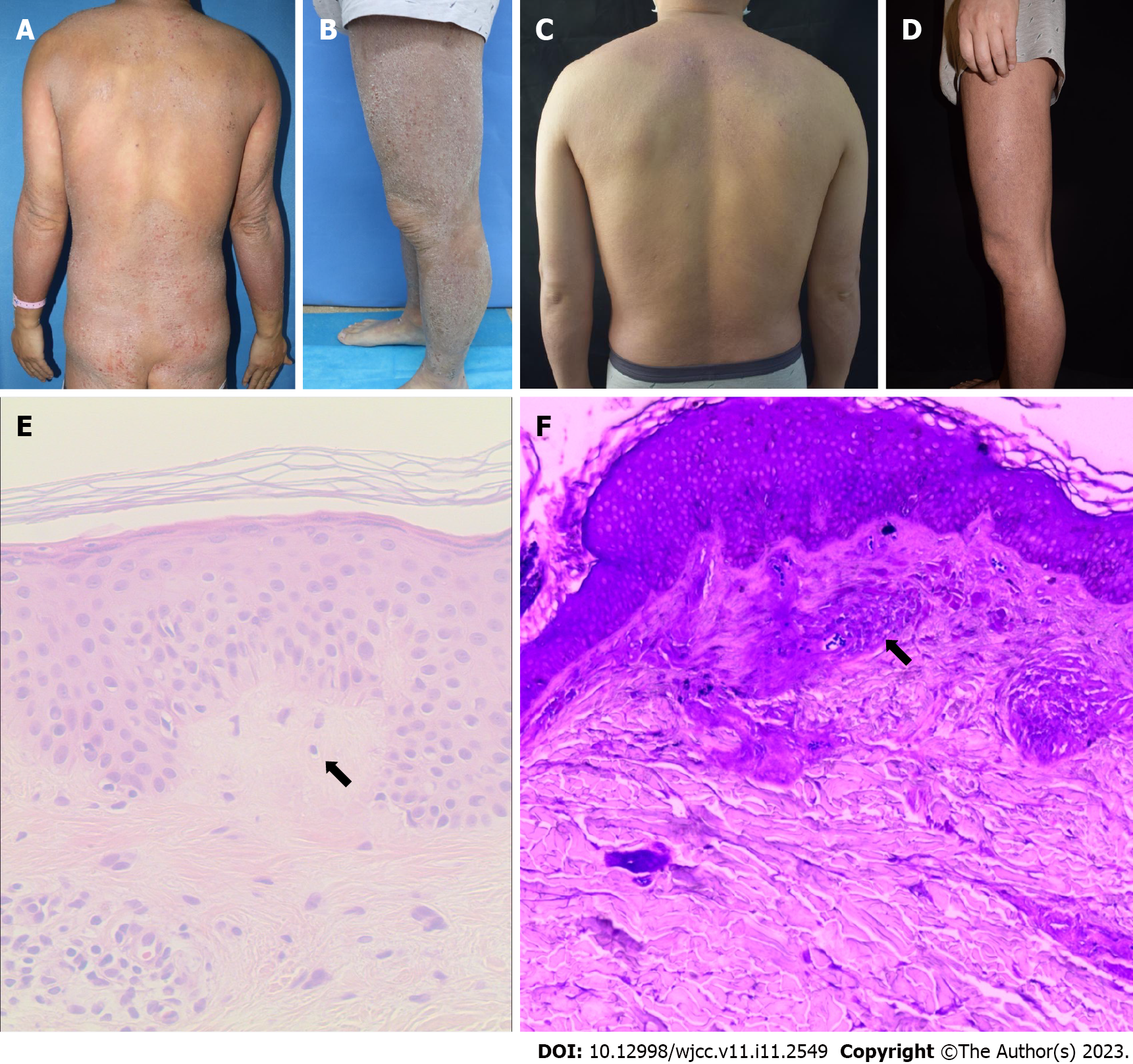
Figure 1 Case 1.
A and B: Dense hard brown plaques were seen on the trunk and limbs at baseline; C and D: After 16 wk of treatment with dupilumab, the trunk rash basically subsided, the lower limb rash markedly improved, and the plaques became flat. The remaining lesions on the thigh after treatment, showed a characteristic rippled appearance indicating lichen amyloidosis as a co-existing condition; E: Histopathological examination revealed mild hyperkeratosis of the epidermis, irregular hyperplasia of the spinous layer, and spongy edema, with the dermal papilla and upper dermis showing a uniformly reddish mass that expanded the dermal papillae and displaced the rete ridges laterally (hematoxylin & eosin staining, × 100); F: Crystal violet staining was positive for amyloid deposits in the dermal papillae and upper dermis with characteristic fissures (crystal violet staining, × 400). The arrow indicates amyloid deposits.
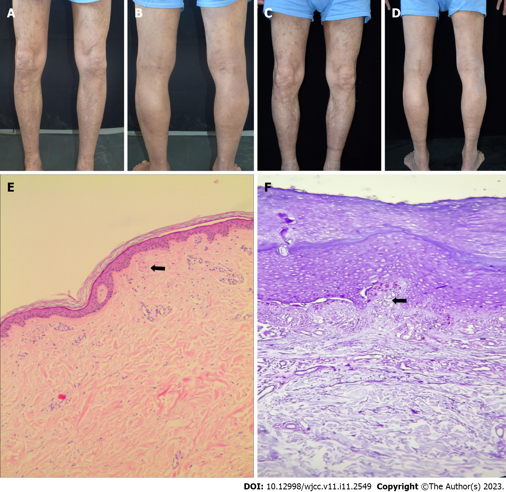
Figure 2 Case 2.
A and B: Dense distribution of hard brown papules was observed on both lower limbs, with scales on the surface and dry and rough skin at baseline. Moreover, post-inflammatory hypopigmentation was evident on the anterior aspects of the calves; C and D: After 16 wk of treatment, the rash on both lower limbs was markedly reduced, the papules became flat, the scales were reduced, and the skin became smooth. Furthermore, the rippled appearance of the lesions of the posterior thighs has considerably diminished after the treatment, suggesting a decreased burden of amyloid deposits; E: Histopathological examination revealed epidermal hyperkeratosis, irregular hyperplasia of the spinous layer, increased basal pigmented cells, and red mass material deposition in the dermal papilla (hematoxylin & eosin staining, × 100); F: Crystal violet staining was positive for amyloid bodies (crystal violet staining, × 400). The arrow indicates amyloid deposits.
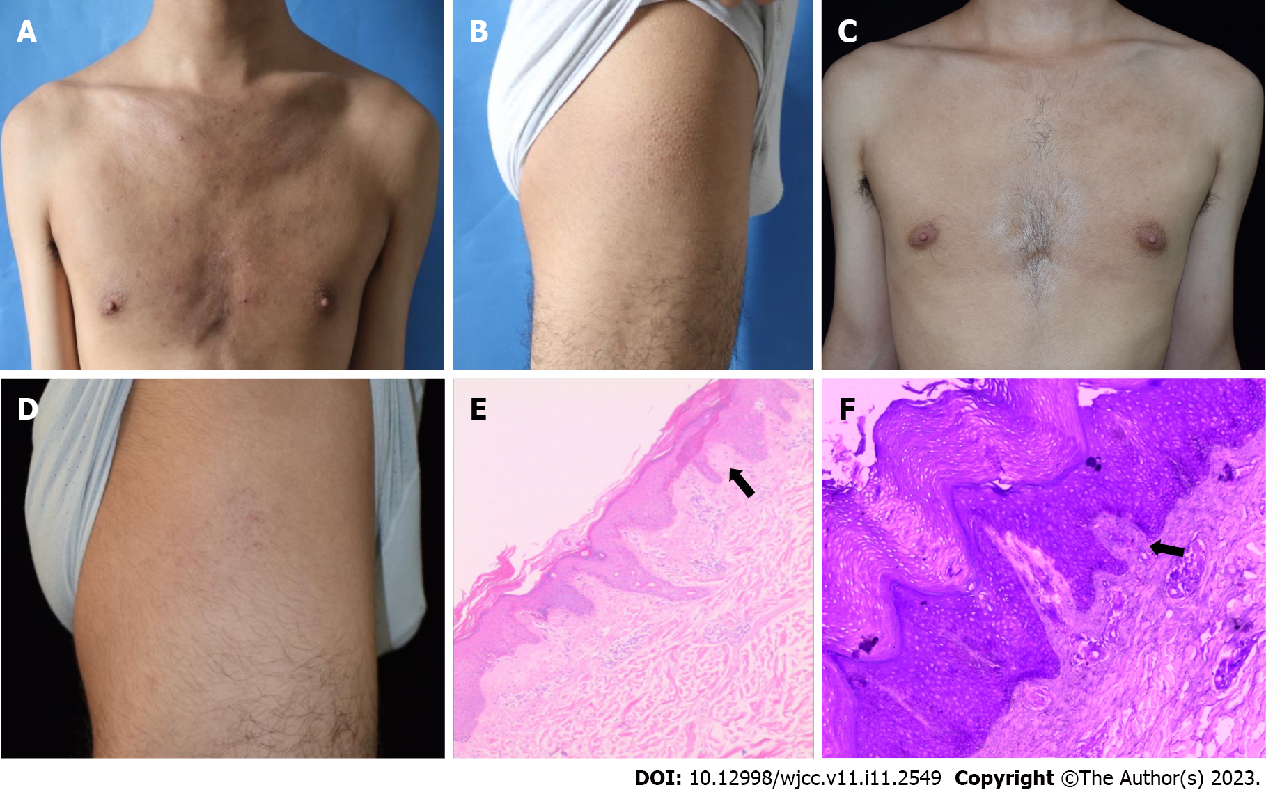
Figure 3 Case 3.
A and B: Dense distribution of hard brown papules was observed on the chest plus papules on the thighs, with a "goose bump" appearance presenting at baseline, suggestive of atopic dermatitis; C and D: After 16 wk of treatment, the rash on the lower limbs markedly improved, and the papules were reduced and became flat; E: Histopathology revealed hyperkeratosis with incomplete keratosis and irregular acanthosis. Further, a pink amorphous material can be seen in the dermal papilla (hematoxylin & eosin staining, × 100); F: Crystal violet staining was positive for amyloid (crystal violet staining, × 400). The arrow indicates amyloid deposits.
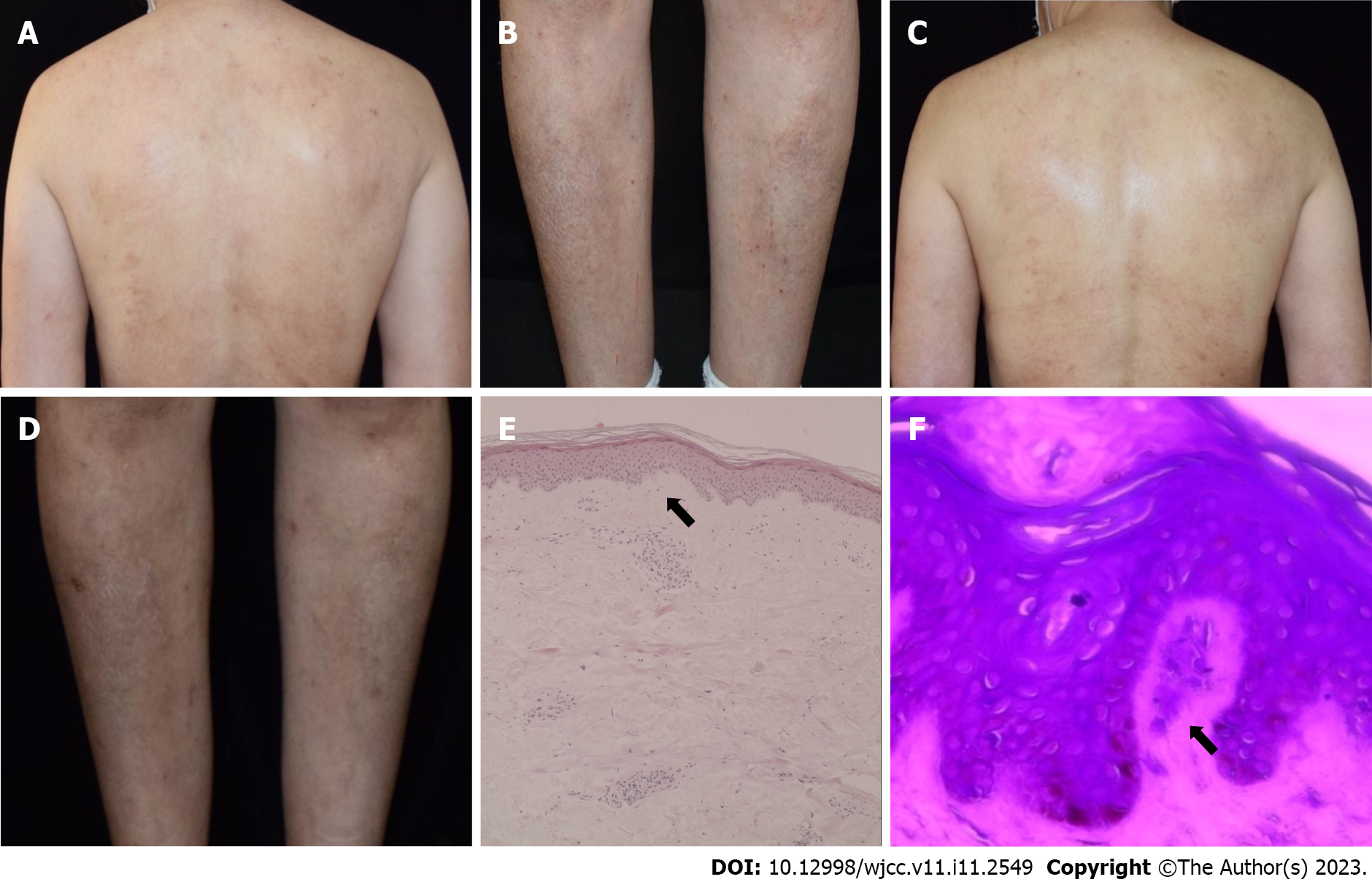
Figure 4 Case 4.
A and B: Erythema and a dense distribution of hard brown papules were seen on the patient’s back. Both calves showed a characteristic rippled pattern, which was most prominent on the anterolateral aspect of the right calf; C and D: After 16 wk of treatment, the papules on both calves became flat and the wavy pattern markedly disappeared, suggestive of diminished amyloid burden; E: Hyperkeratotic and hyperplastic epidermis as well as homogeneous pinkish materials in the dermal papilla were seen (hematoxylin & eosin staining, × 100); F: Crystal violet staining was positive for amyloid deposits (crystal violet staining, × 400). The arrow indicates amyloid deposits.
- Citation: Zhu Q, Gao BQ, Zhang JF, Shi LP, Zhang GQ. Successful treatment of lichen amyloidosis coexisting with atopic dermatitis by dupilumab: Four case reports. World J Clin Cases 2023; 11(11): 2549-2558
- URL: https://www.wjgnet.com/2307-8960/full/v11/i11/2549.htm
- DOI: https://dx.doi.org/10.12998/wjcc.v11.i11.2549


