Copyright
©The Author(s) 2022.
World J Clin Cases. Nov 6, 2022; 10(31): 11617-11624
Published online Nov 6, 2022. doi: 10.12998/wjcc.v10.i31.11617
Published online Nov 6, 2022. doi: 10.12998/wjcc.v10.i31.11617
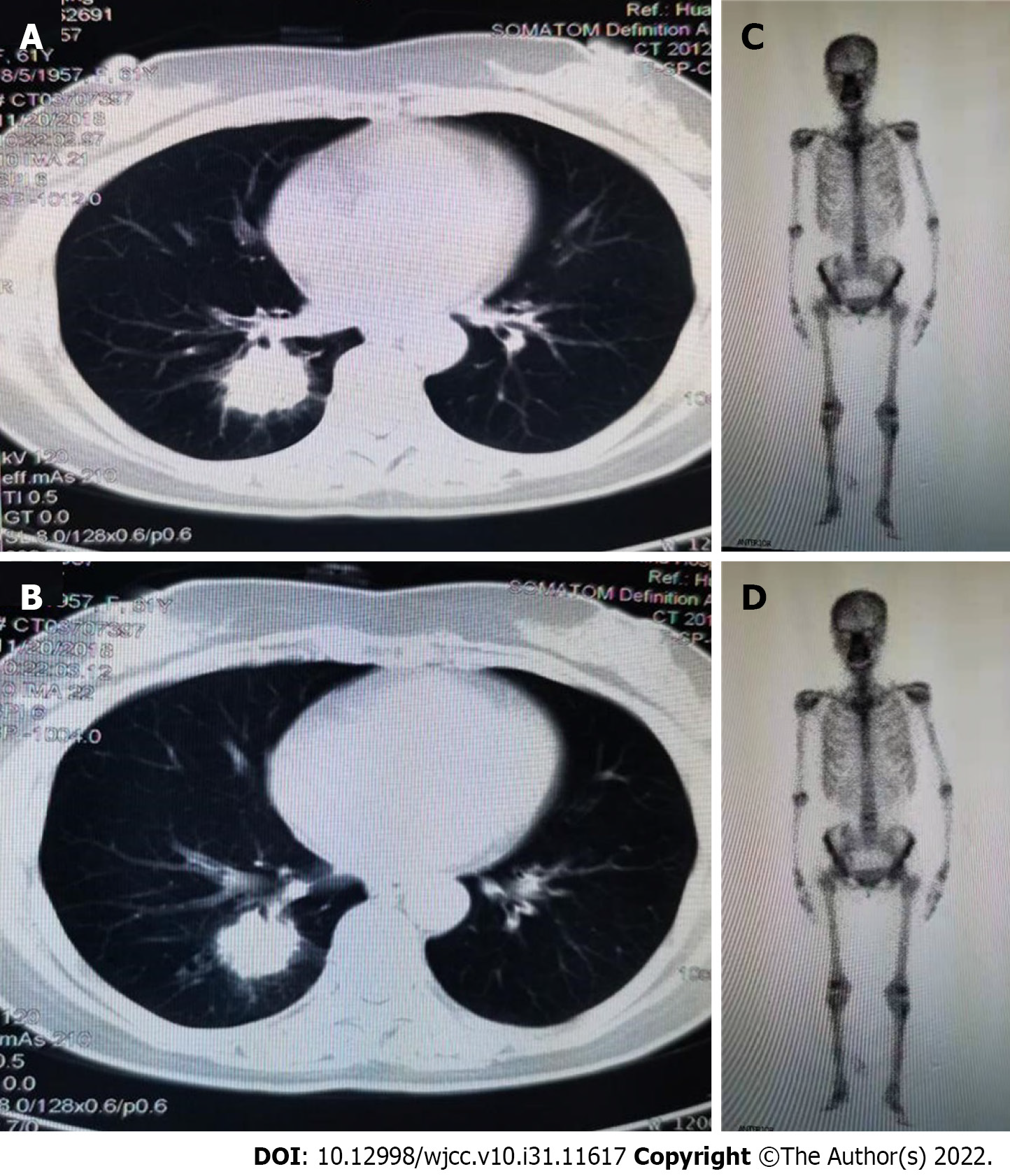
Figure 1 Imaging examinations before surgery.
A and B: Contrast-enhanced computed tomography of the chest showing a 2.8 cm × 2.9 cm mass in the right lower lobe; C and D: Whole body bone scan showed no abnormality.
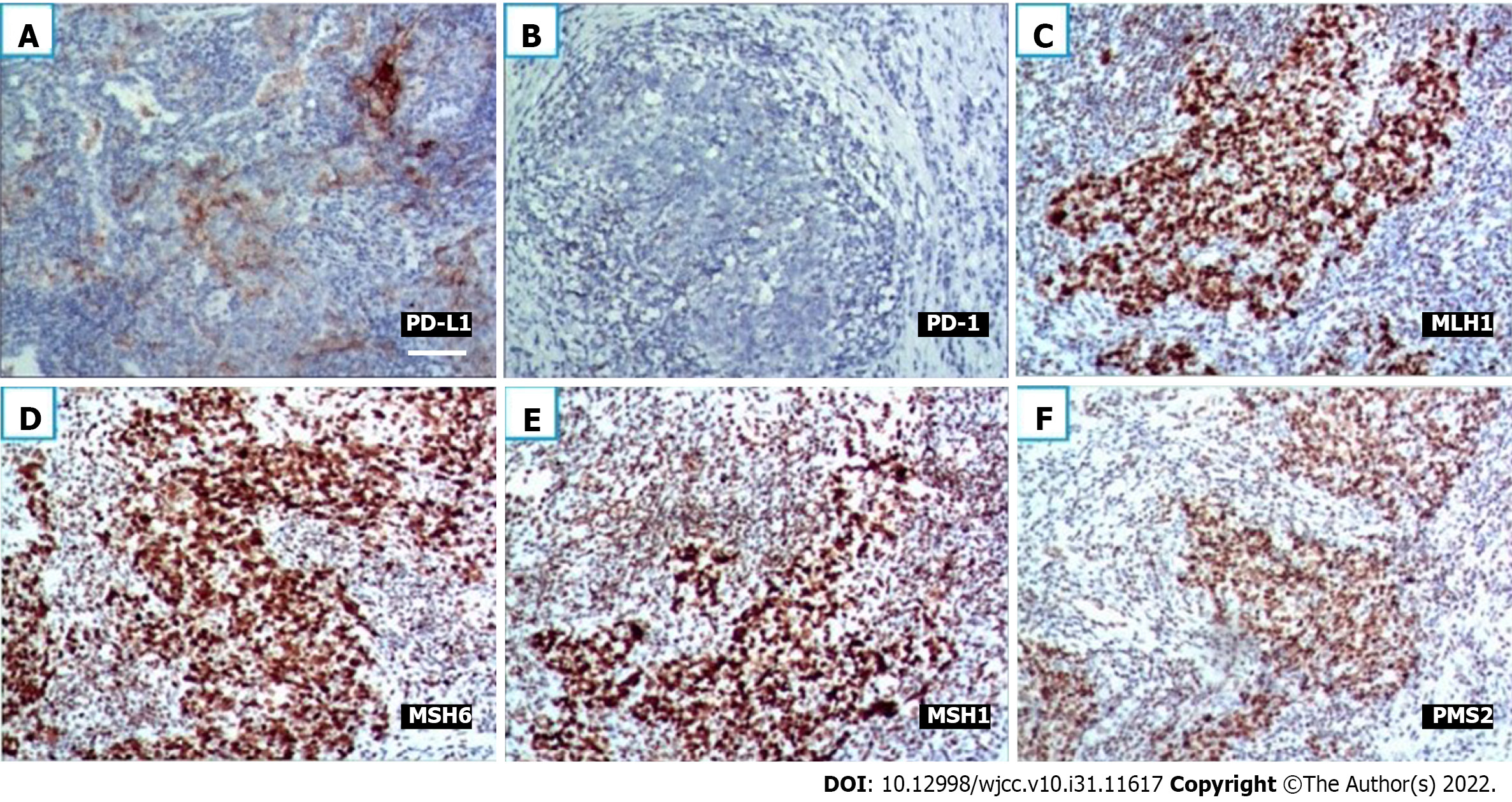
Figure 2 Immunohistochemistry.
A: Immunohistochemical (IHC) staining for programmed death-1 (PD-1) revealed positive neoplastic cells with a membranous pattern (× 200, scale bar: 100 μm); B: IHC staining for PD-1 revealed negative neoplastic cells (× 200, scale bar: 100 μm); C-F: IHC staining for mismatch repair (MMR) proteins revealed integrity of the MMR system (× 200, scale bar: 100 μm).
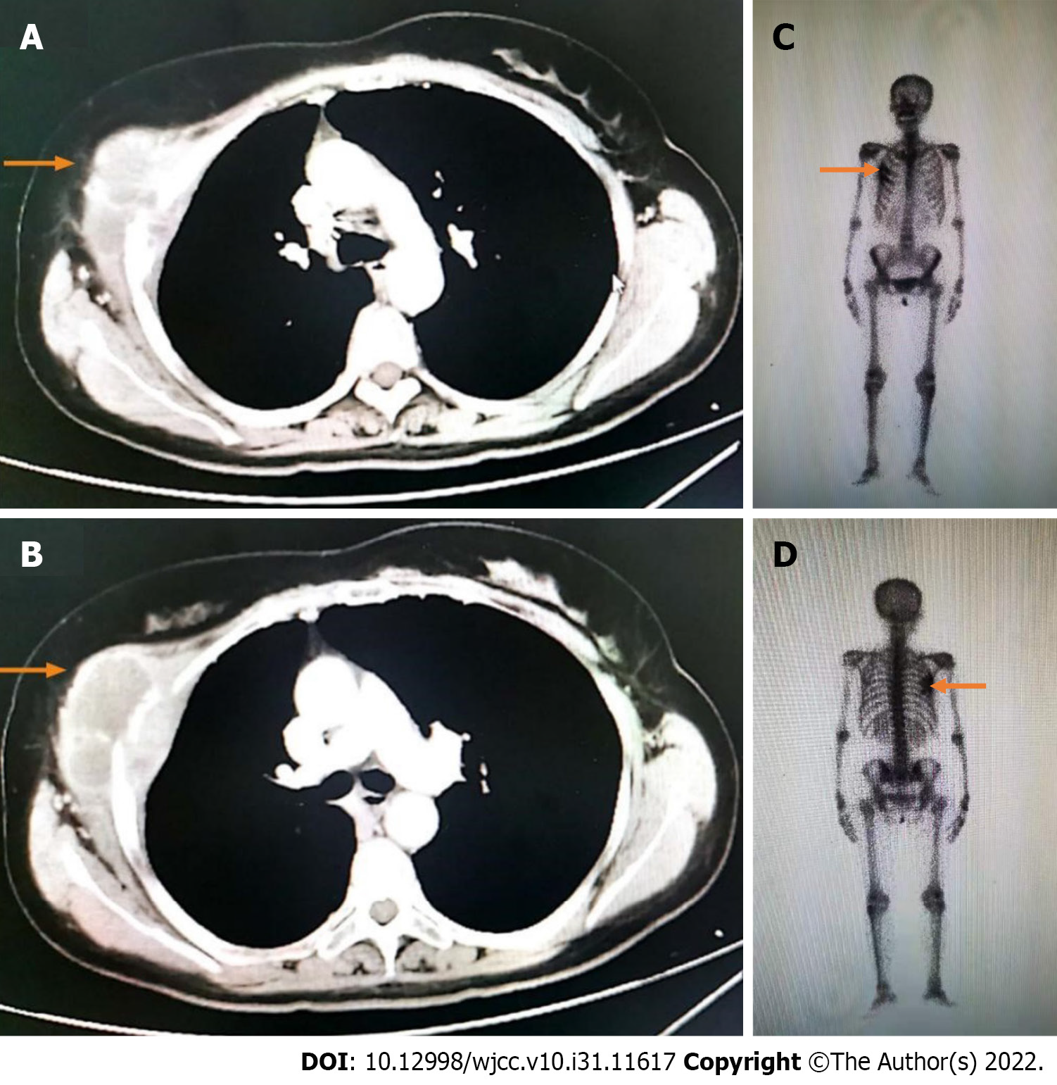
Figure 3 Imaging examinations after recurrence.
A and B: Contrast-enhanced computed tomography of the chest before therapy. The right 4th rib with a soft tissue mass measuring 41 mm × 35 mm, and cortical thickening of the adjacent 5th rib were detected; C and D: Whole-body bone scan revealed that bone metabolism was increased in the 3rd-5th coastal axillary segments on the right side.
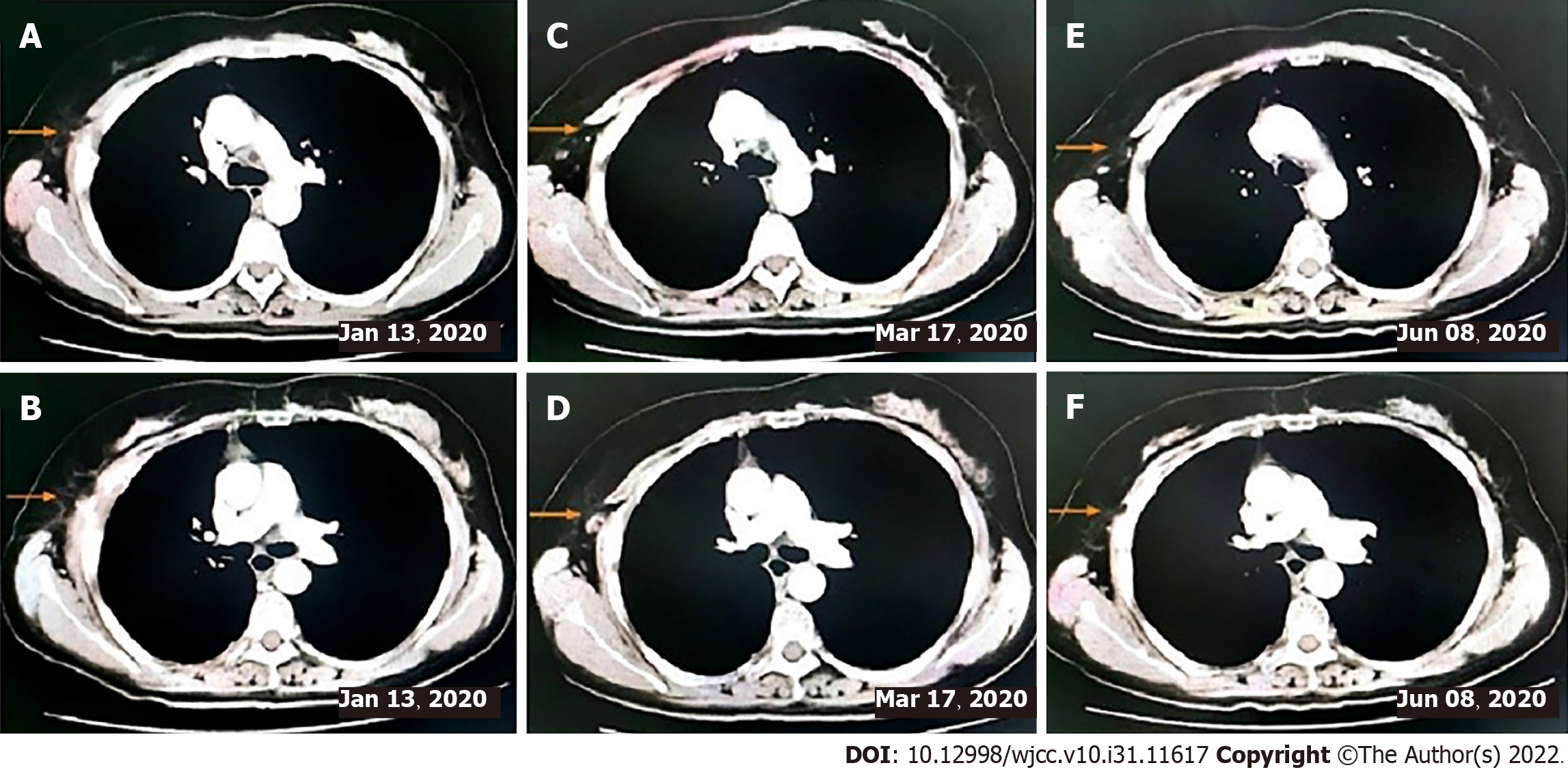
Figure 4 Imaging examination.
A and B: Computed tomography (CT) after two cycles of immunotherapy combined with chemotherapy; C and D: CT scan preformed after six cycles of immunotherapy combined with chemotherapy; E and F: CT revealed a continuous PR state after 6 mo of immunotherapy combined with chemotherapy.
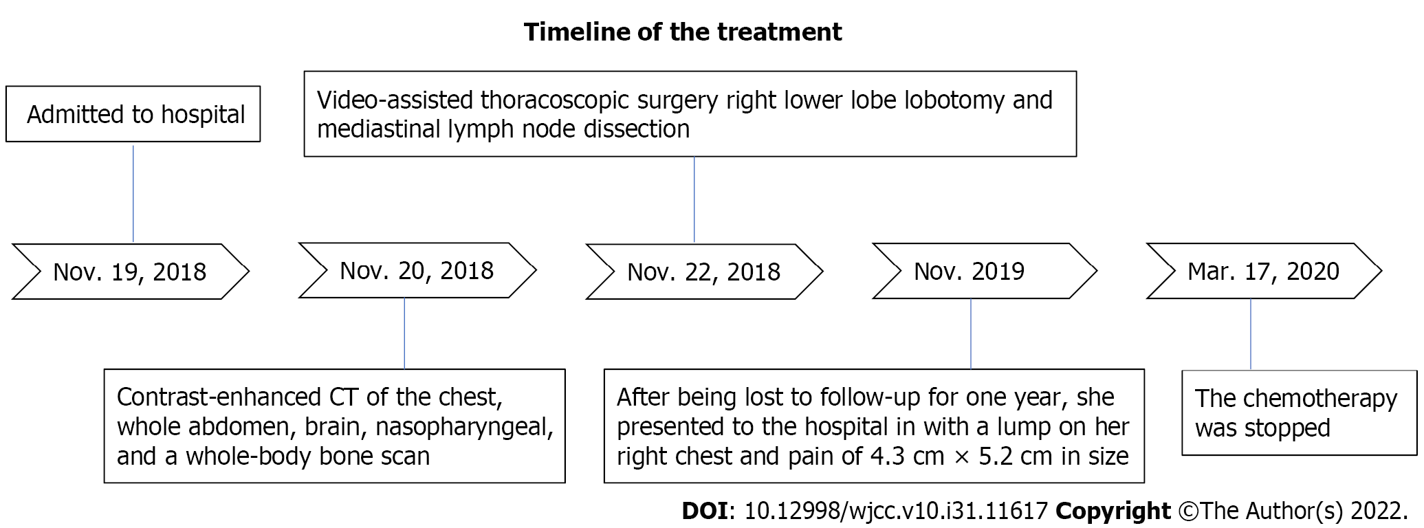
Figure 5 Timeline summarizing main events of this case report.
- Citation: Zeng SY, Yuan J, Lv M. Favorable response of primary pulmonary lymphoepithelioma-like carcinoma to sintilimab combined with chemotherapy: A case report. World J Clin Cases 2022; 10(31): 11617-11624
- URL: https://www.wjgnet.com/2307-8960/full/v10/i31/11617.htm
- DOI: https://dx.doi.org/10.12998/wjcc.v10.i31.11617


