Copyright
©The Author(s) 2022.
World J Clin Cases. Apr 16, 2022; 10(11): 3496-3504
Published online Apr 16, 2022. doi: 10.12998/wjcc.v10.i11.3496
Published online Apr 16, 2022. doi: 10.12998/wjcc.v10.i11.3496
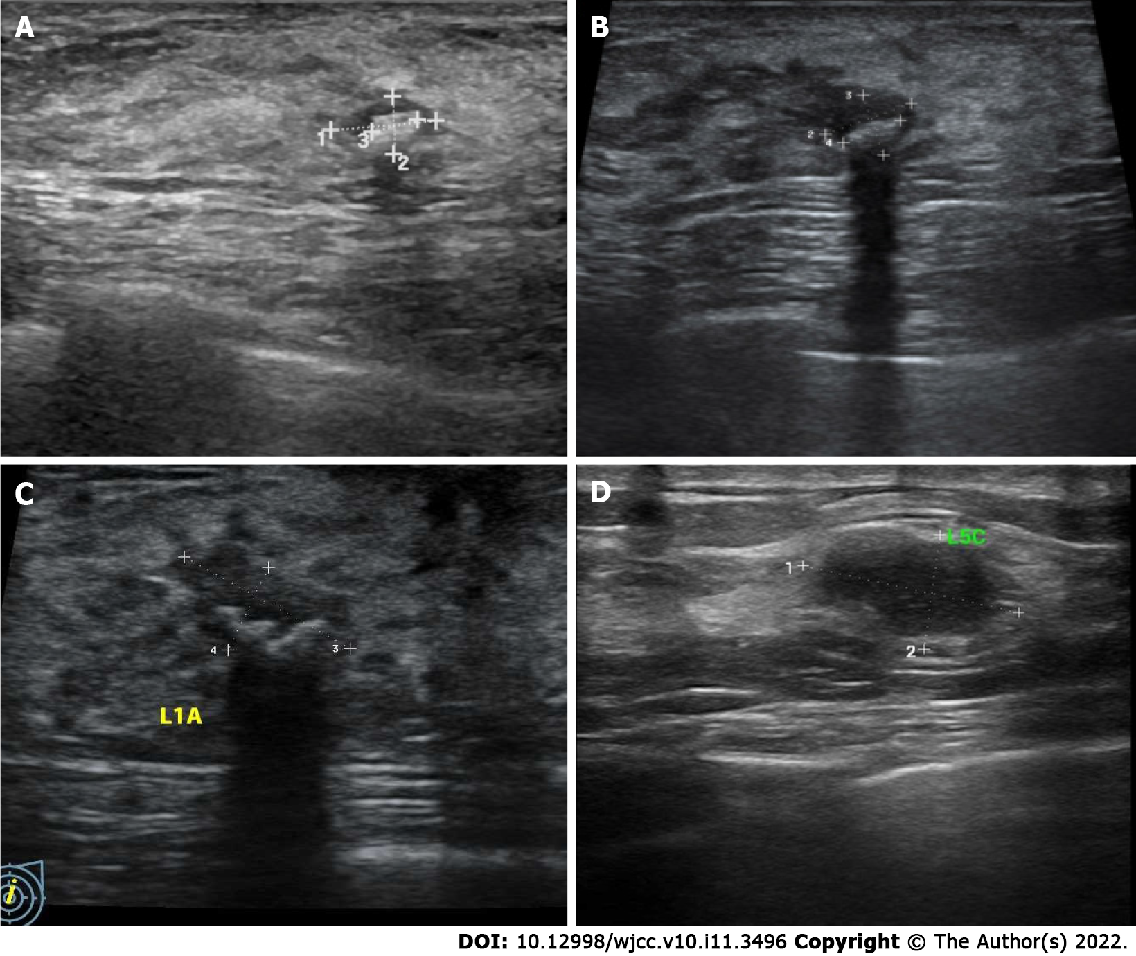
Figure 1 Left breast nodule ultrasonography of case 1 and case 2.
A: May 2018 (case 1); B: March 2019 (case 1); C: June 2021 (case 1); D: Case 2.
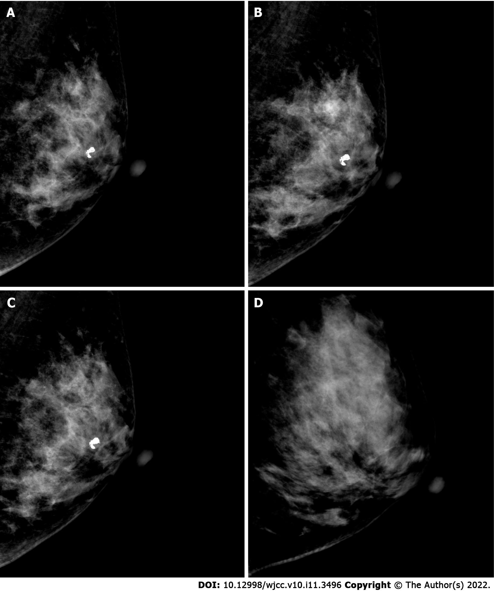
Figure 2 Left breast mammography of case 1 (mediolateral oblique view) and case 2.
A: May 2018 (case 1); B: March 2019 (case 1); C: June 2021 (case 1); D: Case 2.
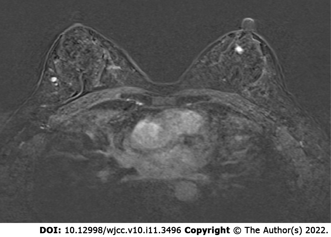
Figure 3 Dynamic contrast-enhanced magnetic resonance imaging of case 1.
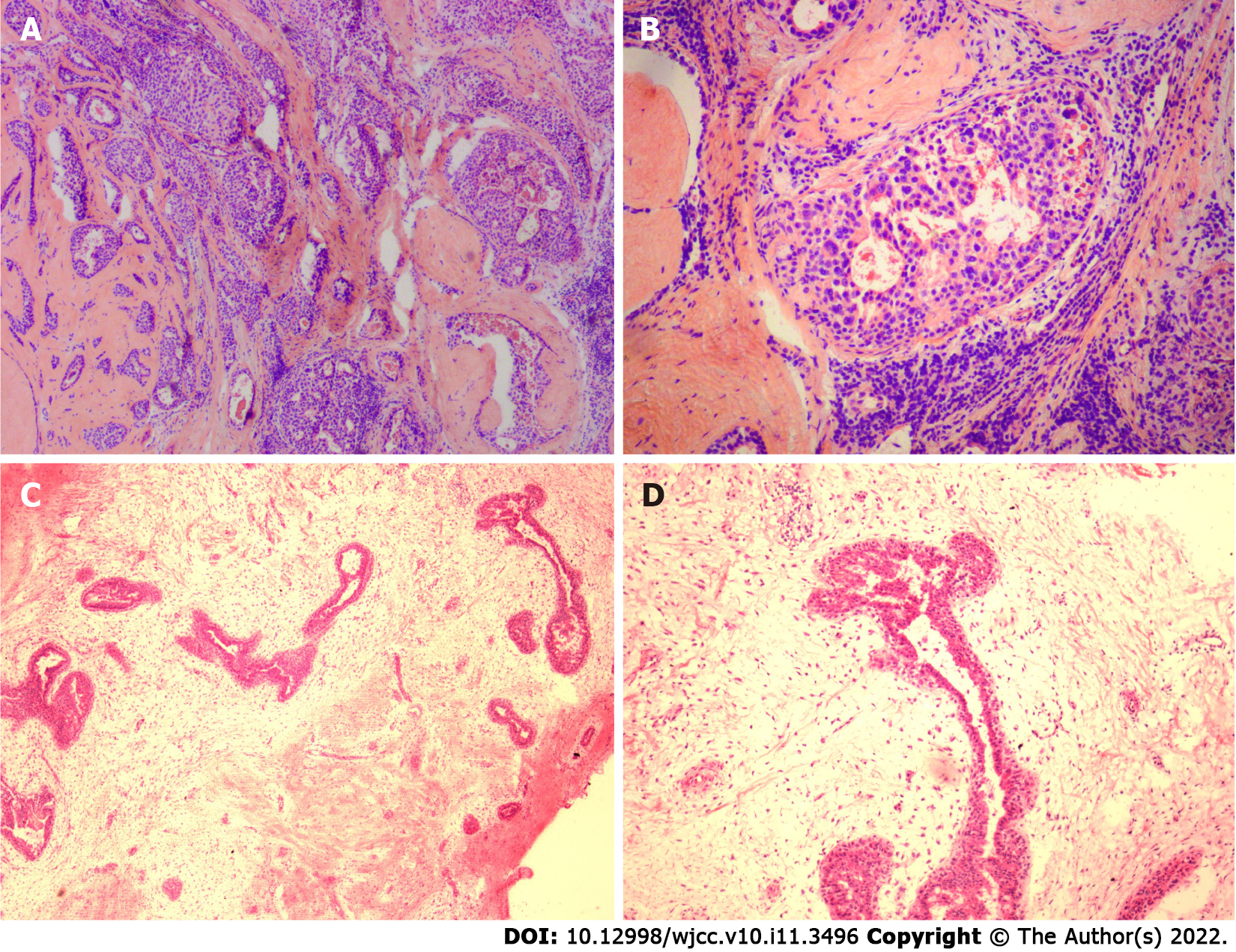
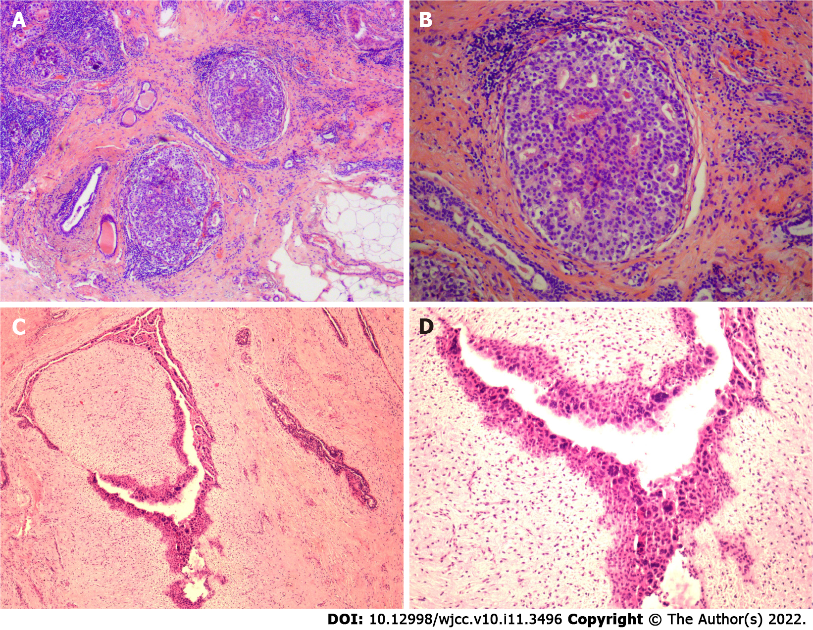
Figure 4 Intraoperative frozen section findings of case 1 and case 2.
Sections were stained with hematoxylin and eosin. A: Low magnification image of case 1 (40 × magnification); B: Medium magnification image of case 1 (100 × magnification); C: Low magnification image of case 2 (40 × magnification); B: Medium magnification image of case 2 (100 × magnification).
Figure 5 Paraffin section findings after operation of case 1 and case 2.
Sections were stained with hematoxylin and eosin. A: Low magnification image of case 1 (40 × magnification); B: Medium magnification image of case 1 (100 × magnification); C: Low magnification image of case 2 (40 × magnification); D: Medium magnification image of case 2 (100 × magnification).
- Citation: Wu J, Sun KW, Mo QP, Yang ZR, Chen Y, Zhong MC. Preoperational diagnosis and management of breast ductal carcinoma in situ arising within fibroadenoma: Two case reports. World J Clin Cases 2022; 10(11): 3496-3504
- URL: https://www.wjgnet.com/2307-8960/full/v10/i11/3496.htm
- DOI: https://dx.doi.org/10.12998/wjcc.v10.i11.3496


