Copyright
©2013 Baishideng Publishing Group Co.
World J Clin Oncol. Nov 10, 2013; 4(4): 91-101
Published online Nov 10, 2013. doi: 10.5306/wjco.v4.i4.91
Published online Nov 10, 2013. doi: 10.5306/wjco.v4.i4.91
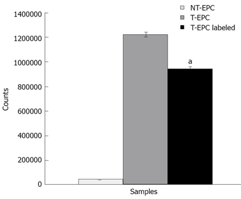
Figure 1 Transgene expression study using TC-99m uptake assay.
Transfected and control endothelial progenitor cells (EPCs) were subjected to Tc-99m uptake assay to determine the transgene expression. Tc-99m uptake assay was performed with three type of conditions: (1) Non transgenic, non-labeled (without iron) EPC (control); (2) transgenic, non-labeled EPC; (3) transgenic and labeled (with iron) EPCs. Transfected EPCs showed higher Tc-99m uptake compared to control non transfected EPCs, which clearly indicates the functional expression of transgene. Data was indicated with Mean ± SD. aP < 0.05 vs control cells.
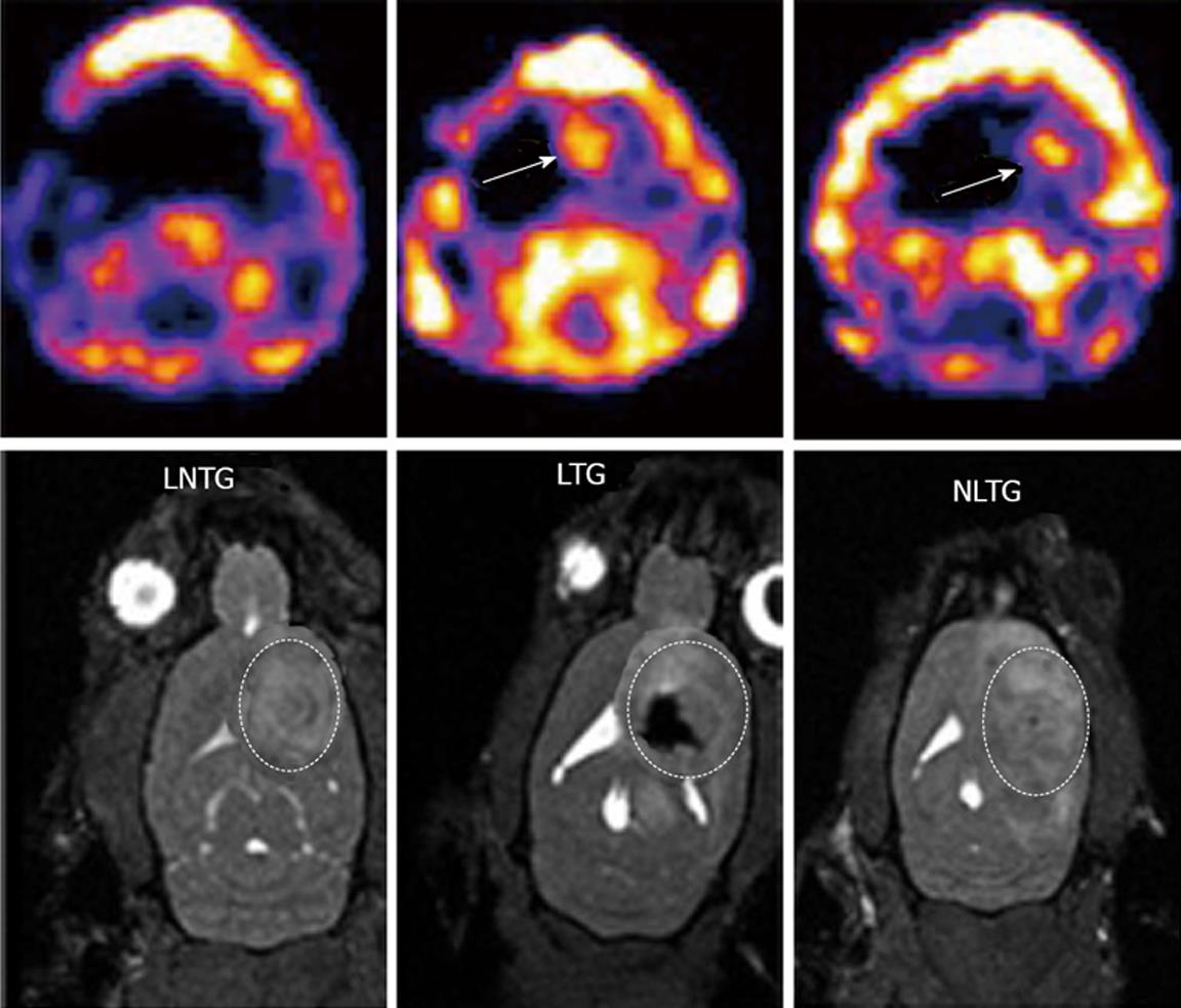
Figure 2 Magnetic resonance imaging and single photon emission computed tomography images for tracking of intratumor injected endothelial progenitor cells and transgene expression.
Expression of transgene (hNIS) was detected by single photon emission computed tomography (SPECT) and magnetic resonance imaging (MRI) was used to detect the cell migration in the tumor. Upper panels (SPECT images): Left: Animals received non transgenic, non-labeled endothelial progenitor cells (EPCs) (control); Middle: Animals received transgenic EPCs labeled with iron; Right: Animals received non labeled transgenic EPCs. Corresponding MRI images are shown in the lower panels. Animals received labeled transgenic EPC or non-labeled transgenic EPC showed higher activities of Tc-99m in the tumors compared to control animals that received non-transgenic EPCs. MRI images clearly indicate the presence of iron labeled transgenic EPCs (lower panel, middle).
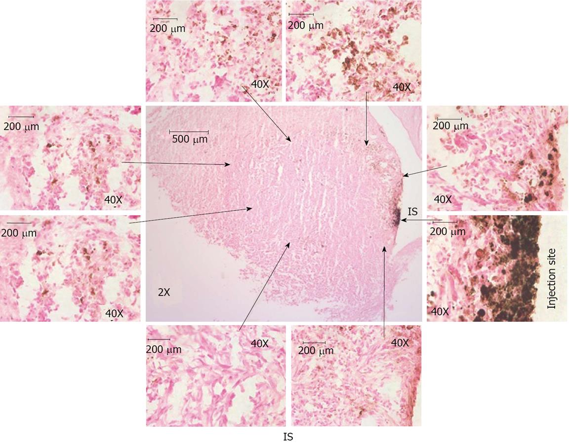
Figure 3 Histological analysis of intratumor injection and migration of cells.
Animal brain sections were stained with Prussian blue and observed under light microscope. Center image (magnification × 2) shows the iron positive cells at injection site and migration around periphery and inside the tumor. Images × 2 shows the whole tumor area and injection sites and images × 40 shows iron labeled cell at injection site and around the tumor. Images × 40 were linked to images × 2 with arrow to show the region × is 40 near to the arrows (not exact match). These images indicate the migration of transgenic endothelial progenitor cells (EPCs) to periphery of the tumor from the injection site. Some of the EPCs were also migrated to the center of the tumor from the injection site. IS: Injection site.
Figure 4 Hematoxylin and eosin staining of brain sections.
Animal brain sections were stained with hematoxylin and eosin, In images × 2 show whole tumor area and images × 10 and images × 40 showing some the necrotic cells at injection site as well as at others regions of the tumor.
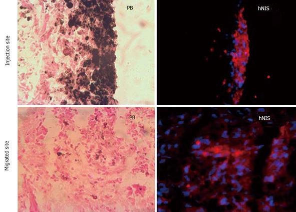
Figure 5 Immunohistochemical analysis of transgene expression at the site of injection and migrated areas.
Red fluorescence (hNIS positive) is observed at site of injection of transgenic iron labeled endothelial progenitor cells (EPCs) and areas where injected EPCs migrated.
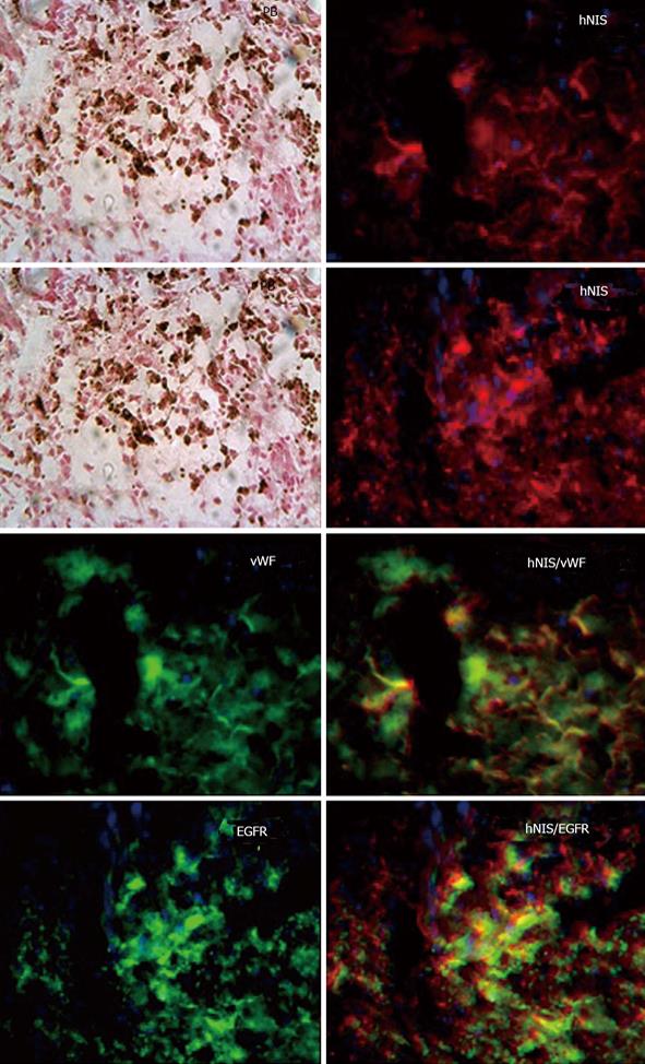
Figure 6 Analysis of transgene delivery to tumor cells by double labeling.
Sections were double with hNIS/vWF or hNIS/EGFR. Sections stain with hNIS/vWF clearly shows some of the red hNIS positive cells also express green vWF positive markers [positive for endothelial progenitor cells (EPC)] indicating the administered EPCs. While hNIS/EGFR double staining clearly shows some of the red positive cells (hNIS positive cells) also express green EGFR positive markers indicating expression of transgenes in glioma cells. Prussian blue staining shows the iron positive cells at the corresponding sites.
- Citation: Varma NRS, Barton KN, Janic B, Shankar A, Iskander A, Ali MM, Arbab AS. Monitoring adenoviral based gene delivery in rat glioma by molecular imaging. World J Clin Oncol 2013; 4(4): 91-101
- URL: https://www.wjgnet.com/2218-4333/full/v4/i4/91.htm
- DOI: https://dx.doi.org/10.5306/wjco.v4.i4.91


