©The Author(s) 2025.
World J Clin Oncol. Sep 24, 2025; 16(9): 110686
Published online Sep 24, 2025. doi: 10.5306/wjco.v16.i9.110686
Published online Sep 24, 2025. doi: 10.5306/wjco.v16.i9.110686
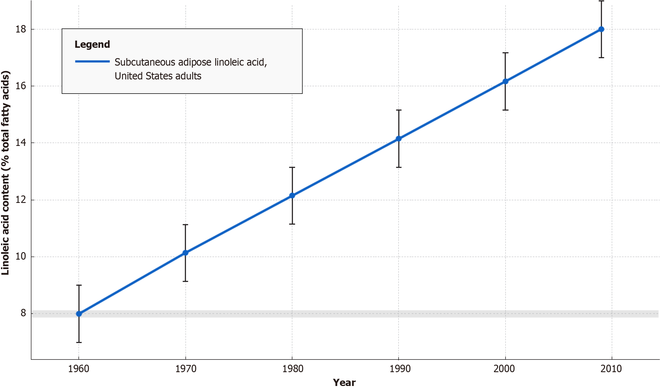
Figure 1 Dramatic four-decade rise in linoleic acid content of United States subcutaneous adipose tissue (1960-2008).
Values are mean ± SD. Data sources by decade (mean ± SD,% total fatty acids): 1960, 8.0% ± 0.9%[257], 1970, 10.1% ± 1.2%[258], 1980, 12.1% ± 1.3%[259], 1990, 14.2% ± 1.0%[260], 2000, 16.2% ± 1.1%[261], 2010, 18.0% ± 1.0%[262]. The proportion of linoleic acid in United States subcutaneous adipose tissue more than doubled, from approximately 8% in 1960 to approximately 18% by 2010, mirroring the progressive incorporation of industrial seed oils into the food supply.
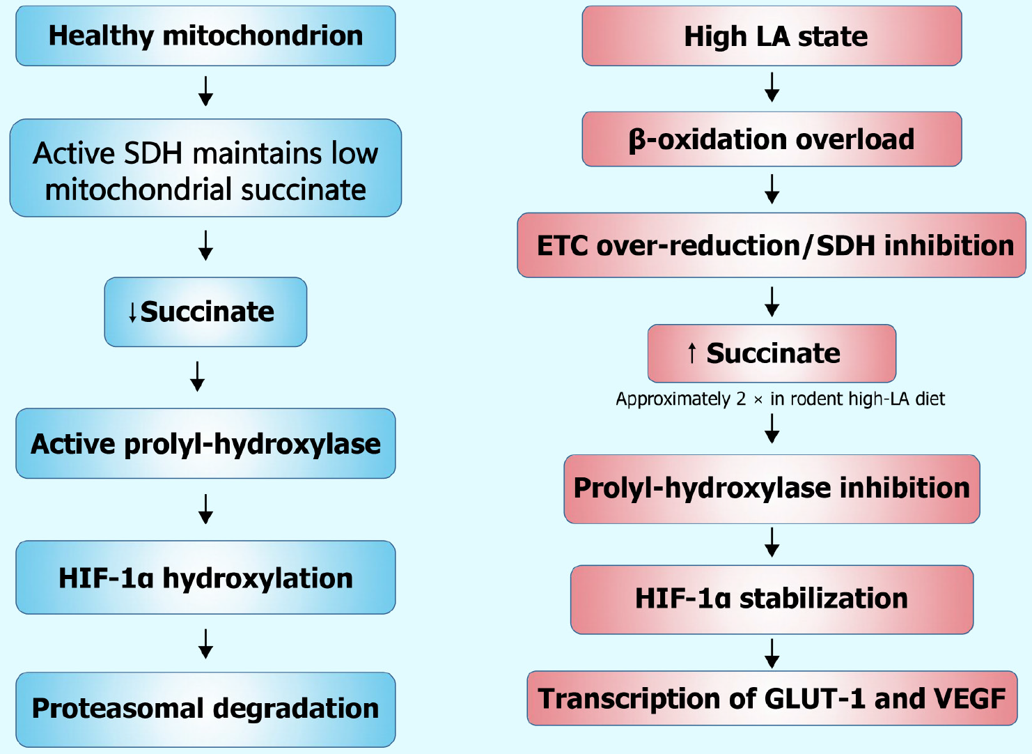
Figure 2 Linoleic acid-induced succinate build-up triggers pseudohypoxic hypoxia-inducible factor-1α stabilization and tumor-promoting glucose transporter type 1/vascular endothelial growth factor signaling.
Excess β-oxidation of linoleic acid overloads succinate dehydrogenase, elevating mitochondrial succinate that inhibits prolyl-hydroxylase, thereby stabilizing hypoxia-inducible factor-1α despite normoxia (“pseudohypoxia”). The resulting transcription of glucose transporter type 1 and vascular endothelial growth factor supports glycolytic flux and neovascularization, hallmarks of aggressive tumor growth. LA: Linoleic acid; 4-HNE: 4-hydroxynonenal; OXLAMs: Oxidized linoleic acid metabolites; ROS: Reactive oxygen species; NF-κB: Nuclear factor kappa-light-chain-enhancer of activated B cells.
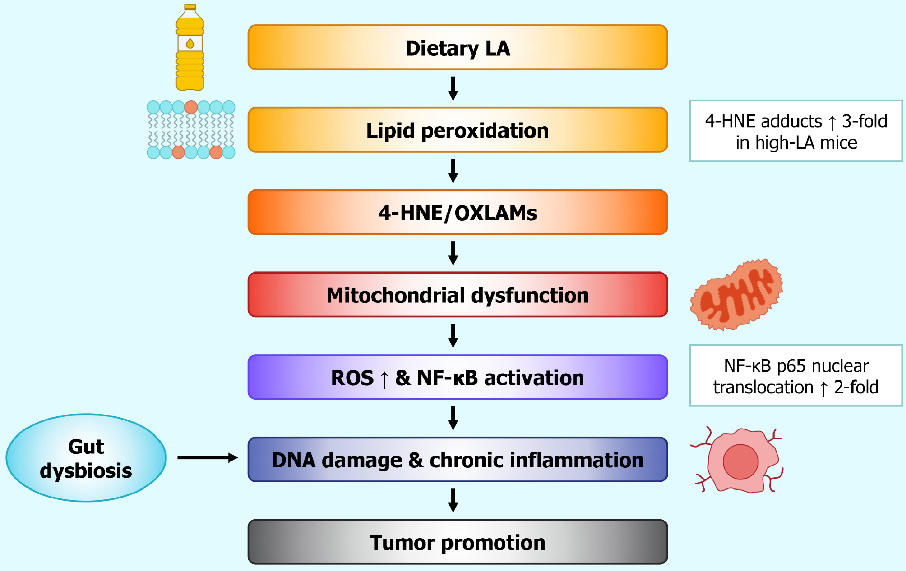
Figure 3 Proposed oxidative cascade linking excess linoleic acid to mitochondrial dysfunction and tumor promotion: Oxidation of membrane-bound linoleic acid yields 4 hydroxynonenal and related oxylipins, which impair mitochondrial respiration, elevate reactive oxygen species, and activate nuclear factor kappa-light-chain-enhancer of activated B cells-dependent inflammatory circuits; concurrent gut dysbiosis amplifies systemic cytokine tone, together fostering DNA damage and tumor promotion.
Quantitative insets reflect representative preclinical data. GLUT-1: Glucose transporter 1; VEGF: Vascular endothelial growth factor; HIF-1α: Hypoxia-inducible factor-1α; ETC: Electron transport chain; SDH: Succinate dehydrogenase; LA: Linoleic acid.
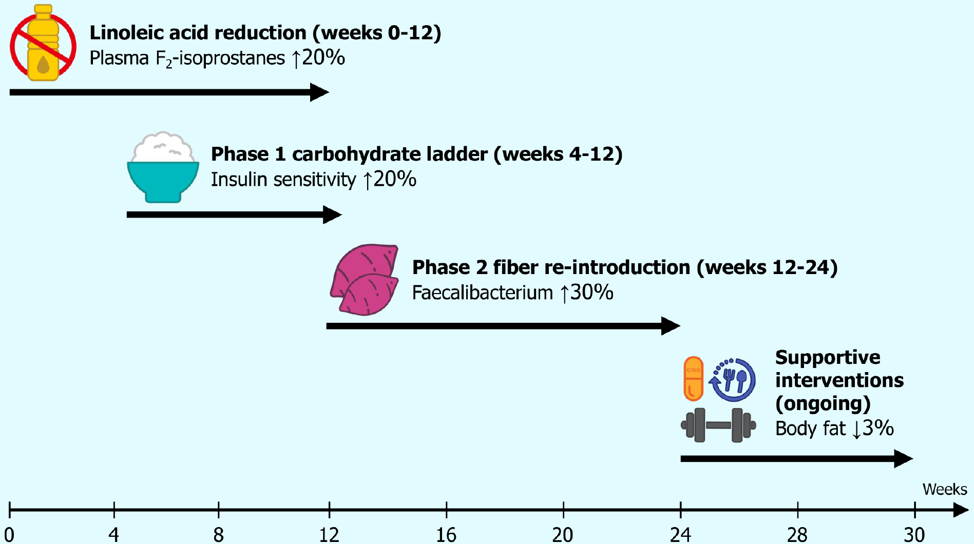
Figure 4 Four-phase dietary-lifestyle protocol for re-establishing a cancer-resistant metabolic terrain sequential dietary-lifestyle program aimed at restoring a cancer-resistant internal terrain: (1) Strict linoleic acid minimization; (2) Low-residue carbohydrate repletion; (3) Graded fermentable-fiber restoration to repopulate butyrogenic taxa; and (4) Adjunctive metabolic supports (intermittent fasting, pentadecanoic acid, exercise).
Quantitative targets reference published human or animal data where available.
- Citation: Mercola J. Historical rise of cancer and dietary linoleic acid: Mechanisms and therapeutic strategies. World J Clin Oncol 2025; 16(9): 110686
- URL: https://www.wjgnet.com/2218-4333/full/v16/i9/110686.htm
- DOI: https://dx.doi.org/10.5306/wjco.v16.i9.110686


