©The Author(s) 2025.
World J Clin Oncol. Sep 24, 2025; 16(9): 108748
Published online Sep 24, 2025. doi: 10.5306/wjco.v16.i9.108748
Published online Sep 24, 2025. doi: 10.5306/wjco.v16.i9.108748
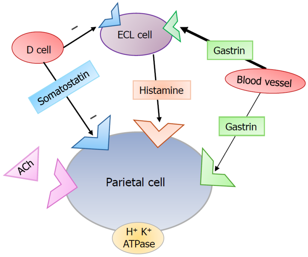
Figure 1 Gastric physiology, interactions between enterochromaffin-like and parietal cell.
This is a simplified schematic of interactions between enterochromaffin-like (ECL) and parietal cells that result in the normal physiologic release of gastric acid. This is an original figure. Ach: Acetylcholine; ECL: Enterochromaffin-like.
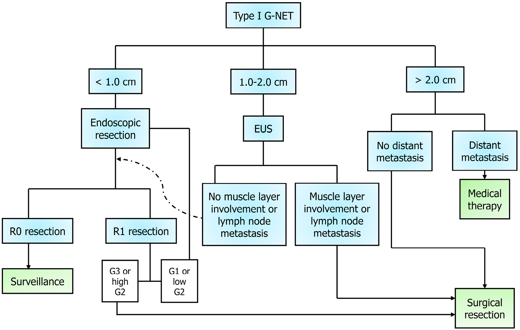
Figure 2 Management of localized gastric neuroendocrine tumors type I tumors.
This diagram provides a roadmap for the management of type I gastric neuroendocrine tumors, which are the most common and typically the most benign subtype. This is an original figure. EUS: Endoscopic ultrasound; G-NET: Gastric neuroendocrine tumor.
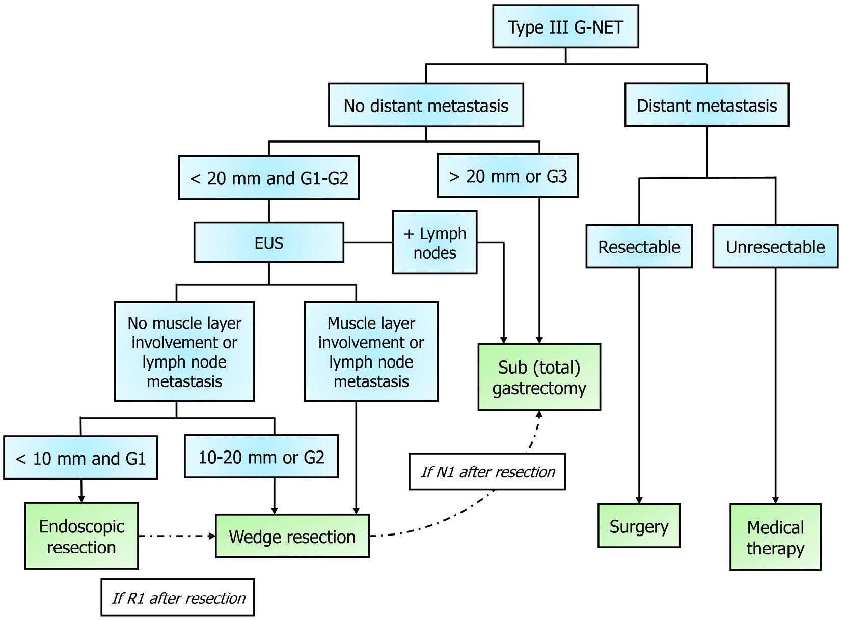
Figure 3 Management of type III gastric neuroendocrine tumors.
This diagram shows the decision-making involved in management of the most aggressive subtype of gastric neuroendocrine tumors, type III. These pathways are adapted from the European Neuroendocrine Tumor Society 2023 Guidelines. This is an original figure. EUS: Endoscopic ultrasound; G-NET: Gastric neuroendocrine tumor.
- Citation: Agathis AZ, Lopez-May M, Brown C, Divino CM. Gastric neuroendocrine tumors: A review of pathology and updated roadmap to surgical management. World J Clin Oncol 2025; 16(9): 108748
- URL: https://www.wjgnet.com/2218-4333/full/v16/i9/108748.htm
- DOI: https://dx.doi.org/10.5306/wjco.v16.i9.108748


