©The Author(s) 2025.
World J Clin Oncol. Oct 24, 2025; 16(10): 108876
Published online Oct 24, 2025. doi: 10.5306/wjco.v16.i10.108876
Published online Oct 24, 2025. doi: 10.5306/wjco.v16.i10.108876
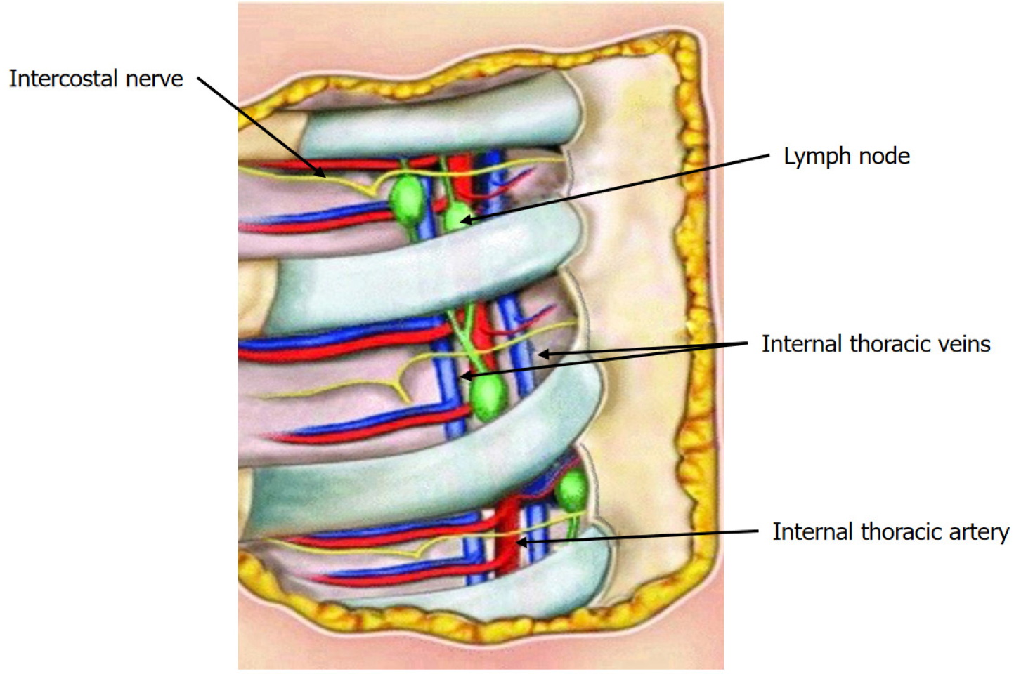
Figure 1 Anterior right exposure of the intercostal spaces[13].
Anatomical relationship of the internal mammary lymph node and internal mammary vessels within mediastinal fat tissue. Citation: Barros AC, Mori LJ, Nishimura D, Jacomo AL. Surgical anatomy of the internal thoracic lymph nodes in fresh human cadavers: Basis for sentinel node biopsy. World J Surg Oncol 2016; 14: 135. Copyright© The Authors 2016. Published by Springer Nature. This article is distributed under the terms of the Creative Commons Attribution 4.0 International License (http://creativecommons.org/Licenses/by/4.0/), which permits unrestricted use, distribution, and reproduction in any medium, provided you give appropriate credit to the original author(s) and the source, provide a link to the Creative Commons license, and indicate if changes were made. The Creative Commons Public Domain Dedication waiver (http://creativecommons.org/publicdomain/zero/1.0/) applies to the data made available in this article, unless otherwise stated.
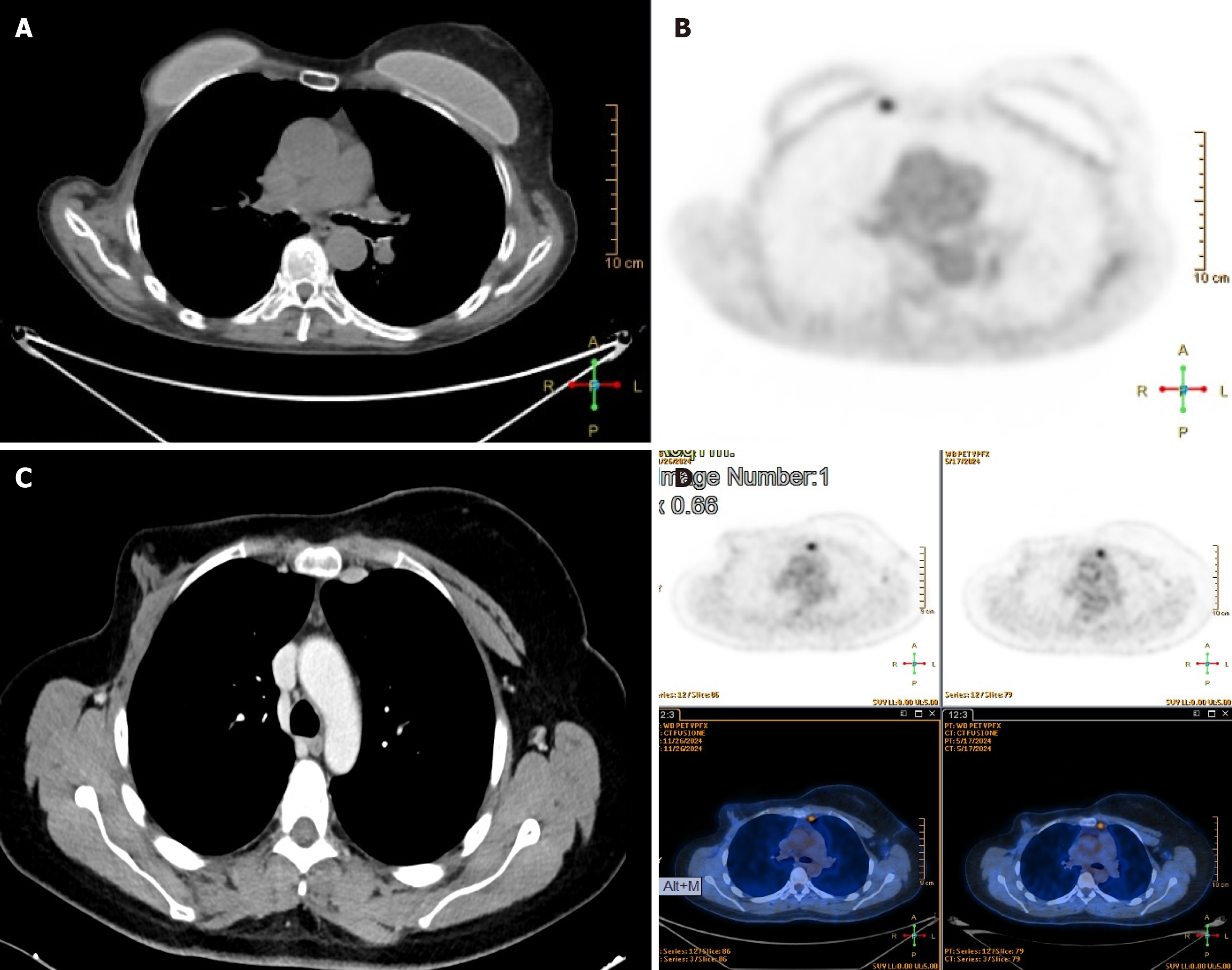
Figure 2 Chest computed tomography scan and positron emission tomography scan.
A and B: Left internal mammary lymphadenopathy; C and D: Right internal mammary lymphadenopathy.
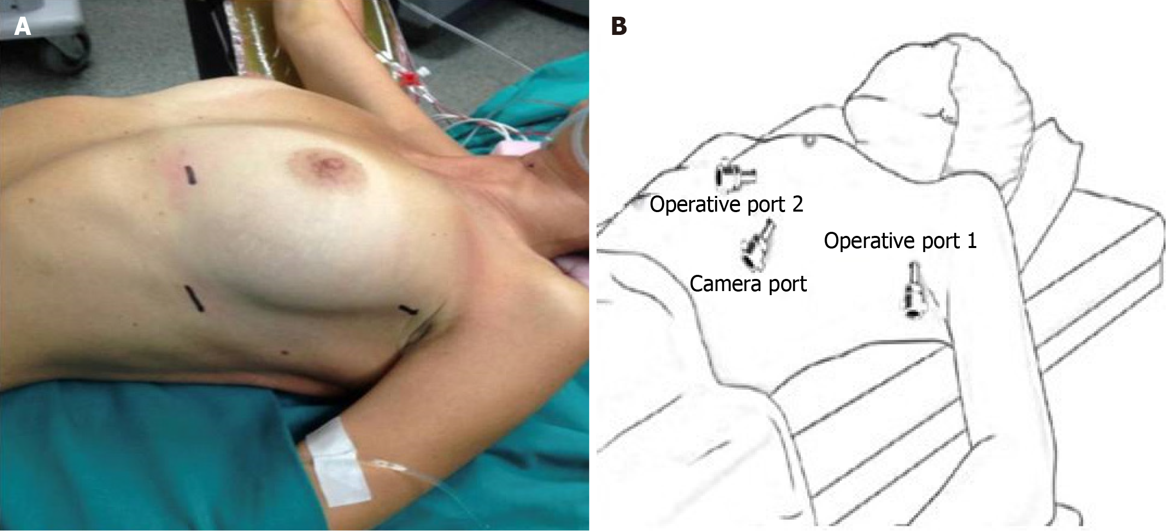
Figure 3 Patient position and port placement.
A: The patient is positioned to the side of the operating room table according to the side of the chest being surgically approached. The ipsilateral arm is fixed below the level of the table to improve the exposure of the chest; B: The first port (camera port) is placed at the fifth intercostal space at the level of the mid-axillary line. Under view guidance, the two operative ports are placed respectively in the third intercostal space (operative port 1) and the fifth intercostal space 3 cm to 4 cm to the parasternal line (operative port 2).
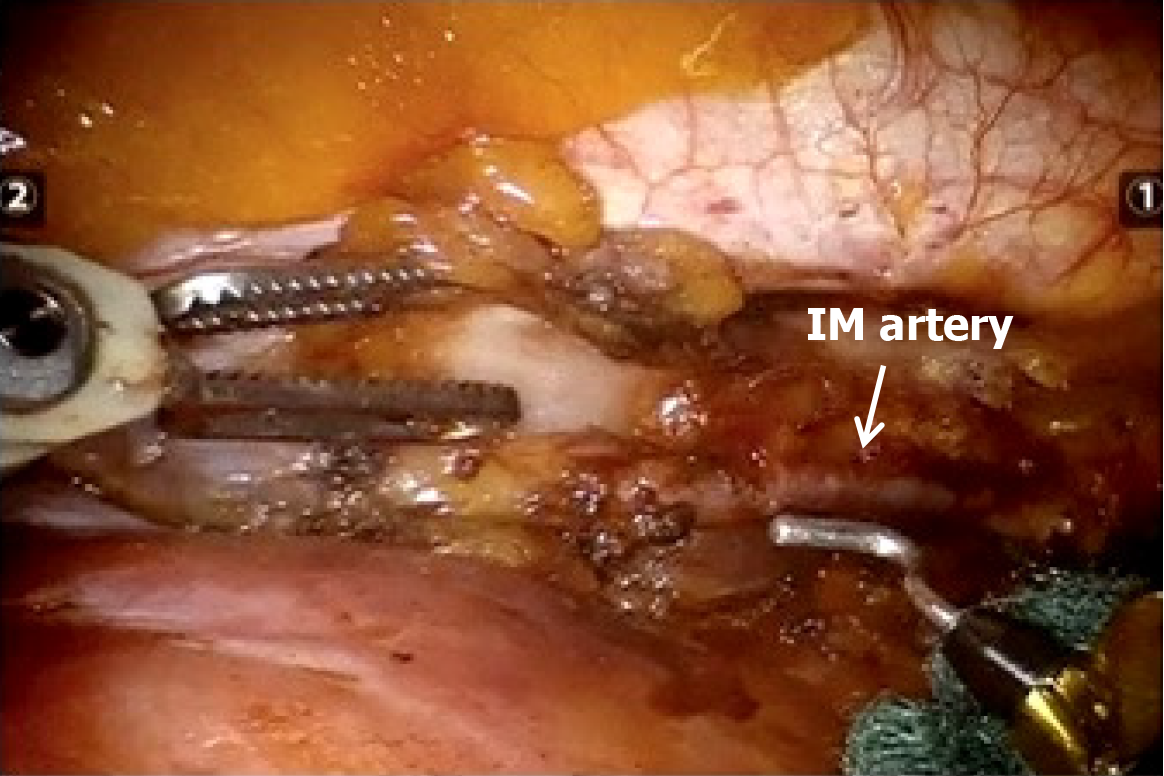
Figure 4 This is an intraoperative view of the left internal mammary chain.
The cautery hook in the rightward arm and the Cadiere forceps in the leftward arm allow performing isolation of the internal mammary artery and dissection of the surrounding fat tissue. IM: Internal mammary.
- Citation: Pardolesi A, Ferrari M, Leuzzi G, Stanzi A, Calderoni M, Uslenghi C, Scarci M, Raveglia F, Cioffi U, Solli P. Robotic approach for lymphadenectomy of internal mammary lymph nodes in breast cancer: Five case reports. World J Clin Oncol 2025; 16(10): 108876
- URL: https://www.wjgnet.com/2218-4333/full/v16/i10/108876.htm
- DOI: https://dx.doi.org/10.5306/wjco.v16.i10.108876


