©The Author(s) 2024.
World J Clin Oncol. Sep 24, 2024; 15(9): 1215-1221
Published online Sep 24, 2024. doi: 10.5306/wjco.v15.i9.1215
Published online Sep 24, 2024. doi: 10.5306/wjco.v15.i9.1215
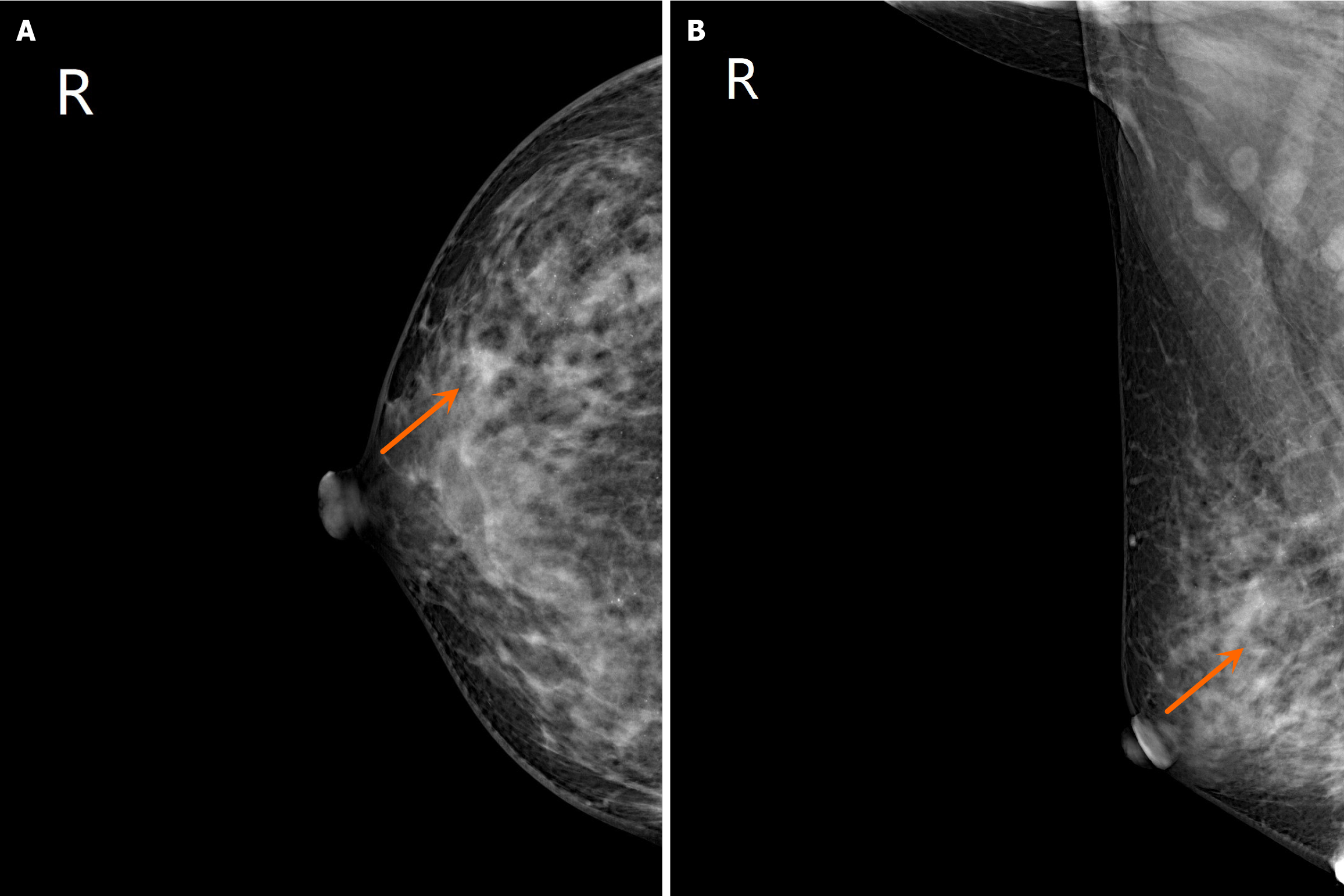
Figure 1 Molybdenum target mammography revealed local fibroid breast lesions around the right breast.
A: Right breast axis (breast cancer-right breast upper outer quadrant); B: Right breast oblique (breast cancer-right upper outer quadrant). The arrow points to the tumour.
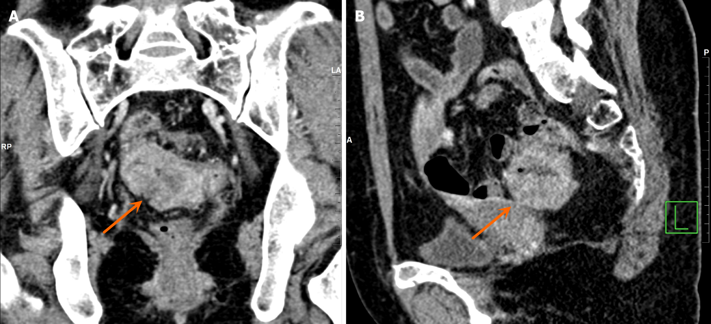
Figure 2 Pelvic magnetic resonance imaging reveals a sigmoid colon mass.
A: Pelvic computed tomography (CT)-coronal position; B: Pelvic CT-sagittal. The arrow points to the tumour.
Figure 3 Immunohistochemical staining of mismatch repair proteins in rectal cancer tissue revealed that the MLH1, MSH2, MSH6 and PMS2 proteins were expressed in the nucleus of cancer cells (100 ×).
A: MLH1 protein expression in the nucleus of cancer cells; B: MSH2 protein expression in the nucleus of cancer cells; C: MSH6 protein expression in the nucleus of cancer cells; D: PMS2 protein expression in the nucleus of cancer cells.
- Citation: Qin PF, Yang L, Hu JP, Zhang JY. Breast cancer and rectal cancer associated with Lynch syndrome: A case report. World J Clin Oncol 2024; 15(9): 1215-1221
- URL: https://www.wjgnet.com/2218-4333/full/v15/i9/1215.htm
- DOI: https://dx.doi.org/10.5306/wjco.v15.i9.1215


