©The Author(s) 2025.
World J Gastrointest Oncol. Sep 15, 2025; 17(9): 110505
Published online Sep 15, 2025. doi: 10.4251/wjgo.v17.i9.110505
Published online Sep 15, 2025. doi: 10.4251/wjgo.v17.i9.110505
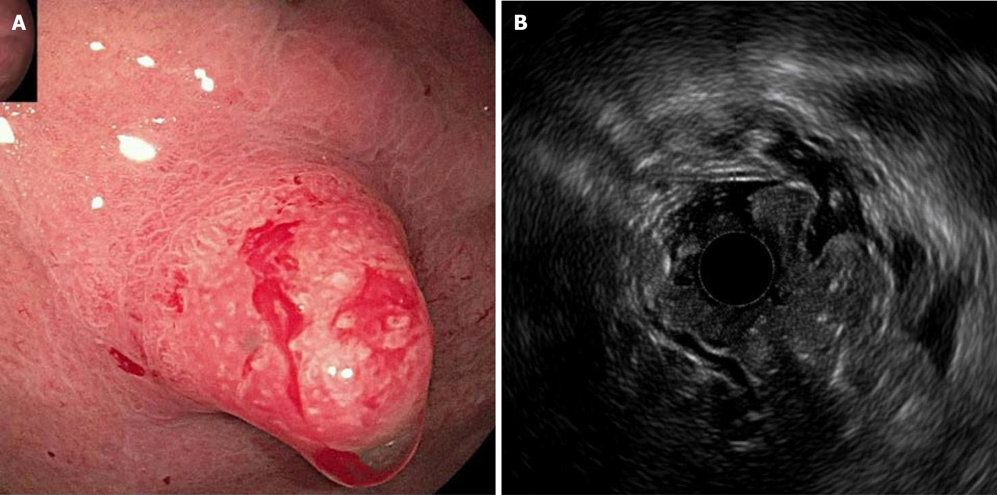
Figure 1 Endoscopic and endoscopic ultrasound findings.
A: Endoscopic findings, on April 14, 2022, endoscopy demonstrated that the surface showed a strawberry-like change and was hard with a tendency to bleed; B: Endoscopic ultrasound findings, endoscopic ultrasound findings indicate thickening of the mucosa and submucosa, interruption and disappearance of the mucosal muscular layer, and unclear layer boundaries.
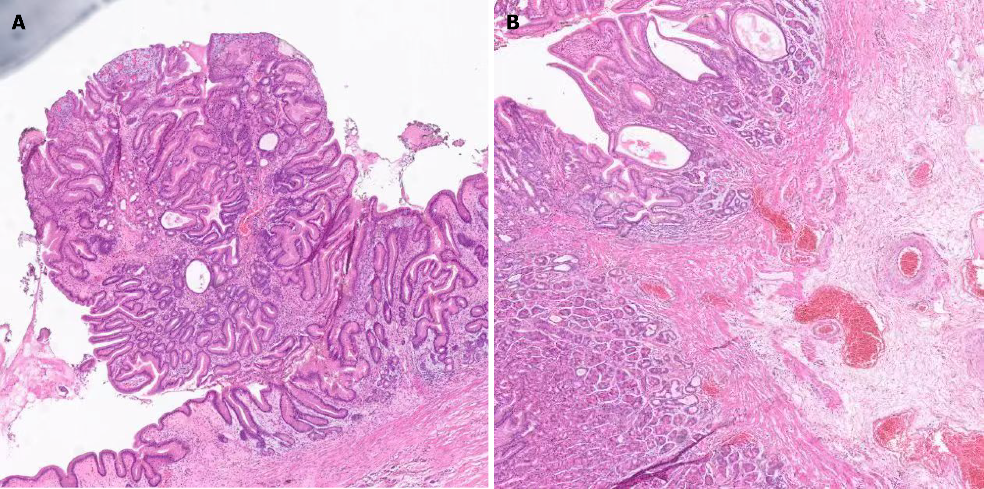
Figure 2 Pathological findings.
A: Hematoxylin-eosin staining (50 ×); B: Hematoxylin-eosin staining (100 ×). Pathology suggested that atypia is not obvious. The epithelium covering is normal and mainly shows the differentiation of parietal cells.
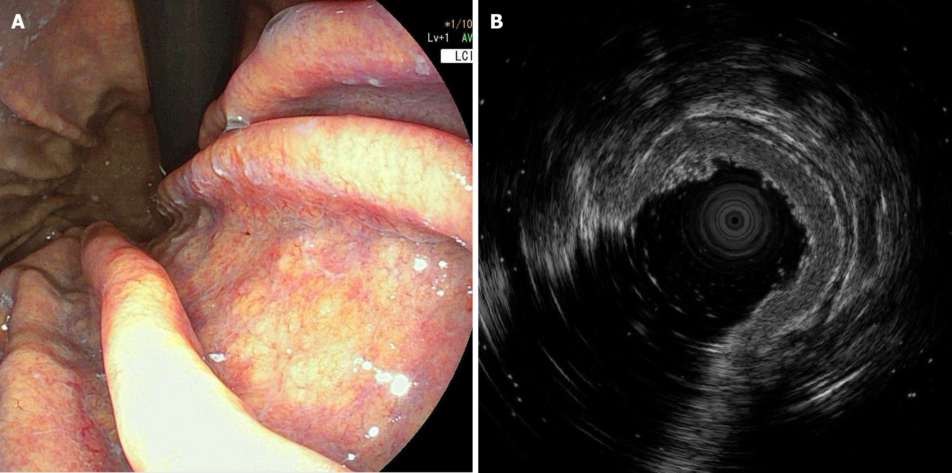
Figure 3 Pathology re-examination.
A and B: Thickening of the muscular layer of the lesser curvature mucosa of the gastric body was revealed after three years.
- Citation: Xue RX, Lu XY, Deng P, Wang LH, Yun YF. Misdiagnosis of gastric oxyntic gland adenoma as hyperplastic polyp: A case report. World J Gastrointest Oncol 2025; 17(9): 110505
- URL: https://www.wjgnet.com/1948-5204/full/v17/i9/110505.htm
- DOI: https://dx.doi.org/10.4251/wjgo.v17.i9.110505


