Dam PTM, Nguyen DD, Ngo ATT, Tran AM, Nguyen TT, Nguyen PV, Nguyen CQ, Nguyen ATT, Pham QTT, Than UTT. Adipose-derived mesenchymal stem cells from solid tissue and lipoaspirates: A comparative study of phenotype, growth, and secretome. World J Stem Cells 2026; 18(1): 110470 [PMID: 41608652 DOI: 10.4252/wjsc.v18.i1.110470]
Corresponding Author of This Article
Uyen Thi Trang Than, PhD, Assistant Professor, Vinmec-VinUni Institute of Immunology, College of Health Sciences, VinUniversity, Ocean Park, Da Ton, Gia Lam, Ha Noi 100000, Viet Nam. uyen.ttt@vinuni.edu.vn
Research Domain of This Article
Cell Biology
Article-Type of This Article
research-article
Open-Access Policy of This Article
This article is an open-access article which was selected by an in-house editor and fully peer-reviewed by external reviewers. It is distributed in accordance with the Creative Commons Attribution Non Commercial (CC BY-NC 4.0) license, which permits others to distribute, remix, adapt, build upon this work non-commercially, and license their derivative works on different terms, provided the original work is properly cited and the use is non-commercial. See: http://creativecommons.org/licenses/by-nc/4.0/
Baishideng Publishing Group Inc, 7041 Koll Center Parkway, Suite 160, Pleasanton, CA 94566, USA
Share the Article
Dam PTM, Nguyen DD, Ngo ATT, Tran AM, Nguyen TT, Nguyen PV, Nguyen CQ, Nguyen ATT, Pham QTT, Than UTT. Adipose-derived mesenchymal stem cells from solid tissue and lipoaspirates: A comparative study of phenotype, growth, and secretome. World J Stem Cells 2026; 18(1): 110470 [PMID: 41608652 DOI: 10.4252/wjsc.v18.i1.110470]
Author contributions: Dam PTM and Than UTT designed the experiments; Dam PTM, Nguyen DD, and Than UTT provided the study materials or patients; Ngo ATT, Tran AM, Nguyen TT, Nguyen PV, Nguyen CQ, Nguyen ATT, and Pham QTT performed the studies; Than UTT reviewed the data and manuscript content; All authors performed the studies, analyzed the data, and wrote the manuscript.
Institutional review board statement: The study was reviewed and approved by the Vinmec International General Hospital Joint Stock Company’s Ethics Committee (Ethical approval number: 01/2023/CN-HĐĐĐ VMEC). Tissues were only harvested when donors signed written informed consent before donating their samples.
Conflict-of-interest statement: The authors have no conflicts of interest to declare.
Data sharing statement: No additional data are available.
Corresponding author: Uyen Thi Trang Than, PhD, Assistant Professor, Vinmec-VinUni Institute of Immunology, College of Health Sciences, VinUniversity, Ocean Park, Da Ton, Gia Lam, Ha Noi 100000, Viet Nam. uyen.ttt@vinuni.edu.vn
Received: June 10, 2025 Revised: July 20, 2025 Accepted: November 18, 2025 Published online: January 26, 2026 Processing time: 227 Days and 12.2 Hours
Abstract
BACKGROUND
Mesenchymal stem cells (MSCs) are considered a promising therapy for various diseases due to their strong potential in regenerative medicine and immunomodulation. The tissue source of MSCs has gained attention for its role in influencing their function, accessibility, and readiness for clinical use.
AIM
To identify the most suitable adipose source for MSC isolation and expansion for further applications.
METHODS
We isolated MSCs from solid adipose tissue and liposuction aspirates using the enzyme method. The MSCs were examined for their expansion using population doubling time, differentiation capacity using multilineage differentiation induction, surface markers using flow cytometry, and stability of chromosomes using the karyotyping method. Growth factors and cytokines in MSC-conditioned media were analyzed using the Luminex assay.
RESULTS
MSCs were isolated from solid adipose tissue and lipoaspirates and expanded from passage 0 to passage 2. All adipose-derived MSCs (AD-MSCs) exhibited the typical elongated, spindle-shaped morphology and comparable proliferation rate. They expressed positive surface markers [cluster of differentiation 73 (CD73): > 97%, CD90: > 98%, and CD105: > 95%], and negative markers (< 1%). All MSCs expressed similar levels of stemness genes (octamer-binding transcription factor 4, SRY-box 2, Krüppel-like factor, and MYC), colony-forming, and trilineage differentiation potential. Karyotyping analysis revealed normal chromosomal patterns in all samples, except one sample exhibiting a polymorphism (1qh+). Furthermore, the growth factors and cytokines of hepatocyte growth factor, vascular endothelial growth factor A, interleukin 6 (IL-6), and IL-8 were detected in all AD-MSC conditioned media; but fibroblast growth factor-2 and keratinocyte growth factor were selectively expressed in conditioned media from solid or lipoaspirate AD-MSCs, respectively.
CONCLUSION
These findings indicate that AD-MSCs from both adipose sources possess all of the characteristic features of MSCs with source-specific secretome differences, which are suitable for further expansion and various clinical applications.
Core Tip: This study compared mesenchymal stem cells (MSC) derived from solid adipose tissue and lipoaspirates, showing that both sources yielded cells with similar morphology, growth, stemness, and differentiation potential. While overall characteristics were comparable, some growth factors were uniquely expressed depending on the source. These findings support the suitability of both adipose sources for MSC isolation and highlight subtle differences that may influence future therapeutic applications.
Citation: Dam PTM, Nguyen DD, Ngo ATT, Tran AM, Nguyen TT, Nguyen PV, Nguyen CQ, Nguyen ATT, Pham QTT, Than UTT. Adipose-derived mesenchymal stem cells from solid tissue and lipoaspirates: A comparative study of phenotype, growth, and secretome. World J Stem Cells 2026; 18(1): 110470
Mesenchymal stem cells (MSCs) are a type of multipotent SC that were first found in the bone marrow by Friedenstein et al[1,2]. Currently, MSCs can be isolated from several tissues, including bone marrow, adipose tissue, umbilical cord, umbilical cord blood, menstrual blood, and even dental pulp; many of them are medical discards and invasive collections, etc[3]. They have characteristics of SCs, including self-renewal and the ability to differentiate into various mesodermal cell types, including those that comprise bone, cartilage, fat, and muscle[3]. MSCs are of great interest in regenerative medicine due to their potential to repair or replace damaged tissues. They are being researched for their role in treating a variety of conditions, including orthopedic injuries, cardiovascular diseases, and autoimmune disorders. Additionally, MSCs also have immunomodulatory properties, meaning they can influence the immune system and may help reduce inflammation or modulate immune responses, which is another area of active research. Thus, MSCs are used to treat diseases or injuries, and in tissue engineering, where MSCs are used to create or repair tissues and organs. It is noted that the origin of MSCs enables different cellular characteristics and their functions, and further applications[4]. For example, MSCs originated from dental tissues have a better proliferation rate, whereas MSCs from adipose and gingival tissue showed a higher clonogenicity[4]. MSCs from bone marrow, widely used in orthopedic regenerative medicine, are preferable for their differentiation ability. Additionally, MSCs from bone marrow, adipose tissues, and dental pulp are considered as potential candidates for oral and dental regeneration[3].
Adipose-derived MSCs (AD-MSCs) are a type of MSC and are typically harvested from liposuction aspirates or abdominal fat biopsies[5]. They are relatively abundant and can be obtained with less invasiveness compared to other sources, such as bone marrow. This cell type has garnered significant interest due to its relatively easy accessibility and its potential for various therapeutic applications. AD-MSCs have reported their ability to differentiate into various cell types, which makes them valuable in treating conditions such as osteoarthritis, cardiovascular diseases, and soft tissue injuries. They can also be used in tissue engineering to create scaffolds or matrices for regenerating tissues or organs. For instance, AD-MSCs can be used to develop bone grafts or cartilage implants[6]. In wound healing areas, AD-MSCs can promote tissue repair, facilitate the healing process, and reduce inflammation by secreting various growth factors and cytokines[7]. This cell type is also being investigated for its potential in treating autoimmune diseases and other conditions where modulation of the immune system is beneficial[8]. Moreover, AD-MSCs are an important therapy for anti-aging due to their regenerative properties, which might help rejuvenate aging tissues and improve skin texture or volume. Overall, AD-MSCs are a promising tool in both clinical and research settings due to their accessibility, versatility, and potential therapeutic benefits. However, ongoing research is needed to fully understand their capabilities and to optimize their use in various medical applications.
It is noted that autologous fat grafting obtained from liposuction has become widely used due to its versatility, safety, and biocompatibility. Clinically, fats harvested through liposuction techniques have been effectively employed for soft tissue augmentation, correction of contour deformities, and rejuvenation of various anatomical regions, including the face, breasts, and buttocks[9,10]. To prepare fats for grafting, donor sites will be selected and injected with an enzyme to facilitate lipoaspiration[10,11]. Next, different centrifugation or filtration methods are used to concentrate fats and remove liquids that contain enzymes[10]. The effectiveness of fat graft largely depends on the quality of fats harvested, which largely depends on donors and sites; despite its widespread acceptance, factors such as fat graft survival, absorption rate variability, and optimal harvesting methods remain areas of ongoing research and debate[12]. Additionally, recent advances have broadened its therapeutic applications, such as scar revision, wound healing enhancement, and even regenerative therapies, as it contains a rich content of AD-MSCs[11]. Therefore, AD-MSCs from liposuction have been an alternative source to expand in vitro for basic research and clinical applications.
Despite the fact that AD-MSCs can be isolated from both solid fat tissue and liposuction aspirates[5], there needs to be more direct comparisons of cells harvested from these two technical samples. Additionally, the viability, proliferation, and stemness of AD-MSCs can be influenced by the type of harvesting procedure. Recently, a comparison of surgical resection, power-assisted liposuction, and laser-assisted liposuction showed that these methods can affect the number of isolated cells, clonogenicity, and doubling time[13]. The author concluded that power-assisted liposuction is the best choice of method for AD-MSCs due to the high proliferation potential and slow senescence of isolated cells[13].
Thus, this study compared AD-MSCs isolated from solid adipose tissue and enzyme-assisted liposuction aspirates in order to select the most appropriate method. In aesthetic surgery, liposuction aspirates are medical waste, enabling them to be a potential source of AD-MSCs for autologous and allogenic applications and material for other applications.
MATERIALS AND METHODS
AD-MSC isolation
The solid fat tissues were obtained from three donors, whereas the liposuction was received from five different donors. All samples were collected from abdominoplasty. The participants were provided informed consent. The first steps of the protocol differ from solid fat to liposuction. From the solid fat, 10 g of adipose tissues were washed with Hanks’ balanced salt solution (HBSS) (Gibco, NY, United States) and minced into small pieces (at a range of 1-3 mm). Then, 10 mL of minced fats were transferred into a 50 mL conical tube. For the liposuction, 10 mL of fat was moved into a 50 mL reaction tube. Both of them were washed three times with 20 mL HBSS to remove the blood fraction. Next, 25 mL of HBSS containing 200 unit/mL collagenase type I (Gibco, Denmark) and 200 g/L human albumin (Baxter, Deerfield, IL, United States) was added to the mixture, which was incubated at 37 °C with agitation 300 rpm/minute for 60 minutes. After the enzymatic digestion, the mixture was passed through a 70 μm filter to remove cell clumps and then centrifuged at 500 × g for 10 minutes to collect the stromal vascular fraction as a pellet. Finally, the stromal vascular fraction containing the MSCs was resuspended in StemMACS MSC Expansion Media (Miltenyi Biotec, Bergisch Gladbach, Germany), supplemented with 1% penicillin/streptomycin (Gibco, United States). Cells were incubated in a humidified environment at 37 °C and 5% CO2. To remove non-adherent cells, the medium was exchanged after 3 days. After reaching a confluence of 80%-90%, the cells were detached by incubation with TrypLE™ Select Enzyme (Gibco, United States), harvested, and washed with the corresponding completed medium. Cells were counted after staining with trypan blue (Life Technologies, Carlsbad, CA, United States). Cells were expanded and cryopreserved at the end of passage 0 (P0) and P1.
Population doubling time
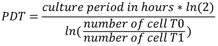
MSCs isolated from solid fat tissues and liposuction aspirates were examined for their proliferation rate through population doubling time (PDT) in each passage. PDT was identified following the formula:
T0 is the seeding cell number and T1 is the cell number at harvest. Cells were stained with trypan blue and counted using the hemocytometer.
Colony-forming unit assay
To test the capacity of AD-MSCs to form fibroblast colony-forming units, AD-MSCs at P1 were seeded at a density of 4 cells/cm2 in the 24-well plate in different culture media. The culture medium was maintained for 2 weeks, and fresh media were replaced every 3 days. Then, the cells were fixed in absolute ethanol and stained with Giemsa 1 × for 15 minutes. Then cells were washed with distilled water to remove excess Giemsa, and the number of colonies was identified and quantified under a microscope.
Multilineage differentiation
AD-MSCs were seeded in 96-well plates to evaluate their multilineage differentiation capacity into adipocytes, osteocytes, and chondrocytes. After the initial culture of 2 days, the medium was replaced with an induction medium. For adipogenic differentiation, cells were maintained for 20 days before being fixed in 4% formaldehyde for 30 minutes and washed with phosphate-buffered saline (PBS). Subsequently, cells were stained with a 0.5% Oil Red O (Sigma Aldrich, St. Louis, MO, United States) diluted in distilled water at room temperature for 15 minutes and washed with PBS to observe the lipid-rich adipocytes. For osteogenic differentiation, AD-MSCs were maintained for 25 days before being fixed in 4% formaldehyde for 30 minutes and washed with PBS. Then the cells were stained with a 2% Alizarin Red S (Sigma Aldrich) and rewashed twice with distilled water to visualize the mineralized matrix produced by osteoblasts. Regarding chondrogenic differentiation, AD-MSCs were incubated with the chondrogenic differentiation medium for 22 days. Then, they were fixed in 4% formaldehyde for 30 minutes, washed with PBS, and stained with Alcian Blue (Sigma-Aldrich) in 3% acetic acid. Finally, cells were washed with HCL 0.1 N and distilled water twice. Images of differentiated cells were captured using an inverted microscope.
Flow cytometry for marker analysis
AD-MSC surface markers were examined at P1 and P2 by flow cytometry using a Human MSC Analysis Kit (BD Biosciences, San Diego, CA, United States). Cells were stained with positive control [anti-cluster of differentiation 73 (CD73) APC, anti-CD90 PerCP-Cy5.5, and anti-CD105 FITC] and negative cocktail control (anti-CD45 PE, anti-CD34 PE, anti-CD11b/CD19 PE, and HLA-DR PE). The results were captured and analyzed using a Beckman Coulter flow cytometer (Beckman Coulter, Brea, CA, United States) with Navios software 3.2.
Gene expression analysis using quantitative reverse transcription polymerase chain reaction
Total RNA was extracted using TRIzol™ reagent (Thermo Fisher Scientific, Waltham, MA, United States) at a 9:1 ratio of TRIzol to cell suspension. The lysis mixture was supplemented with magnesium chloride and chloroform, then incubated at room temperature. The aqueous phase was collected and incubated with isopropanol overnight at -20 °C. RNA was centrifuged and washed twice with RNase-free 75% ethanol, followed by air drying and resuspended in RNase-free water. Complementary DNA was synthesized using SuperScript™ IV Reverse Transcriptase (Thermo Fisher Scientific), following the manufacturer’s protocol. The resulting complementary DNA was used for quantitative polymerase chain reaction with primers specifically designed for stemness markers [octamer-binding transcription factor 4 (OCT4), SRY-box 2 (SOX2), Krüppel-like factor (KLF), and MYC], and differentiation markers [alkaline phosphatase (ALP), parathyroid hormone-related protein (PTHR), and collagen type I alpha 1 chain (COL1A1) for osteogenesis; SOX9 and COL3A1 for chondrogenesis; and LEPTIN and peroxisome proliferator-activated receptor (PPAR) for adipogenesis]. Gene expression levels were analyzed using the ∆Ct method, with glyceraldehyde 3-phosphate dehydrogenase (GAPDH) serving as the internal control.
Karyotyping test
To evaluate the stability of chromosomes in the cells during the culture, we used the karyotyping technique to analyze chromosomes when the cells at P2 were in the metaphase stage. When the cell was in the growth phase (coverage density 60%-80%), the culture medium was removed, and the cell was incubated with KaryoMax Colcemid (Life Technology) (1:100) at 37 °C and 5% CO2 for 30 minutes. The medium was discarded, and then 2 mL of 0.25% trypsin-EDTA solution (Gibco, United States) was added and incubated at 37 °C for 3 minutes for cellular detachment. The cell suspension was collected and centrifuged at 500 × g/5 minutes/20 ℃ before 9 mL of 0.5% potassium chloride warmed at 37 °C was added to the tube containing cell pellets and incubated for 30 minutes. The cells were then washed with Carnoy’s solution (3 methanol: 1 acetic acid). The cell mixture was transferred onto a slide and immediately placed on a drying table at 60-70 °C, and then the slides were dried overnight or dried at 80-90 °C for 4 hours. The dried slides were immersed in the newly prepared 2.5% trypsin solution warmed up to 37 °C for 3 to 5 seconds, removed and the reaction was inactivated by immersion in Gurr Buffer (Gibco, United States). The cells were then stained with Giemsa (3 mL of Giemsa in 50 mL of Gurr Buffer) for 8 minutes. Rinse cells with water and allow them to dry until photographing. Chromosomes were captured under the microscope Metafer Msearch4 (Heidelberg, Germany) and analyzed with Metaphase Analysis software.
Growth factor analysis using an immunoassay
MSCs were cultured to P2 and reached 90% confluency, and conditioned media were collected for growth factor and cytokine analysis. A Luminex assay was performed using the ProcartaPlex™ Multiplex Immunoassays (Human Custom ProcartaPlex 7-Plex Kit) (Thermo Fisher Scientific) to quantify seven growth factors and cytokines in MSC supernatants, including fibroblast growth factor-2 (FGF-2), hepatocyte growth factor, platelet-derived growth factor BB (PDGF-BB), vascular endothelial growth factor A (VEGF-A), interleukin 6 (IL-6), IL-7, and IL-8. Briefly, capture beads were added to each well, followed by the addition of the supernatant, which was incubated for 2 hours. Then the wells were washed with washing buffer to remove unbound reagents. All procedures were carried out according to the manufacturer’s instructions. The luminescent signal was detected using the Luminex™ 100/200™ system with xPONENT 3.1 software.
Additionally, the level of keratinocyte growth factor (KGF) in the MSC supernatant was measured using an enzyme-linked immunosorbent assay (Abcam, Cambridge, United Kingdom). In brief, the supernatant was added to each well and incubated for 2.5 hours. After removing unbound components by washing, a biotinylated secondary antibody was added, followed by streptavidin-conjugated horseradish peroxidase. The KGF concentration was determined based on absorbance measured at 450 nm using a SpectraMax M5 plate reader (Molecular Devices, Silicon Valley, CA, United States), and quantified against a standard curve.
Statistical analyses
The statistical analyses were performed on GraphPad Prism 10 (GraphPad Software, CA, United States) using the t-test. The statistical significance was defined as P < 0.05. All data are shown as the mean ± SD of at least three biological replicates.
RESULTS
Isolation efficacy of AD-MSCs from solid tissues and liposuction
AD-MSCs were isolated from adipose tissues and liposuction aspirates (Table 1) using enzymatic methods and seeded for the expansion of cells at P0. When cells reached 80% confluency, we harvested and counted cells for the analysis of isolation efficacy. The results showed that despite the number of AD-MSCs at P0 from adipose tissue being higher than liposuction (Table 1), the difference is not significant (P = 0.237). These data indicate that the enzymatic method is appropriate for both solid adipose tissues and liposuction aspirates. We evaluated the proliferation of primary AD-MSCs from P0 to P1 through PDT, where the lower PDT is faster proliferation. Data showed that there was no difference between PDTs of the AD-MSCs from adipose tissue and liposuction (Figure 1A). Observation showed that AD-MSCs, both from tissue and liposuction, grew fast when PDT was 15.909 ± 3.07 hours and 16.074 ± 3.782 hours, respectively. Regarding cell morphology, all MSCs isolated from solid adipose tissues and liposuction aspirates were heterogeneous cell populations. Most MSCs expressed spindle shapes; several cells expressed cuboidal or polygonal shapes (Figure 1B). These are typical morphologies of human MSCs in 2D cultures.
Figure 1 Population doubling times and morphology of mesenchymal stem cells isolated from adipose tissue and liposuction aspirates.
A: There was no difference in the population doubling times (PDTs) of primary adipose-derived mesenchymal stem cells at passage 2 (P2) originating from solid adipose tissue (AD) and liposuction aspirates, and all cells grew fast with PDTs < 20 hours; B: Similar morphology of mesenchymal stem cells at P0 and P1 from solid AD and liposuction aspirates.
Table 1 Characteristics of fat samples for cell harvest and isolation efficacy.
We analyzed the expression of stemness genes in AD-MSCs isolated from solid tissue and liposuction aspirates. The results showed that there was no significant differences in the expression levels of OCT4, SOX2, KLF, and MYC in the MSCs from two cell sources (Figure 2A). Using GAPDH as an internal control, all four stemness genes were normalized to this gene; the expression of KLF was the highest (∆Ct < 0) compared to the three others (∆Ct > 0). Additionally, results from marker analysis of AD-MSCs at P1 and P2 showed that all positive markers, including CD73, CD90, and CD105, were greater than 95%, and the negative marker panel (CD45, CD34 PE, CD11b/CD19, and HLA-DR) was smaller than 2% (Figure 2B). These data indicate that AD-MSCs from both solid tissue and liposuction aspirates express a typical characteristic of MSCs in terms of immunophenotype.
Figure 2 Expression of stemness gene and surface markers of primary adipose-derived mesenchymal stem cells.
A: No differences in the expression level of stemness genes, including octamer-binding transcription factor 4 (OCT4), SRY-box 2 (SOX2), Krüppel-like factor (KLF), and MYC, were observed between primary mesenchymal stem cells (MSCs) at passage 2 (P2) isolated from adipose tissue (AD) and liposuction aspirates; B: Marker expression of primary adipose-derived MSCs at P1 and P2.
Capacity of AD-MSCs in colony formation and multilineage differentiation
Human AD-MSCs derived from solid adipose tissues and liposuction aspirates were further examined for colony formation and trilineage differentiation. Regarding colony-forming units, AD-MSCs were seeded with low cell density (4 cells/1 cm2), and cells proliferated into large numbers and formed colony units after 14 days (Figure 3A). Additionally, every human AD-MSC lineage derived from solid adipose tissues and liposuction aspirates was positive for adipogenesis, osteogenesis, and chondrogenesis. They expressed a great capacity for differentiation by forming lipid droplets, mineralized nodules, and micromass in vitro in the presence of an inducing medium (Figure 3B). Then we further examined the expression of gene markers for trilineage differentiation at the time before and after differentiation induction (at the time of trilineage differentiation data collection). ∆Ct data indicate that there were no significant differences in the gene expression, including ALP, PTHR, and COL1A1, which are osteogenesis markers; SOX9 and COL3A1 are chondrogenesis markers, and PPAR is an adipogenesis marker in MSCs isolated from solid adipose tissues and liposuction aspirates at points before or after differentiation induction (Figure 3C). The only differences belong to LEPTIN, which is a marker gene for adipogenesis, expressed differently in MSCs isolated from liposuction aspirate, where a lower level of LEPTIN gene was detected before induction (∆Ct = 16.5 ± 0.13) and a higher level of the gene in adipogenesis-induced MSCs (∆Ct = 13.25 ± 0.75).
Figure 3 Similar capacity of colony-forming and multilineage differentiation of primary adipose-derived mesenchymal stem cells.
A: Ability to form colony units by human primary adipose-derived mesenchymal stem cells; B: Capacity of multi-lineage differentiation of adipocytes, osteocytes, and chondrocytes by human primary adipose-derived mesenchymal stem cells; C: Expression of differentiated marker genes before and after differentiation induction, where alkaline phosphatase (ALP), parathyroid hormone-related protein (PTHR), and collagen type I alpha 1 chain (COL1A1) are markers for osteogenesis; SRY-box 9 (SOX9) and COL3A1 are markers for chondrogenesis; and LEPTIN and peroxisome proliferator–activated receptor (PPAR) are markers for adipogenesis. aP < 0.05. AD: Adipose tissue; CFU: Colony-forming unit.
Karyotyping examination
We also examined the stability of chromosomes of primary AD-MSCs during the expansion using the karyotyping technique. The results showed that all cell lines expressed typical genetic characteristics of chromosomes, including chromosome number, size, shape, and structural changes (Figure 4A). There was only a polymorphism (1qh+) in an AD-MSC lineage, occupied for 10% of all cell samples analyzed (Figure 4B).
Figure 4 Karyotype characteristics of primary adipose-derived mesenchymal stem cells during cultures.
A: Normal chromosomes from liposuction-derived mesenchymal stem cells; B: Chromosomes with polymorphisms 1qh+ from adipose tissue-derived mesenchymal stem cells.
Expression levels of growth factors and cytokines in the MSC conditioned media
To examine the secretome of MSCs, we examined the levels of several growth factors and cytokines in the supernatant after 5 days of MSC cultures (conditioned media). Results showed that among six factors analyzed, two growth factors of PDGF-BB and VEGF-A and two cytokines of IL-6 and IL-8 expressed with similar levels in conditioned media from MSCs isolated from solid adipose tissues or liposuction aspirates (Figure 5). Furthermore, PDGF-BB and IL-7 were not detected in all samples investigated in this study. Interestingly, KGF was only detected in the MSC supernatant related to solid adipose tissue, whereas FGF-2 was only detected in the MSC supernatant related to liposuction aspirates (Figure 5). This indicates that there may be different components in the secretome of MSCs related to solid adipose tissues or liposuction aspirates.
Figure 5 Levels of growth factors and cytokines in the conditioned media of adipose-derived mesenchymal stem cell cultures at passage 2.
There was no significant difference in the expression level of hepatocyte growth factor (HGF), vascular endothelial growth factor A (VEGF-A), interleukin 6 (IL-6), and IL-8 from mesenchymal stem cell (MSC) supernatants associated with solid adipose tissue (AD) and liposuction aspirates. Keratinocyte growth factor (KGF) was only detected in the MSC supernatant related to solid adipose tissue, while fibroblast growth factor-2 (FGF-2) was only detected in the MSC supernatant related to liposuction aspirates.
DISCUSSION
MSCs have been emerging as a potential candidate for regenerative medicines and chronic and incurable diseases. Despite tissue sources of MSCs can affect the treatment efficacy, but scientists are also interest in MSCs harvested from medical wastes including birth-derived tissues, dental pulp, menstrual blood, and liposuction aspirates[3]. In this study, we isolated AD-MSCs using enzymatic methods from solid adipose tissues and liposuction aspirates from abdominal aesthetic surgery and expanded them up to P2. Previously, MSCs could be isolated from the infranatant layer of the suction canister after liposuction; however, the harvested cell number appears to be significantly lower than the number of cells in solid tissues digested by collagenase. Additionally, methods for isolating AD-MSCs typically rely on either enzymatic digestion or mechanical processes; each technique exhibits advantages and limitations. For example, enzymatic digestion, most commonly using collagenase, effectively disrupts the extracellular matrix to generate a high yield of viable MSCs having robust proliferative and differentiation potentials[14-16]. This approach is highly reproducible and standardized, allowing consistent isolation of cells; however, the enzymatic method may introduce variability due to enzyme source, concentration, digestion time, and temperature, which can potentially influence cell viability and quality[14,16]. Several studies have compared two common methods for isolating AD-MSCs: Enzymatic digestion (typically using collagenase) and mechanical processes such as shear force, centrifugation, or specialized devices. Evidence shows that both methods provide comparable results in terms of cell yield and cell viability, although mechanical processes are generally faster and more cost-effective compared with enzymatic digestion[17]. Specifically, enzymatic methods typically achieve cell viability in the range of 70%-99%, while mechanical isolation yields similar viability values, ranging from 46%-97.5%[17]. Additionally, a study using the “microlyzer” system, which is a mechanical and enzyme-free method, demonstrated that this approach produced a comparable number of progenitor cells, with viability and proliferative capacity similar to enzymatic methods, and even microlyzer-derived cells exhibited faster differentiation and stronger gene marker expression[18]. Despite the fact that mechanical processes are simpler and enzyme-free alternatives that can reduce procedure costs. However, cells harvested from these techniques are often fewer in cell number and potentially lower in purity, viability, and proliferative capacity compared to enzymatic approaches due to increased cellular trauma and incomplete tissue disruption[14,16]. The lack of standardized protocols for outcome measurement makes it difficult to definitively identify the superior method in clinical contexts. The enzymatic digestion remains the gold standard for AD-MSC isolation due to its consistent efficiency and high cell viability; on the other hand, mechanical methods appear to be a viable alternative, offering advantages in cost, time, and point-of-care feasibility[17-20]. Therefore, research groups have to develop their own protocol for AD-MSC isolation. Thus, in this study, we used collagenase to support the digestion of fat fragments and stromal vascular fraction. The AD-MSCs expressed typical morphology, immunotypes, capacity for colony forming, and trilineage differentiation. Karyotype tests show nine of 10 have normal chromosomes. These characteristics of AD-MSCs have been reported previously and are consistent with our results[6,7,21].
Importantly, AD-MSCs isolated in this study expressed a great proliferative rate, as indicated by the low PDT values (15.909 hours and 16.074 hours). The PDTs of MSCs derived from human adipose tissues have been reported to be greater than 40 hours from P1 to P7 or even up to P11[22,23]. The PDTs increase following the larger passages of the cells. For example, Izadpanah et al[24] and Zhu et al[25] reported that PDTs of human AD-MSCs are 48-60 hours at P20 and 110 hours at P30, and 36 hours at P3 and 96 hours at P25, respectively. The PDTs might be affected by the characteristics of donors, such as age[26]. For this aspect, the better PDTs from our study may be due to the tissues and liposuctions harvested from healthy and young people who were under aesthetic surgery. There are only reports on the short PDTs on canine-derived AD-MSCs (15.8 hours)[27] and female sheep AD-MSCs (29.41 hours to 51.61 hours)[28]. However, the application of animal SCs on human is a serious ethical issue that are in argument.
MSCs derived from both solid adipose tissue and liposuction aspirates secreted comparable levels of hepatocyte growth factor, VEGF-A, IL-6, and IL-8 in their supernatants. However, KGF was detected only in the supernatant of MSCs derived from liposuction aspirates, while FGF-2 was exclusively found in the supernatant of MSCs from solid adipose tissue. Previous studies have reported the presence of PDGF-BB, VEGF, IL-6, IL-7, and IL-8 in the conditioned media of MSCs derived from various sources, including solid adipose tissue, umbilical cord, placenta, and gingival tissue[29]. Additionally, VEGF and FGF-2 have also been detected in AD-MSC conditioned media in other studies[30,31]. However, PDGF-BB and IL-7 were not detected in this study. This indicates that the composition of the MSC secretome is influenced not only by the tissue origin but also by donor-specific, cell density, or methodological factors. The absence of PDGF-BB and IL-7, despite being reported in other studies, suggests potential variability in cytokine and growth factor secretion profiles depending on cell source, culture conditions, or assay sensitivity. Such differences emphasize the need for standardized protocols and thorough profiling of MSC-derived secretomes when considering their application in regenerative medicine or therapeutic development.
The observation that KGF was only detected in the supernatant of AD-MSCs derived from liposuction aspirates, whereas FGF-2 was uniquely present in AD-MSCs isolated from solid adipose tissue, may suggest distinct secretory profiles influenced by the method of tissue harvesting and processing. However, the effect of the processing method on MSC’s secretome profile has not been well investigated. Notably, in our previous work, we identified FGF-2 in exosomes derived from AD-MSCs[32]. This FGF-2 has also been detected in MSC secretome associated with bone marrow, adipose tissue, and umbilical cord[33-35]. Interestingly, this factor expressed higher in secretome of MSCs in 3D cultures and spheroids; and in a combination with other factors in the secretome, FGF-2 play a positive effects in the treatment of various pathology, including wound healing, inducing angiogenesis, neurological diseases of traumatic brain injury and Alzheimer’s disease, cardiac diseases and bone tissue regeneration, as reviewed by Trigo et al[36]. Regarding KGF, this factor has been detected previously in MSC secretomes originating from the umbilical cord and mouse bone marrow[37,38]. Mouse bone marrow MSC-derived KGF increased the level of Gab1 protein, inhibited the activation of extracellular signal-regulated kinase/nuclear factor kappa B signaling, and suppressed p65 nuclear translocation in lipopolysaccharide-treated AT2 cells in vitro; and alleviated lung injury and pulmonary edema in an acute lung injury mouse model[38]. These findings suggest that variations in molecular secretion, particularly of KGF and FGF-2, may be influenced by the tissue source of the MSCs. Such differences underscore the importance of carefully profiling the MSC secretome, as these factors could significantly impact the therapeutic potential and efficacy of MSC-derived secretomes in future applications.
Our study aims to understand the two types of fat tissues used for MSC isolation and expansion. These adipose sources are from aesthetic surgery, which is medical waste, and do not face ethical issues. Data from this study showed great results on AD-MSC expansion up to P2. However, in order to apply AD-MSCs from adipose tissues and lipoaspirates from this study, further investigations are required on the longer passages to expand more cells sufficient for further experiments and clinical uses. Additionally, more examination is needed for the cellular senescence as well. The development of cell senescence may reflect the responsiveness of cells to external stimuli and culture conditions by impacting not only the ability to generate large numbers of cells but also altering phenotype and gene expression patterns of the cells[39,40]. Finally, to reveal the mechanism of AD-MSC secretome and its components, functional assays will be examined.
CONCLUSION
AD-MSCs isolated from solid adipose tissues and liposuction aspirates using enzyme methods with high efficacy. These AD-MSCs expressed characteristics of MSCs from P0 to P2 and typical chromosomes. Thus, these AD-MSC lineages could be used for further applications. In order to use these cells for other purposes, it requires expanding AD-MSCs to higher passages to obtain enough number of cells. Therefore, it also needs to repeat the stemness tests for these AD-MSCs. Additionally, the liposuction aspirate is a medical waste source that can be used for AD-MSC harvest. These results support the suitability of both sources for therapeutic and translational research, with source-specific secretome differences offering targeted application potential.
Ben-Porath I, Weinberg RA. When cells get stressed: an integrative view of cellular senescence.J Clin Invest. 2004;113:8-13.
[PubMed] [DOI] [Full Text]
Footnotes
Provenance and peer review: Unsolicited article; Externally peer reviewed.
Peer-review model: Single blind
Specialty type: Cell and tissue engineering
Country of origin: Viet Nam
Peer-review report’s classification
Scientific Quality: Grade B, Grade B, Grade B
Novelty: Grade B, Grade B, Grade B
Creativity or Innovation: Grade B, Grade B, Grade B
Scientific Significance: Grade B, Grade B, Grade B
Open Access: This article is an open-access article that was selected by an in-house editor and fully peer-reviewed by external reviewers. It is distributed in accordance with the Creative Commons Attribution NonCommercial (CC BY-NC 4.0) license, which permits others to distribute, remix, adapt, build upon this work non-commercially, and license their derivative works on different terms, provided the original work is properly cited and the use is non-commercial. See: https://creativecommons.org/Licenses/by-nc/4.0/
P-Reviewer: Gentile P, MD, PhD, Professor, Senior Researcher, Italy; Khurram MF, MD, PhD, Professor, India S-Editor: Wang JJ L-Editor: Filipodia P-Editor: Zhao YQ