BPG is committed to discovery and dissemination of knowledge
Correction Open Access
Copyright ©The Author(s) 2025. Published by Baishideng Publishing Group Inc. All rights reserved.
World J Stem Cells. Oct 26, 2025; 17(10): 111792
Published online Oct 26, 2025. doi: 10.4252/wjsc.v17.i10.111792
Correction to “ADSC-Exos outperform BMSC-Exos in alleviating hydrostatic pressure-induced injury to retinal ganglion cells by upregulating nerve growth factors”
Min Dai, Department of Ophthalmology, Affiliated Hospital of Yunnan University/Yunnan Eye Hospital, Kunming 650021, Yunnan Province, China
ORCID number: Min Dai (0000-0002-5203-5585).
Author contributions: Dai M was the sole contributor to this paper.
Conflict-of-interest statement: The author reports no relevant conflicts of interest for this article.
Open Access: This article is an open-access article that was selected by an in-house editor and fully peer-reviewed by external reviewers. It is distributed in accordance with the Creative Commons Attribution NonCommercial (CC BY-NC 4.0) license, which permits others to distribute, remix, adapt, build upon this work non-commercially, and license their derivative works on different terms, provided the original work is properly cited and the use is non-commercial. See: https://creativecommons.org/Licenses/by-nc/4.0/
Corresponding author: Min Dai, PhD, Department of Ophthalmology, Affiliated Hospital of Yunnan University/Yunnan Eye Hospital, No. 176 Qingnian Road, Wuhua District, Kunming 650021, Yunnan Province, China. eyedm9024@outlook.com
Received: July 15, 2025
Revised: July 22, 2025
Accepted: September 22, 2025
Published online: October 26, 2025
Processing time: 107 Days and 22.1 Hours

Abstract

This is an erratum to the published paper titled “ADSC-Exos outperform BMSC-Exos in alleviating hydrostatic pressure-induced injury to retinal ganglion cells by upregulating nerve growth factors”. The original images had various issues including a limited field of view (Figure 1B), abnormal cell adhesion (Figure 2A), inadequate sample dehydration resulting in ice crystal artifacts (Figure 2B), excessive magnification causing localized signal saturation (Figure 3), and cellular overlap (Figure 5). This correction is being requested to ensure the accuracy of the information provided in this article.

Key Words: World Journal of Stem Cells; Correction

Core Tip: This is a correction to “ADSC-Exos outperform BMSC-Exos in alleviating hydrostatic pressure-induced injury to retinal ganglion cells by upregulating nerve growth factors”. The initial images presented several technical limitations, including a restricted field of view (Figure 1B), irregular cell aggregation patterns (Figure 2A), suboptimal dehydration resulting in cryoartifacts (Figure 2B), oversaturation of localized signals due to excessive magnification (Figure 3), and cellular overlap (Figure 5). As the corresponding author, I am submitting corrected figures.


TO THE EDITOR

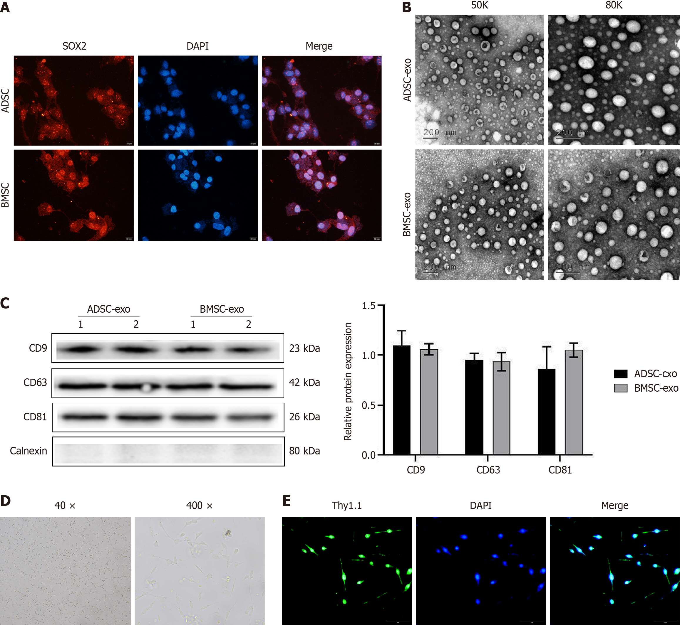
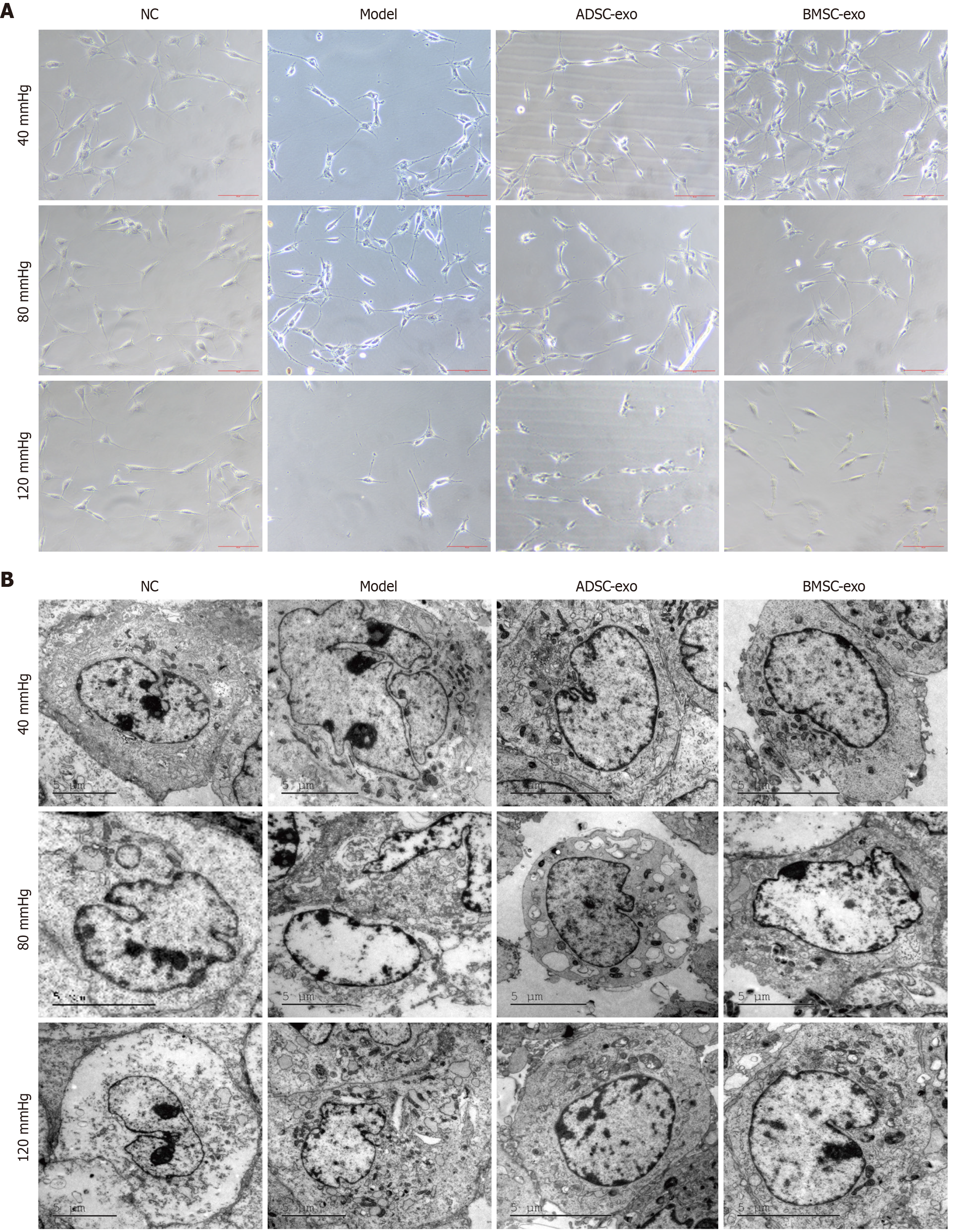
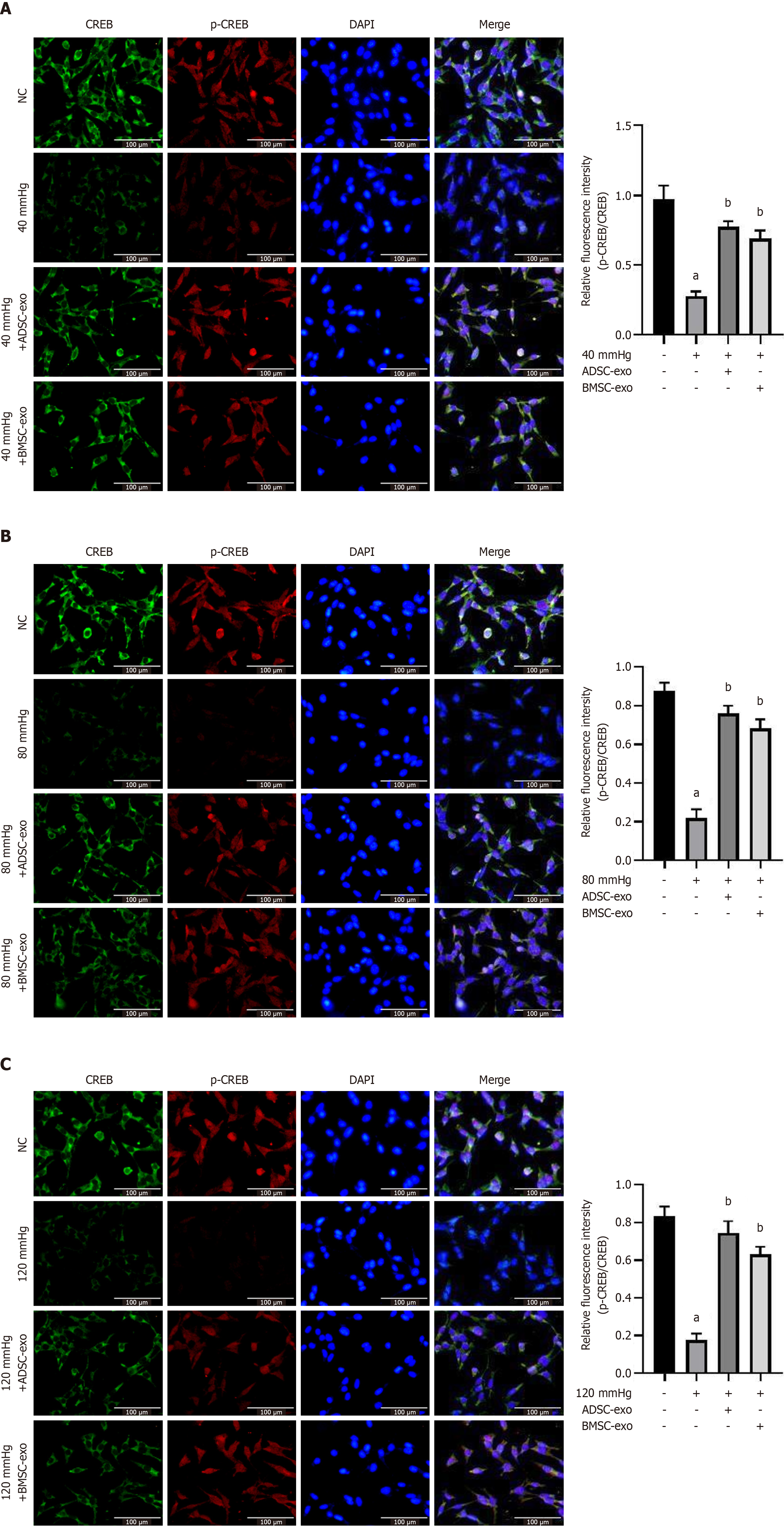
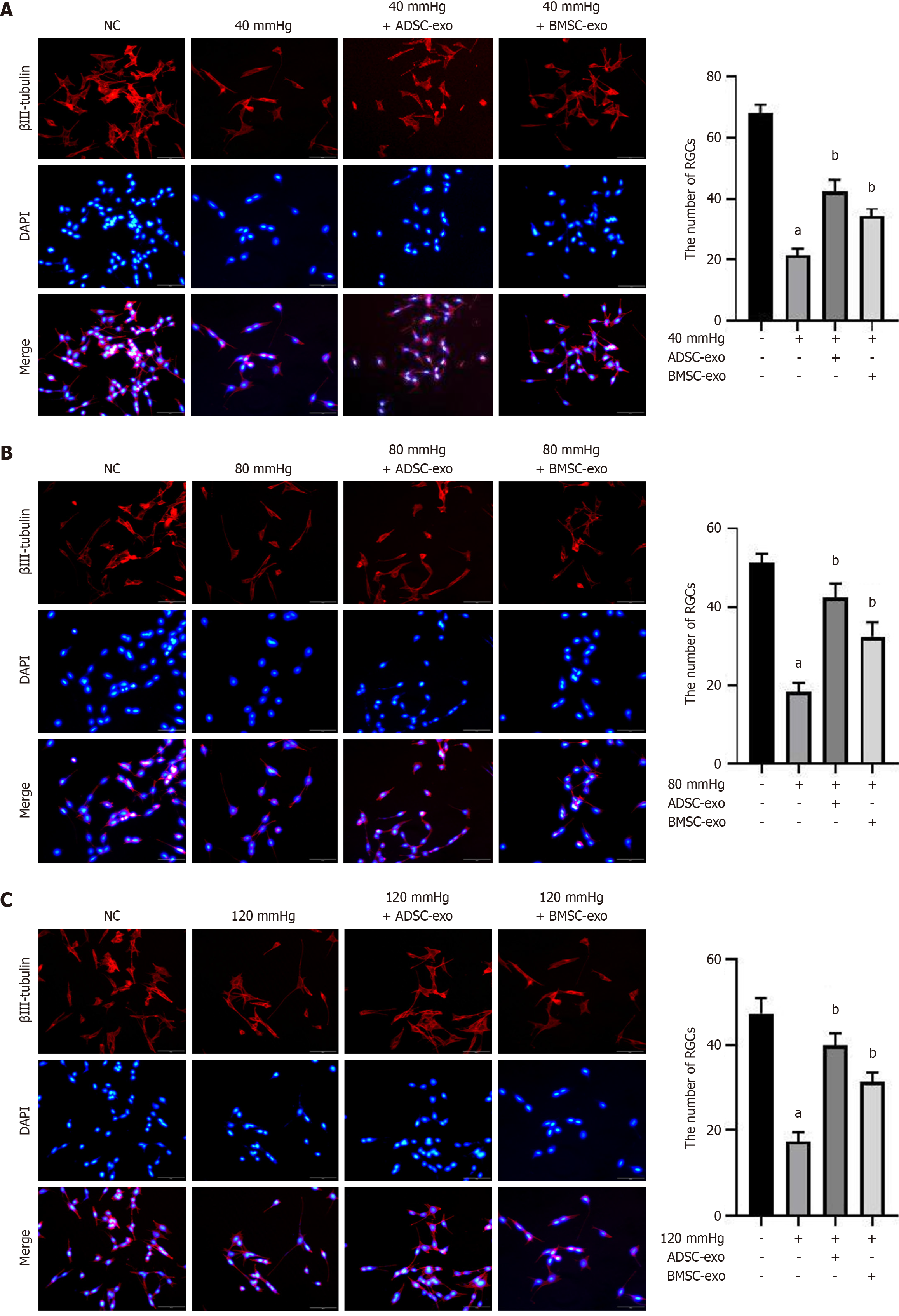
This is a correction to “Zheng ZK, Kong L, Dai M, Chen YD, Chen YH. ADSC-Exos outperform BMSC-Exos in alleviating hydrostatic pressure-induced injury to retinal ganglion cells by upregulating nerve growth factors. World J Stem Cells 2023; 15: 1077-1092 [PMID: 38179214 DOI: 10.4252/wjsc.v15.i12.1077]”. This article was published in 2023[1]. Several challenges were observed in the original images: A narrow imaging area [Figure 1B (original Figure 1B)], aberrant cell-cell adhesion [Figure 2A (original Figure 2A)], ice crystal formation due to insufficient dehydration [Figure 2B (original Figure 2B)], signal oversaturation in highly magnified regions [Figure 3 (original Figure 3)], and overlapping cellular structures [Figure 4 (original Figure 5)]. As the corresponding author, I am submitting the corrected figures [Figures 1, 2, 3, and 4 (original Figures 1, 2, 3, and 5)]. This correction is being requested to ensure the accuracy of the published data in the original paper. This correction does not impact the conclusion of the manuscript.

Figure 1
Figure 1 Adipose-derived stem cells, bone marrow-derived stem cell-exosomes, and retinal ganglion cells were isolated and characterized. A: Immunofluorescence staining of the stem cell marker SOX2 (scale bar: 20 μm); B: Electron microscopy images of the morphology of adipose-derived stem cell-exosomes and bone marrow-derived stem cell-exosomes (scale bar: 200 nm); C: Adipose-derived stem cell-exosomes and bone marrow-derived stem cell-exosomes marker molecules (CD9, CD63, and CD81) were detected by western blotting; D: Light microscopy images of retinal ganglion cells (magnification: 40 × and 400 × ); E: Isolated retinal ganglion cells were examined by Thy1.1 immunofluorescence (scale bar: 50 μm). ADSC: Adipose-derived stem cell; BMSC: Bone marrow-derived stem cell; Exo: Exosome.
Figure 2
Figure 2 Growth of retinal ganglion cells. A: Phase contrast microscopy images of the morphology and growth of retinal ganglion cells (scale bar: 50 μm); B: Transmission electron microscopy images of retinal ganglion cell morphology (scale bar: 5 μm). ADSC: Adipose-derived stem cell; BMSC: Bone marrow-derived stem cell; Exo: Exosome; NC: Normal control.
Figure 3
Figure 3 Immunofluorescence detection of β-III tubulin. A-C: Immunofluorescence staining of β-III tubulin (red) and nuclear staining with DAPI (blue) (left panel) and the number of β-III tubulin-positive retinal ganglion cells (right panel) in different groups after exposure to 40 mmHg (A), 80 mmHg (B), and 120 mmHg (C). aP < 0.05, compared with the normal control group (scale bar: 100 μm); bP < 0.05, compared with the model (40, 80, and 120 mmHg) group. ADSC: Adipose-derived stem cell; BMSC: Bone marrow-derived stem cell; Exo: Exosome; NC: Normal control.
Figure 4
Figure 4 Immunofluorescence detection of p-CREB and CREB. A-C: Immunofluorescence staining of CREB (green) and p-CREB (red) and nuclear staining with DAPI (blue) (left panel) and quantification of the p-CREB/CREB ratio (right panel) in different groups after exposure to (A) 40 mmHg, (B) 80 mmHg, and (C) 120 mmHg (scale bar: 20 μm). aP < 0.05, compared with the normal control group; bP < 0.05, compared with the model (40, 80, and 120 mmHg) group. ADSC: Adipose-derived stem cell; BMSC: Bone marrow-derived stem cell; Exo: Exosome; NC: Normal control.
Footnotes

Provenance and peer review: Unsolicited article; Externally peer reviewed.

Peer-review model: Single blind

Specialty type: Cell and tissue engineering

Country of origin: China

Peer-review report’s classification

Scientific Quality: Grade B

Novelty: Grade B

Creativity or Innovation: Grade B

Scientific Significance: Grade C

P-Reviewer: Kasban H, PhD, Chairman, Chief, Full Professor, Head, Professor, Egypt S-Editor: Wang JJ L-Editor: A P-Editor: Wang CH

References
1.  Zheng ZK, Kong L, Dai M, Chen YD, Chen YH. ADSC-Exos outperform BMSC-Exos in alleviating hydrostatic pressure-induced injury to retinal ganglion cells by upregulating nerve growth factors. World J Stem Cells. 2023;15:1077-1092.  [RCA]  [PubMed]  [DOI]  [Full Text]  [Full Text (PDF)]  [Cited by in RCA: 7]  [Reference Citation Analysis (0)]