©The Author(s) 2025.
World J Stem Cells. Nov 26, 2025; 17(11): 112393
Published online Nov 26, 2025. doi: 10.4252/wjsc.v17.i11.112393
Published online Nov 26, 2025. doi: 10.4252/wjsc.v17.i11.112393
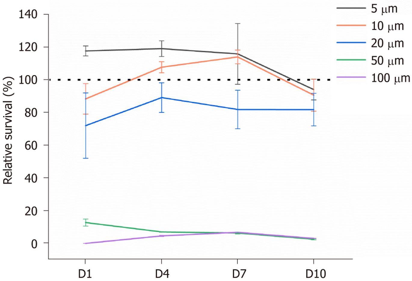
Figure 1 The cytotoxicity assay of thymoquinone.
The cytotoxic effects of thymoquinone were evaluated by assessing the relative viability of tendon-derived stem cells, following exposure to varying concentrations of thymoquinone (5, 10, 20, 50, and 100 μmol/L), over a 10-day period. Cellular viability was quantified using the Cell Counting Kit-8 assay.
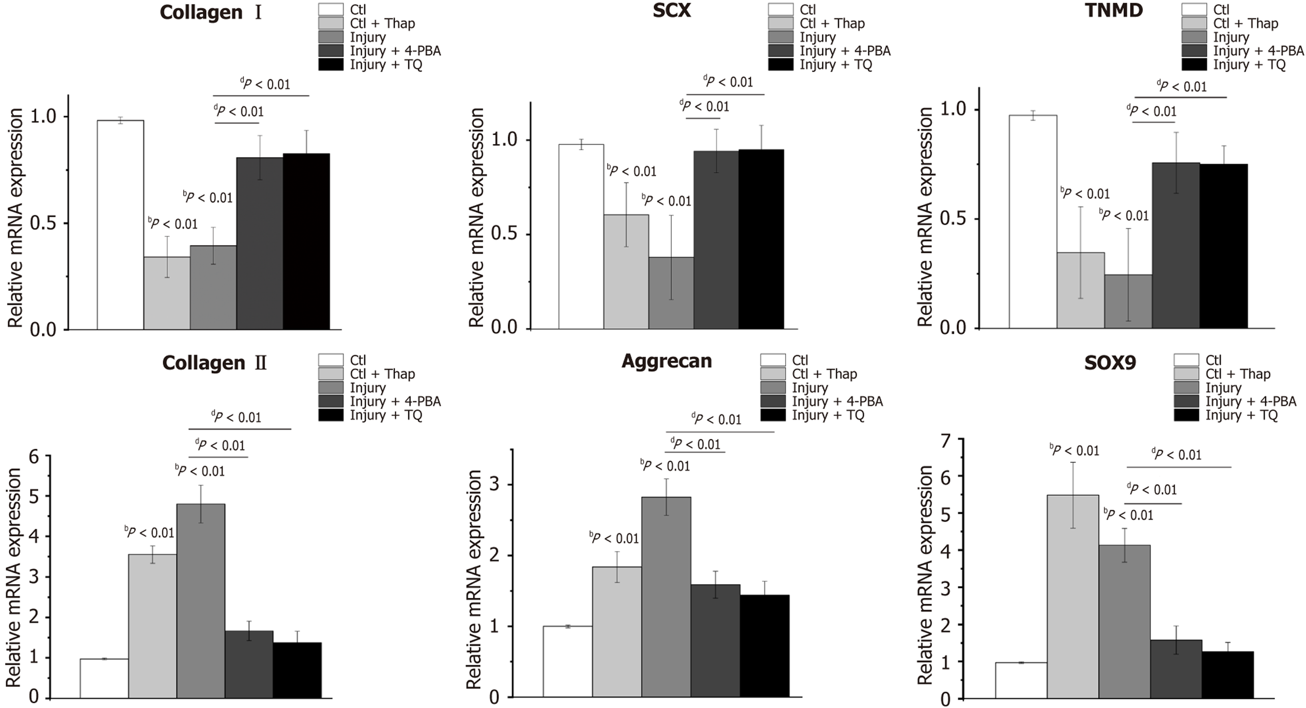
Figure 2 Thymoquinone inhibited the chondrogenic differentiation of tendon-derived stem cells subjected to injury conditions.
Reverse transcription-quantitative polymerase chain reaction was utilized to assess the expression levels of tenocyte markers (collagen I, scleraxis, tenomodulin), and chondrocyte markers (collagen II, aggrecan, SOX9), in tendon-derived stem cells (TDSCs) across various experimental groups. The control group refers to TDSCs cultured on decellularized tendon slices with 0% strain; the control + thapsigargin group refers to TDSCs treated with thapsigargin on decellularized tendon slices with 0% strain; the injury group refers to TDSCs cultured on decellularized tendon slices with 6.4% strain; the injury + 4-phenylbutyric acid group refers to TDSCs treated with 4-phenylbutyric acid on decellularized tendon slices with 6.4% strain; the injury + thymoquinone group refers to TDSCs treated with thymoquinone on decellularized tendon slices with 6.4% strain. bP < 0.01, compared to the control group; dP < 0.01, comparisons between the experimental groups. SCX: Scleraxis; TNMD: Tenomodulin; Ctl: Control; Ctl + Thap: Control + thapsigargin; 4-PBA: 4-phenylbutyric acid; TQ: Thymoquinone.
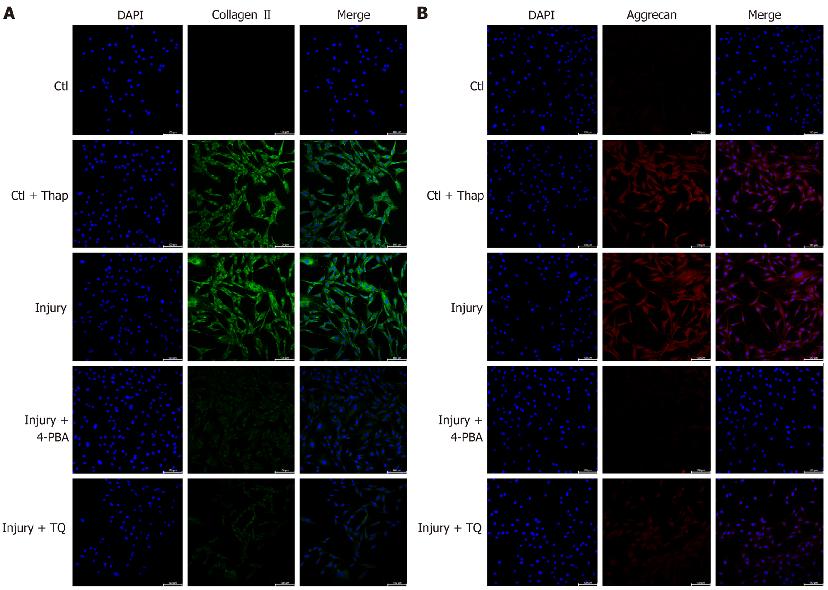
Figure 3 Thymoquinone decreased chondrogenic markers, including collagen II and aggrecan, in tendon-derived stem cells subjected to injury conditions.
A: Immunofluorescence staining of collagen II, detected using a CoraLite® Plus 488-conjugated antibody, alongside nuclear staining with Hoechst 33342, across different experimental groups; B: Immunofluorescence staining of aggrecan, labeled with a CoraLite 594-conjugated antibody, together with nuclear staining by Hoechst 33342 in the same groups. Bar: 100 μm. The control group refers to tendon-derived stem cells (TDSCs) cultured on decellularized tendon slices with 0% strain; the control + thapsigargin group refers to TDSCs treated with thapsigargin on decellularized tendon slices with 0% strain; the injury group refers to TDSCs cultured on decellularized tendon slices with 6.4% strain; the injury + 4-phenylbutyric acid group refers to TDSCs treated with 4-phenylbutyric acid on decellularized tendon slices with 6.4% strain; the injury + thymoquinone group refers to TDSCs treated with thymoquinone on decellularized tendon slices with 6.4% strain. Ctl: Control; Ctl + Thap: Control + thapsigargin; 4-PBA: 4-phenylbutyric acid; TQ: Thymoquinone.
Figure 4 Thymoquinone attenuated endoplasmic reticulum stress of tendon-derived stem cells subjected to injury conditions.
A: Reverse transcription-quantitative polymerase chain reaction analysis of the expression levels of endoplasmic reticulum stress-related markers, including glucose-regulated protein 78, protein kinase RNA-like endoplasmic reticulum kinase, eukaryotic initiation factor 2α, activating transcription factor 4, and CCAAT/enhancer-binding protein homologous protein. bP < 0.01, compared to the control group; dP < 0.01, comparisons between the experimental groups; B: Western blot analysis assessing the protein expression of glucose-regulated protein 78, SOX9, and components of the protein kinase RNA-like endoplasmic reticulum kinase/eukaryotic initiation factor 2α/activating transcription factor 4/CCAAT/enhancer-binding protein homologous protein signaling pathway. The control group refers to tendon-derived stem cells (TDSCs) cultured on decellularized tendon slices with 0% strain; the control + thapsigargin group refers to TDSCs treated with thapsigargin on decellularized tendon slices with 0% strain; the injury group refers to TDSCs cultured on decellularized tendon slices with 6.4% strain; the injury + 4-phenylbutyric acid group refers to TDSCs treated with 4-phenylbutyric acid on decellularized tendon slices with 6.4% strain; the injury + thymoquinone group refers to TDSCs treated with thymoquinone on decellularized tendon slices with 6.4% strain. Ctl: Control; Ctl + Thap: Control + thapsigargin; 4-PBA: 4-phenylbutyric acid; TQ: Thymoquinone; GRP78: Glucose-regulated protein 78; PERK: Protein kinase RNA-like endoplasmic reticulum kinase; EIF2α: Eukaryotic initiation factor 2α; ATF4: Activating transcription factor 4; CHOP: CCAAT/enhancer-binding protein homologous protein.
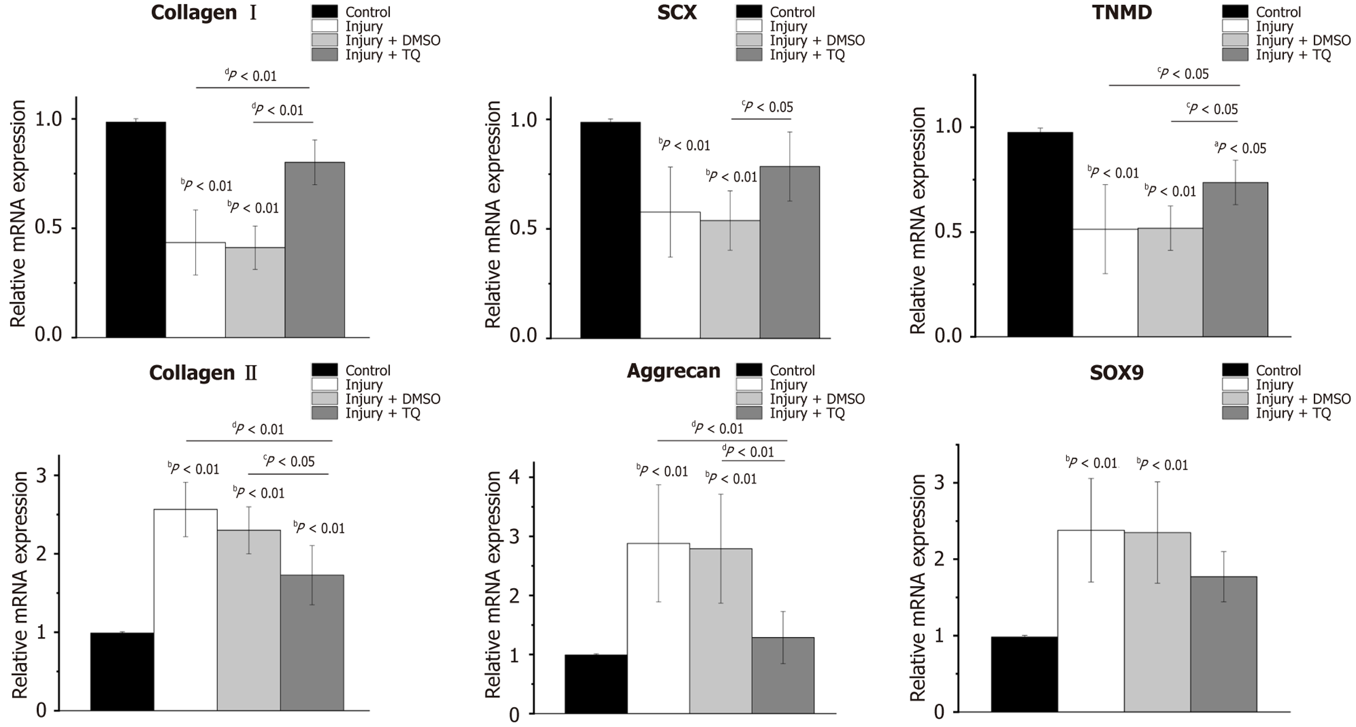
Figure 5 Thymoquinone treatment reinstated the expression of tenocyte markers, while concurrently diminishing the expression of chondrocyte markers in injured rat tendons.
Reverse transcription-quantitative polymerase chain reaction was employed to assess the expression levels of tenocyte markers (collagen I, scleraxis, tenomodulin), and chondrocyte markers (collagen II, aggrecan, SOX9), across different experimental groups. aP < 0.05, compared to the control group; bP < 0.01, compared to the control group; cP < 0.05, comparisons between the experimental group; dP < 0.01, comparisons between the experimental groups. The control group refers to tendons from rats maintained under standard cage activity conditions; the injury group refers to tendons from rats subjected to treadmill running without subsequent treatment; the injury + dimethyl sulfoxide group refers to tendons from rats subjected to treadmill running that subsequently received injections of saline solution containing dimethyl sulfoxide; the injury + thymoquinone group refers to tendons from rats subjected to treadmill running that subsequently received injections of thymoquinone. DMSO: Dimethyl sulfoxide; TQ: Thymoquinone; SCX: Scleraxis; TNMD: Tenomodulin.
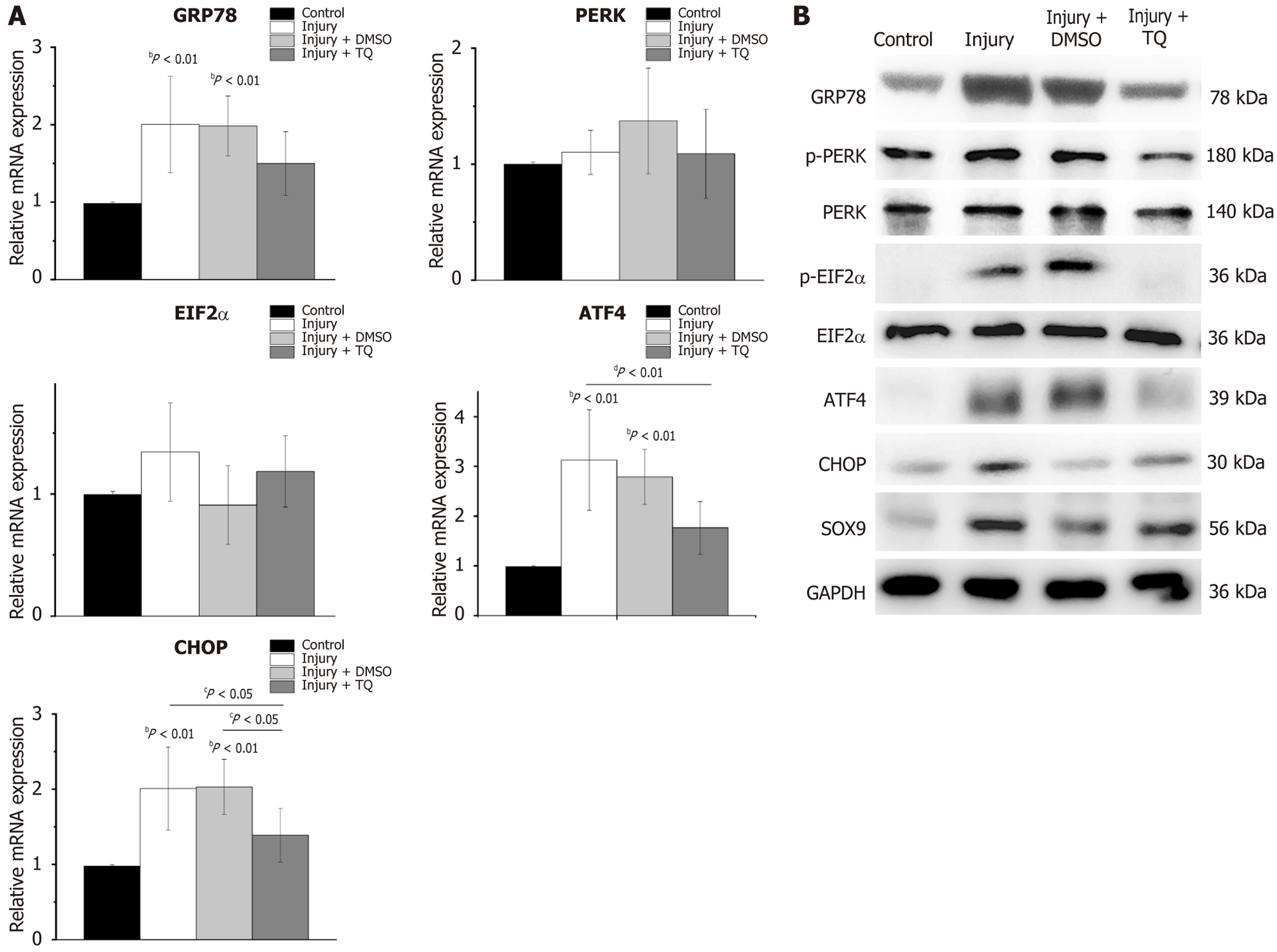
Figure 6 Thymoquinone mitigated endoplasmic reticulum stress in injured rat tendons.
A: Reverse transcription-quantitative polymerase chain reaction analysis of the expression levels of endoplasmic reticulum stress-related markers, including glucose-regulated protein 78, protein kinase RNA-like endoplasmic reticulum kinase, eukaryotic initiation factor 2α, activating transcription factor 4, and CCAAT/enhancer-binding protein homologous protein. bP < 0.01, compared to the control group; cP < 0.05, comparisons between the experimental groups; dP < 0.01, comparisons between the experimental group; B: Western blot analyses evaluating the protein expression of glucose-regulated protein 78, SOX9, and components of the protein kinase RNA-like endoplasmic reticulum kinase/eukaryotic initiation factor 2α/activating transcription factor 4/CCAAT/enhancer-binding protein homologous protein signaling pathway. The control group refers to tendons from rats maintained under standard cage activity conditions; the injury group refers to tendons from rats subjected to treadmill running without subsequent treatment; the injury + dimethyl sulfoxide group refers to tendons from rats subjected to treadmill running that subsequently received injections of saline solution containing dimethyl sulfoxide; the injury + thymoquinone group refers to tendons from rats subjected to treadmill running that subsequently received injections of thymoquinone. DMSO: Dimethyl sulfoxide; TQ: Thymoquinone; GRP78: Glucose-regulated protein 78; PERK: Protein kinase RNA-like endoplasmic reticulum kinase; EIF2α: Eukaryotic initiation factor 2α; ATF4: Activating transcription factor 4; CHOP: CCAAT/enhancer-binding protein homologous protein.
- Citation: Tu YJ, Liu YQ, Pan YY, Cai HY, Liu C. Thymoquinone inhibited the chondrogenic differentiation of tendon-derived stem cells caused by tendon injury. World J Stem Cells 2025; 17(11): 112393
- URL: https://www.wjgnet.com/1948-0210/full/v17/i11/112393.htm
- DOI: https://dx.doi.org/10.4252/wjsc.v17.i11.112393


