©The Author(s) 2025.
World J Gastroenterol. Nov 14, 2025; 31(42): 112107
Published online Nov 14, 2025. doi: 10.3748/wjg.v31.i42.112107
Published online Nov 14, 2025. doi: 10.3748/wjg.v31.i42.112107
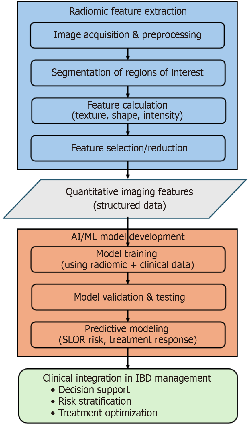
Figure 1 Workflow illustrating the distinction between radiomic feature extraction and artificial intelligence/machine learning model development in inflammatory bowel disease management.
The diagram highlights two sequential stages. The radiomics workflow (blue cluster) begins with image acquisition and preprocessing (e.g., computed tomography enterography, magnetic resonance enterography), followed by segmentation of regions of interest, calculation of quantitative features (such as shape, intensity, and texture), and feature selection or reduction. This process yields structured quantitative imaging features, which serve as standardized data inputs. The AI/machine learning workflow (orange cluster) uses these features – often combined with clinical and laboratory data – for model training, validation, and predictive modeling. Outputs from the artificial intelligence/machine learning stage include individualized predictions, such as risk of secondary loss of response or likelihood of treatment response. Finally, these outputs feed into clinical integration (green cluster), where predictions inform decision support, risk stratification, and optimization of therapeutic strategies in Crohn’s disease and ulcerative colitis. AI: Artificial intelligence; ML: Machine learning; SLOR: Secondary loss of response; IBD: Inflammatory bowel disease.
- Citation: Liu ZG, Xie SS. Expanding the role of radiomics and artificial intelligence in the management of inflammatory bowel disease: Insights, opportunities, and challenges. World J Gastroenterol 2025; 31(42): 112107
- URL: https://www.wjgnet.com/1007-9327/full/v31/i42/112107.htm
- DOI: https://dx.doi.org/10.3748/wjg.v31.i42.112107


