©The Author(s) 2017.
World J Gastroenterol. Apr 7, 2017; 23(13): 2308-2317
Published online Apr 7, 2017. doi: 10.3748/wjg.v23.i13.2308
Published online Apr 7, 2017. doi: 10.3748/wjg.v23.i13.2308
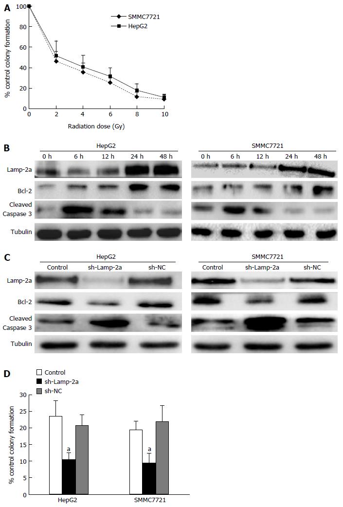
Figure 1 Irradiation-induced chaperone-mediated autophagy pathway activation regulated the apoptosis in hepatocellular carcinoma cells.
A: HepG2 and SMMC7721 cells were irradiated at different doses and the ability of proliferation was detected by clone formation assay; B: HepG2 and SMMC7721 cells were irradiated at doses of 6 Gy; the levels of Lamp-2a, Caspase 3 (cleaved) and Bcl-2 were determined by western blot at different post-irradiation times; C: sh-Lamp-2a HepG2 and sh-Lamp-2a SMMC7721 cells were irradiated at doses of 6 Gy; the levels of Lamp-2a, Caspase 3 (cleaved) and Bcl-2 were detected at 48 h by western blot; D: The ability of propagation in sh-Lamp-2a HepG2 and sh-Lamp-2a SMMC7721 cells was detected by clone formation assay at 48 h after 6 Gy irradiation. aP < 0.05, vs control groups or sh-NC groups. Each experiment was repeated three times and similar results were obtained.
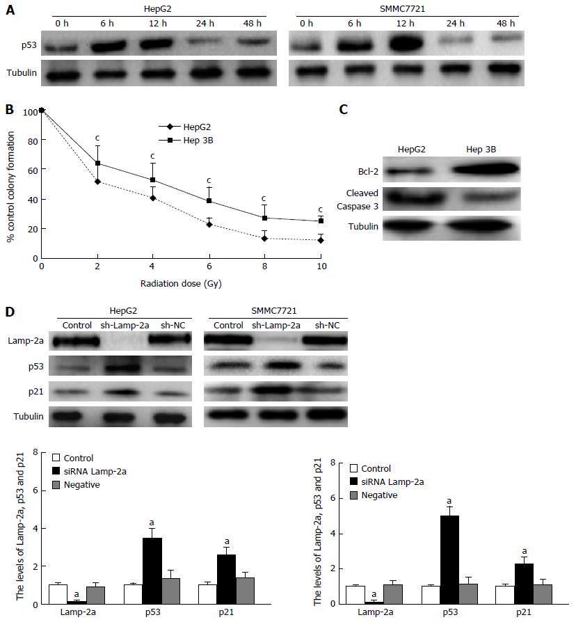
Figure 2 p53 was regulated through chaperone-mediated autophagy pathway activation in hepatocellular carcinoma cells on irradiation.
A: HepG2 and SMMC7721 cells were irradiated with doses of 6 Gy. At different post-irradiation times, the levels of p53 were determined by western blot; B: HepG2 and Hep3B (p53-/-) cells were irradiated at different doses and the ability of proliferation was detected by clone formation assay; C: HepG2 and Hep 3B (p53-/-) cells were irradiated at doses of 6 Gy; the levels of Caspase 3 (cleaved) and Bcl-2 were detected at 48 h by western blot; D: sh-Lamp-2a HepG2 and sh-Lamp-2a SMMC7721 cells were irradiated at doses of 6 Gy; the levels of p53 and p21 were determined after 48 h. aP < 0.05 vs control group or sh-NC group; cP < 0.05 vs HepG2 groups. Each experiment was repeated three times and similar results were obtained.
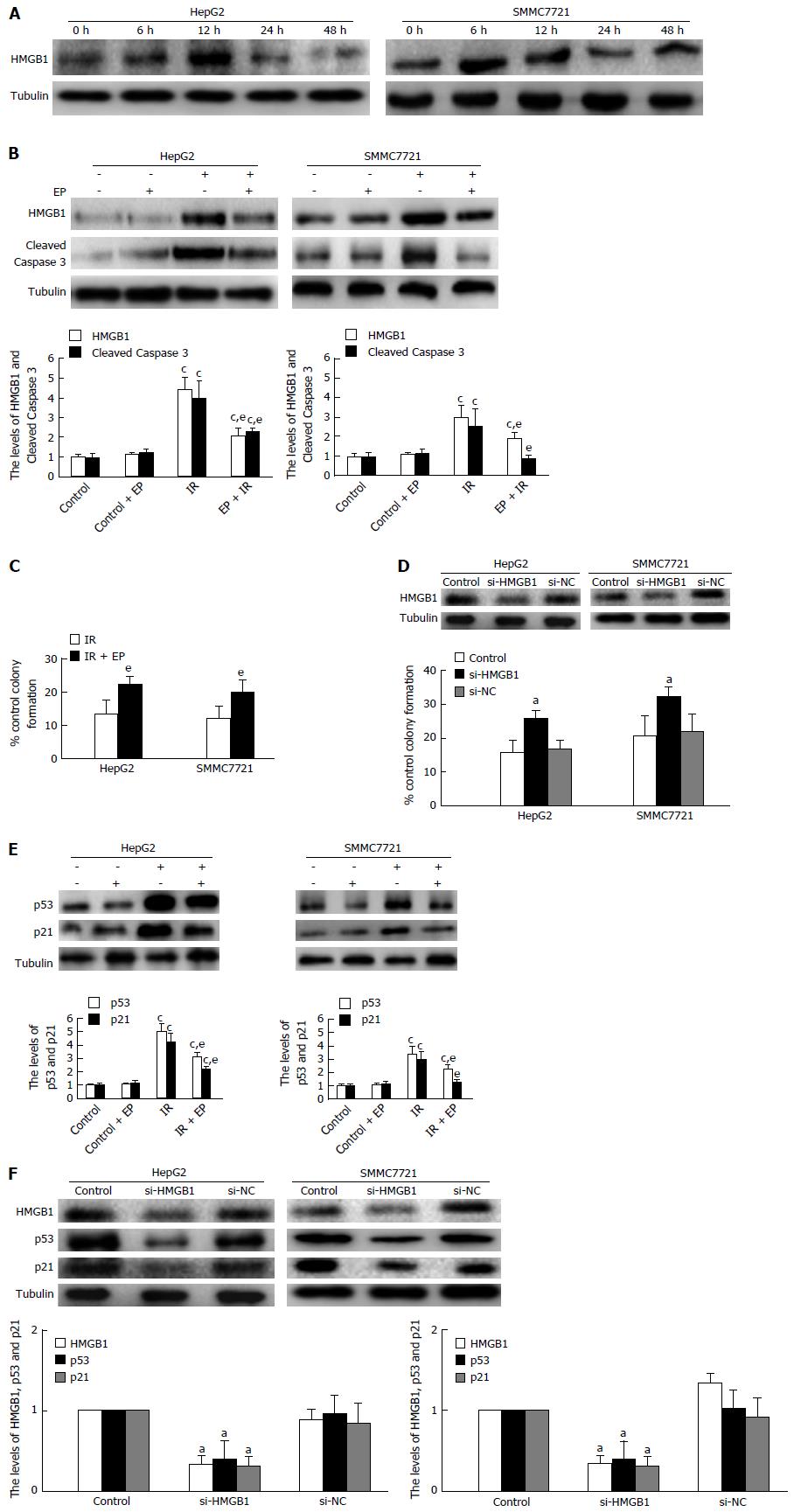
Figure 3 p53 expression was regulated by HMGB1 in hepatocellular carcinoma cells on irradiation.
A: HepG2 and SMMC7721 cells were irradiated with doses of 6 Gy. At different post-irradiation times, the levels of HMGB1 were determined by western blot; B: In the presence of EP (10 μg/mL). HMGB1, Caspase3 (cleaved) were detected by western blot in representative cells irradiated at 6 Gy after 48 h; C: HepG2 and SMMC7721 were exposed to EP(10 μg/mL) and the ability of proliferation was detected by clone formation assay; D: The ability of proliferation in HepG2 and SMMC7721 cells affected by HMGB1 siRNA was detected by clone formation assay after 6 Gy irradiation; E: In the presence of EP (10 μg/mL), the levels of p53 and p21 were detected in HepG2 and SMMC7721 cells irradiated at 6 Gy after 48 h; F: After si-HMGB1 HepG2 and si-SMMC7721 cells were irradiated at doses of 6 Gy, the levels of p53 and p21 were determined after 48 h. aP < 0.05, vs control group or si-NC group; cP < 0.05, vs control group or control + EP group; eP < 0.05, vs IR group. Each experiment was repeated three times and similar results were obtained.
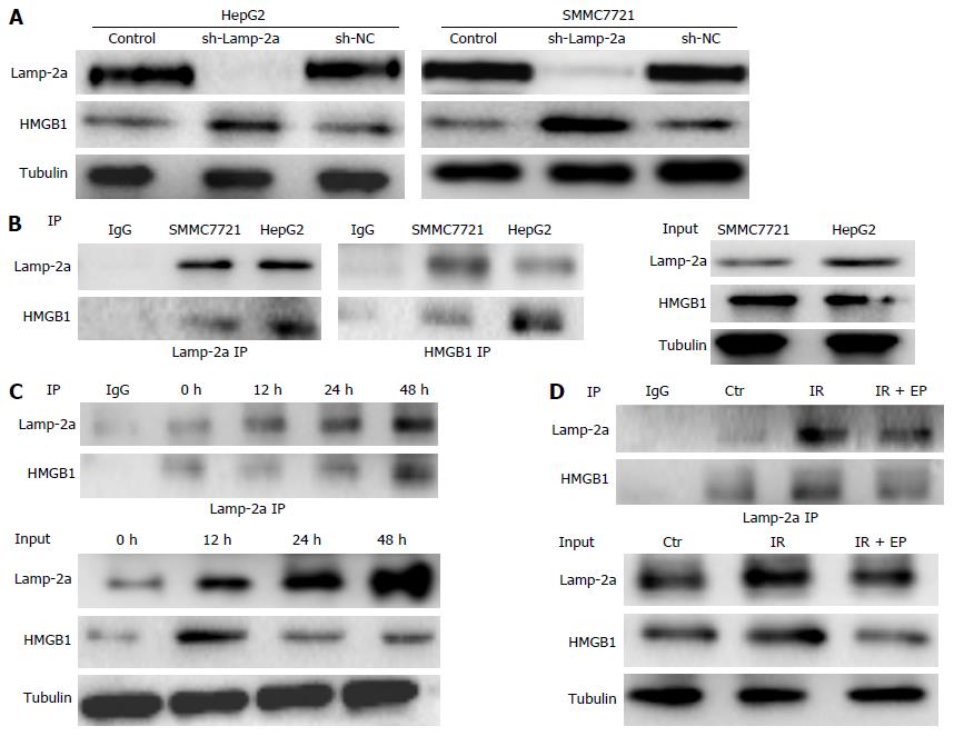
Figure 4 HMGB1 was degraded through chaperone-mediated autophagy pathway in hepatocellular carcinoma cells.
sh-Lamp-2a HepG2 and sh-Lamp-2a SMMC7721 cells were irradiated at doses of 6 Gy, (A) the levels of HMGB1 were determined after 48 h by western blot and (B) the interaction of Lamp-2a with HMGB1 was detected by immunoprecipitation (IP); C: Lamp-2a and HMGB1 were detected at different post-irradiation times by IP; D: In the presence of EP (10 μg/mL), the levels of HMGB1 were detected after 48 h by IP. Each experiment was repeated three times and similar results were obtained.
- Citation: Wu JH, Guo JP, Shi J, Wang H, Li LL, Guo B, Liu DX, Cao Q, Yuan ZY. CMA down-regulates p53 expression through degradation of HMGB1 protein to inhibit irradiation-triggered apoptosis in hepatocellular carcinoma. World J Gastroenterol 2017; 23(13): 2308-2317
- URL: https://www.wjgnet.com/1007-9327/full/v23/i13/2308.htm
- DOI: https://dx.doi.org/10.3748/wjg.v23.i13.2308


