Published online Nov 26, 2020. doi: 10.12998/wjcc.v8.i22.5611
Peer-review started: July 21, 2020
First decision: August 8, 2020
Revised: August 19, 2020
Accepted: October 1, 2020
Article in press: October 1, 2020
Published online: November 26, 2020
Processing time: 127 Days and 3.5 Hours
Schwannoma is a rare benign, encapsulated tumor of the nerve sheath under the tongue, mostly occurring as solitary tumors with classical histological pattern and several common morphological variants. To our knowledge, multiple schwannomas with pseudoglandular element synchronously occurring under the tongue are rare; we report herein the first such case.
A 53-year-old man had first noticed an isolated asymptomatic mass under the tongue, and as the mass grew, the tongue was elevated. Physical examination showed multiple oval neoplasms, and the overlying mucosa was normal. Computed tomography showed three low-density oval neoplasms under the tongue, which were cystic-solid with unclear boundary. The patient has no cutaneous tumors, VIII nerve tumors, or lens opacities and no history of neurofibromatosis 2 or confirmed schwannomatosis in any first-degree relative. Magnetic resonance imaging showed no evidence of vestibular schwannoma. The preoperative diagnosis was mucoepidermoid carcinoma. During hospitalization, all neoplasms were completely excised by surgeons through an intraoral approach under general anesthesia. The diagnosis of the multiple schwannomas with pseudoglandular element was made by histopathology after surgery. At the 15-mo follow-up visit, the patient had no sign of recurrence or development of other peripheral nerve tumors.
Although rare, multiple schwannomas with pseudoglandular element do exist in patients presenting with masses under the tongue. Oral surgeons should be aware of the existence of multiple schwannomas with pseudoglandular element when considering masses under the tongue due to the different prognosis between multiple schwannomas with pseudoglandular element and mucoepidermoid carcinoma.
Core Tip: Schwannoma is a rare benign, encapsulated tumor of the nerve sheath under the tongue and mostly occurs as solitary tumors with classical histological pattern and several common morphological variants. Multiple schwannomas with pseudoglandular element synchronously occurring under the tongue are of great rarity. Here, we present the first report of a case of multiple schwannomas with pseudoglandular element synchronously occurring under the tongue.
- Citation: Chen YL, He DQ, Yang HX, Dou Y. Multiple schwannomas with pseudoglandular element synchronously occurring under the tongue: A case report. World J Clin Cases 2020; 8(22): 5611-5617
- URL: https://www.wjgnet.com/2307-8960/full/v8/i22/5611.htm
- DOI: https://dx.doi.org/10.12998/wjcc.v8.i22.5611
Schwannoma is a rare benign, encapsulated tumor of the nerve sheath under the tongue, and it mostly occurs as solitary tumors with classical histological pattern and several common morphological variants. Multiple schwannomas with pseudoglandular element synchronously occurring under the tongue are of great rarity. Here, we present the first report of a case of multiple schwannomas with pseudoglandular element synchronously occurring under the tongue.
A 53-year-old man had first noticed an isolated asymptomatic mass under the tongue, and then the mass grew, causing the tongue to be elevated.
The patient has no cutaneous tumors, VIII nerve tumors, or lens opacities.
The patient has no history of neurofibromatosis 2 or confirmed schwannomatosis in any first-degree relative.
Physical examination showed multiple oval neoplasms, and the overlying mucosa was normal. We considered mucoepidermoid carcinoma as our main differential diagnosis.
All neoplasms were completely excised by surgeons through an intraoral approach under general anesthesia. There was no communication between the neoplasms and nerve bundles. Gross examinations showed three separated oval encapsulated masses with smooth surface. The biggest tumor was 4 cm × 3 cm × 3 cm, and the smallest was 2.2 cm × 1.8 cm × 1.3 cm. The sectioned surface was grayish-white in color and cystic-solid lesion with moderate hardness (Figure 1). Microscopic examination showed a lesion composed of bland spindle cells and demonstrated typical Antoni A and Antoni B areas with scattered pseudoglandular and microcystic foci. These pseudoglandular and microcystic areas were lined by flat to cuboidal cells (Figure 2). Some cystic areas showed hemorrhage. There were some hyalinized blood vessels elsewhere. No mitotic figure was found in tumor cells. The tumor cells and lining cells were positive for the S-100 protein and negative for Ckp protein by immunohistochemistry (IHC) staining (Figure 3).
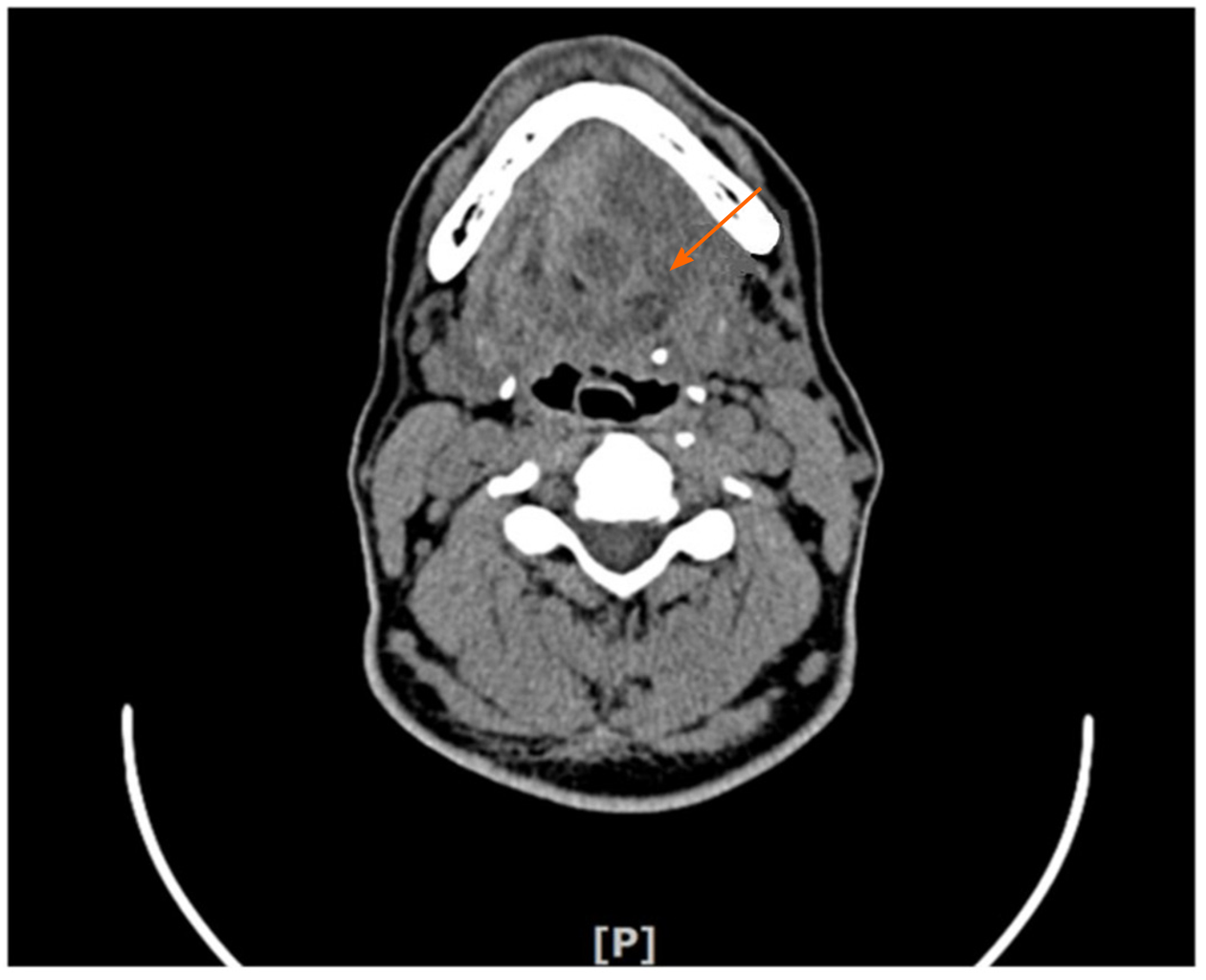
Computed tomography showed three low-density oval neoplasms under the tongue, which were cystic-solid lesion and unclear boundary (Figure 4). Magnetic resonance imaging scan showed no evidence of vestibular schwannoma.
The timeline of case reports is shown in Table 1.
| Events | Timeline | Description |
| Consultation | 2018-01-03 | First outpatient |
| Physical exam | 2018-01-10 | Gross and Microscopic examinations, CT |
| Surgical operation | 2018-02-07 | An intraoral approach under general anesthesia |
| Postoperative examination | 2018-02-10 | 3 d after the operation |
| Follow-up | 2019-07-21 | 15-mo follow-up visit, no recurrence |
Consequently, the diagnosis of the multiple schwannomas with pseudoglandular element under the tongue was established.
During hospitalization, the all neoplasms were completely excised by surgeons through an intraoral approach under general anesthesia. Three days after the operation, the patient recovered well and discharged.
The diagnosis of the multiple schwannomas with pseudoglandular element was made by histopathology after surgery. At the 15-mo follow-up visit, the patient had no sign of recurrence or no other peripheral nerve tumors had developed.
Schwannomas are benign neoplasms derived from Schwann cells[1]. They mostly occur as solitary tumors[2]. Multiple schwannomas developing in individual nerves are very rare[3]. Ogose et al[4] reported multiple schwannomas were in 4.6% of all patients with schwannoma. Their presence may be one of the symptoms indicative of neurofibromatosis 2, which is an autosomal dominant inherited disorder, or schwannomatosis, which is recognized as the third main form of NF[5].
Apart from the classic biphasic pattern, schwannomas may show several common morphologic variants including cellular, plexiform, epithelioid, ancient, and glandular variants[6]. A very rare pseudoglandular variant that has gland-like structure or cystic spaces that sometimes contain secretion-like material was first described by Ferry and Dickersin in 1988[7]. Since then, this extremely rare variant has been reported in a few case reports. The frequency of pseudoglandular element was 6.3% of schwannomas[8]. Most cases of schwannomas with pseudoglandular element have shown a predilection for location in the spinal nerve roots. Ud Din et al[8] and Robinson et al[15] reported that 56 or 61 cases (91.8%) and 13 of 16 cases (81%), respectively, showed pseudoglandular spaces located in the spinal nerve roots. Other schwannomas with pseudoglandular elements have been described only in single case reports and involved the right forearm, the right index finger, the retrobulbar region, submandibular region, soft tissue of shoulder, the parotid gland, the scalp, the retroperitoneum, thigh, popliteal fossa, and toe (Table 2)[6,8-12]. However, to date, schwannomas with pseudoglandular element located under the tongue have not been described previously in the English literature. In order to broaden further the clinicopathological spectrum of schwannomas with pseudoglandular element, we present the first report of a case of multiple schwannomas with pseudoglandular element under the tongue.
| No. | Age/sex | Location of tumors and number | Size in cm | Follow-up | Ref. |
| 1 | 60/F | Right forearm, one | 1.1 | 6 mo, no recurrence | Deng et al[9] |
| 2 | 34/F | Right index, one | Not described | Not described | Lisle et al[6] |
| 3 | 37/M | Retrobulbar mass, one | 1.5 | 10 yr, no recurrence | Chan et al[10] |
| 4 | 31/F | Submandibular region, one | 5.8 | Not described | Chan et al[10] |
| 5 | 24/F | Soft tissue of shoulder, one | 2.5 | Not described | Chan et al[10] |
| 6 | 27/M | Parotid gland, one | 3.5 | Not described | Ide et al[11] |
| 7 | 33/M | Cauda equine, one | 3 | 18 mo, no recurrence | Ruggeri et al[12] |
| 8 | Not described | Scalp, one | Not described | Not described | Ud Din et al[8] |
| 9 | Not described | Retroperitoneum, one | Not described | Not described | Ud Din et al[8] |
| 10 | Not described | Thigh, one | Not described | Not described | Ud Din et al[8] |
| 11 | Not described | Popliteal fossa, one | Not described | Not described | Ud Din et al[8] |
| 12 | Not described | Toe, one | Not described | Not described | Ud Din et al[8] |
| 13 | 53/M | Under the tongue, multiple (three) | The biggest was 4, and the smallest was 2.2 | 15 mo, no recurrence | Chen et al (the present case) |
The gland-like structure or cystic spaces in the pseudoglandular variant of schwannomas must be different from those true glandular structures in schwannomas and mucoepidermoid carcinoma[13]. These pseudoglandular structures are lined by Schwann cells, and these lining cells were positive for the S-100 protein and negative for Ckp protein by IHC staining[14]. Robison et al[15] suggested that the pseudoglandular element schwannomas likely represented a type of response to degenerative changes, perhaps reflecting the propensity of the tumors to form palisading structures. However, the true glandular structures in schwannomas may line intestinal and respiratory type epithelium[16], representing true epithelial differentiation, and IHC stains are negative for S-100 and positive for epithelial membrane antigen and Ckp. The theory is that glandular schwannomas are derived from multipotential neural crest cells that can develop into various phenotypes. This would explain the different types of elements found in schwannomas. Another conjecture is that tumorigenesis may involve stem cells with the potential to produce both neural and heterologous elements[17].
Mucoepidermoid carcinoma (MEC) is characterized by variable components of squamoid, mucin-producing, and intermediate-type cells, with a cystic and solid growth pattern[18]. However, it is usually difficult to distinguish MEC based on computed tomography. IHC stains are negative for S-100 and positive for epithelial membrane antigen and Ckp. MECs are characterized by gene translocation and fusion, but their diagnostic and clinical implications in the pathological evaluation remain uncertain.
We suggest that multiple schwannomas with pseudoglandular element may affect a wider range of body locations than previously reported. It is important to deepen our understanding of the clinicopathological spectrum of multiple schwannomas with pseudoglandular element so as to avoid its misdiagnosis.
Manuscript source: Unsolicited manuscript
Specialty type: Medicine, research and experimental
Country/Territory of origin: China
Peer-review report’s scientific quality classification
Grade A (Excellent): 0
Grade B (Very good): B, B
Grade C (Good): C
Grade D (Fair): 0
Grade E (Poor): 0
P-Reviewer: Gordon L, Parikh ND, Shimizu Y S-Editor: Gao CC L-Editor: Filipodia P-Editor: Zhang YL
| 1. | Gosk J, Gutkowska O, Urban M, Wnukiewicz W, Reichert P, Ziółkowski P. Results of surgical treatment of schwannomas arising from extremities. Biomed Res Int. 2015;2015:547926. [RCA] [PubMed] [DOI] [Full Text] [Full Text (PDF)] [Cited by in Crossref: 25] [Cited by in RCA: 36] [Article Influence: 3.3] [Reference Citation Analysis (0)] |
| 2. | Leverkus M, Kluwe L, Röll EM, Becker G, Bröcker EB, Mautner VF, Hamm H. Multiple unilateral schwannomas: segmental neurofibromatosis type 2 or schwannomatosis? Br J Dermatol. 2003;148:804-809. [RCA] [PubMed] [DOI] [Full Text] [Cited by in Crossref: 38] [Cited by in RCA: 23] [Article Influence: 1.0] [Reference Citation Analysis (0)] |
| 3. | Shao X, Zhang X, Su X. Multiple schwannomas of the ulnar nerve. J Plast Surg Hand Surg. 2014;48:281-282. [RCA] [PubMed] [DOI] [Full Text] [Cited by in Crossref: 2] [Cited by in RCA: 2] [Article Influence: 0.2] [Reference Citation Analysis (0)] |
| 4. | Ogose A, Hotta T, Morita T, Otsuka H, Hirata Y. Multiple schwannomas in the peripheral nerves. J Bone Joint Surg Br. 1998;80:657-661. [RCA] [PubMed] [DOI] [Full Text] [Cited by in Crossref: 8] [Cited by in RCA: 12] [Article Influence: 0.4] [Reference Citation Analysis (0)] |
| 5. | Baser ME, Friedman JM, Evans DG. Increasing the specificity of diagnostic criteria for schwannomatosis. Neurology. 2006;66:730-732. [RCA] [PubMed] [DOI] [Full Text] [Cited by in Crossref: 105] [Cited by in RCA: 100] [Article Influence: 5.0] [Reference Citation Analysis (0)] |
| 6. | Lisle A, Jokinen C, Argenyi Z. Cutaneous pseudoglandular schwannoma: a case report of an unusual histopathologic variant. Am J Dermatopathol. 2011;33:e63-e65. [RCA] [PubMed] [DOI] [Full Text] [Cited by in Crossref: 12] [Cited by in RCA: 15] [Article Influence: 1.0] [Reference Citation Analysis (0)] |
| 7. | Ferry JA, Dickersin GR. Pseudoglandular schwannoma. Am J Clin Pathol. 1988;89:546-552. [RCA] [PubMed] [DOI] [Full Text] [Cited by in Crossref: 26] [Cited by in RCA: 25] [Article Influence: 0.7] [Reference Citation Analysis (0)] |
| 8. | Ud Din N, Ahmad Z, Ahmed A. Schwannomas with pseudoglandular elements: clinicopathologic study of 61 cases. Ann Diagn Pathol. 2016;20:24-28. [RCA] [PubMed] [DOI] [Full Text] [Cited by in Crossref: 7] [Cited by in RCA: 9] [Article Influence: 0.8] [Reference Citation Analysis (0)] |
| 9. | Deng A, Petrali J, Jaffe D, Sina B, Gaspari A. Benign cutaneous pseudoglandular schwannoma: a case report. Am J Dermatopathol. 2005;27:432-435. [RCA] [PubMed] [DOI] [Full Text] [Cited by in Crossref: 16] [Cited by in RCA: 17] [Article Influence: 0.8] [Reference Citation Analysis (0)] |
| 10. | Chan JK, Fok KO. Pseudoglandular schwannoma. Histopathology. 1996;29:481-483. [RCA] [PubMed] [DOI] [Full Text] [Cited by in Crossref: 24] [Cited by in RCA: 23] [Article Influence: 0.8] [Reference Citation Analysis (0)] |
| 11. | Ide F, Obara K, Mishima K, Saito I. Intraparotid pseudoglandular schwannoma. J Oral Pathol Med. 2006;35:379-381. [RCA] [PubMed] [DOI] [Full Text] [Cited by in Crossref: 4] [Cited by in RCA: 4] [Article Influence: 0.2] [Reference Citation Analysis (0)] |
| 12. | Ruggeri F, De Cerchio L, Bakacs A, Orlandi A, Lunardi P. Pseudoglandular schwannoma of the cauda equina. Case report. J Neurosurg Spine. 2006;5:543-545. [RCA] [PubMed] [DOI] [Full Text] [Cited by in Crossref: 4] [Cited by in RCA: 4] [Article Influence: 0.2] [Reference Citation Analysis (0)] |
| 13. | Sundarkrishnan L, Bradish JR, Oliai BR, Hosler GA. Cutaneous Cellular Pseudoglandular Schwannoma: An Unusual Histopathologic Variant. Am J Dermatopathol. 2016;38:315-318. [RCA] [PubMed] [DOI] [Full Text] [Cited by in Crossref: 5] [Cited by in RCA: 6] [Article Influence: 0.6] [Reference Citation Analysis (0)] |
| 14. | Gómez-Mateo Mdel C, Compañ-Quilis A, Monteagudo C. Microcystic pseudoglandular plexiform cutaneous neurofibroma. J Cutan Pathol. 2015;42:884-888. [RCA] [PubMed] [DOI] [Full Text] [Cited by in Crossref: 3] [Cited by in RCA: 4] [Article Influence: 0.4] [Reference Citation Analysis (0)] |
| 15. | Robinson CA, Curry B, Rewcastle NB. Pseudoglandular elements in schwannomas. Arch Pathol Lab Med. 2005;129:1106-1112. [PubMed] |
| 16. | Uri AK, Witzleben CL, Raney RB. Electron microscopy of glandular Schwannoma. Cancer. 1984;53:493-497. [RCA] [PubMed] [DOI] [Full Text] [Cited by in RCA: 1] [Reference Citation Analysis (0)] |
| 17. | Ducatman BS, Scheithauer BW. Malignant peripheral nerve sheath tumors with divergent differentiation. Cancer. 1984;54:1049-1057. [RCA] [PubMed] [DOI] [Full Text] [Cited by in RCA: 1] [Reference Citation Analysis (0)] |
| 18. | Jhuang JY, Chou YH, Hua SF, Hsieh MS. Mixed lung mucoepidermoid carcinoma and adenocarcinoma with identical mutations in an epidermal growth factor receptor gene. Ann Thorac Surg. 2014;98:695-697. [RCA] [PubMed] [DOI] [Full Text] [Cited by in Crossref: 3] [Cited by in RCA: 4] [Article Influence: 0.3] [Reference Citation Analysis (0)] |


