Published online Aug 26, 2020. doi: 10.12998/wjcc.v8.i16.3440
Peer-review started: April 8, 2020
First decision: April 24, 2020
Revised: May 7, 2020
Accepted: July 18, 2020
Article in press: July 18, 2020
Published online: August 26, 2020
Processing time: 138 Days and 22.9 Hours
Medical robot is a promising surgical tool, but no specific one has been designed for interventional treatment of chronic pain. We developed a computed tomography-image based navigation robot using a new registration method with binocular vision. This kind of robot is appropriate for minimal invasive interventional procedures and easy to operate. The feasibility, accuracy and stability of this new robot need to be tested.
To assess quantitatively the feasibility, accuracy and stability of the binocular-stereo-vision-based navigation robot for minimally invasive interventional procedures.
A box model was designed for assessing the accuracy for targets at different distances. Nine (three sets) lead spheres were embedded in the model as puncture goals. The entry-to-target distances were set 50 mm (short-distance), 100 mm (medium-distance) and 150 mm (long-distance). Puncture procedure was repeated three times for each goal. The Euclidian error of each puncture was calculated and statistically analyzed. Three head phantoms were used to explore the clinical feasibility and stability. Three independent operators conducted foramen ovale placement on head phantoms (both sides) by freehand or under the guidance of robot (18 punctures with each method). The operation time, adjustment time and one-time success rate were recorded, and the two guidance methods were compared.
On the box model, the mean puncture errors of navigation robot were 1.7 ± 0.9 mm for the short-distance target, 2.4 ± 1.0 mm for the moderate target and 4.4 ± 1.4 mm for the long-distance target. On the head phantom, no obvious differences in operation time and adjustment time were found among the three performers (P > 0.05). The median adjustment time was significantly less under the guidance of the robot than under free hand. The one-time success rate was significantly higher with the robot (P < 0.05). There was no obvious difference in operation time between the two methods (P > 0.05).
In the laboratory environment, accuracy of binocular-stereo-vision-based navigation robot is acceptable for target at 100 mm depth or less. Compared with freehand, foramen ovale placement accuracy can be improved with robot guidance.
Core tip: We developed a computed tomography image-based navigation robot using new registration method with binocular vision. The objective of this study was to evaluate the feasibility, accuracy and stability of this new robot. Our results showed that the accuracy of this kind of navigation robot is acceptable for interventional treatment with target depth of 100 mm or less. Compared with freehand operation, the robot navigation can improve the accuracy and stability when conducting foramen ovale placement. The binocular-stereo-vision based navigation robot is a promising tool, and our research provides key validation results for its clinical application.
- Citation: Wang R, Han Y, Luo MZ, Wang NK, Sun WW, Wang SC, Zhang HD, Lu LJ. Accuracy study of a binocular-stereo-vision-based navigation robot for minimally invasive interventional procedures. World J Clin Cases 2020; 8(16): 3440-3449
- URL: https://www.wjgnet.com/2307-8960/full/v8/i16/3440.htm
- DOI: https://dx.doi.org/10.12998/wjcc.v8.i16.3440
Recently, minimal invasive surgery has been an irresistible trend in clinical treatment. In the field of chronic pain management, interventional therapy has also become the main approach for its advantages of exact therapeutic effect and minimal invasion[1]. The percutaneous puncture is a vital process of minimally invasive surgery, and its successful operation requires the accurate location of the target and the precise placement of the interventional tool. Although many imaging technologies such as fluoroscopy, computed tomography (CT) and ultrasound have been utilized to enhance accuracy and visualization[2-4], complex spatial puncture is still an intractable problem. For this reason, the clinical need for guidance has increased along with the trend of minimal invasion[1,5].
Apart from basic imaging equipment, many other navigation tools have been designed for clinical demand[6-8]. Among these techniques, the medical robot is the most promising one. This technology has shown great potential in increasing the accuracy and consistency of needle punctures in many kinds of interventional therapies. For spinal surgery, the reported accuracy rate of a pedicle screw under the guidance of a robot can be up to 96.7%-98.9%[9-11]. For neurological surgery, the navigation robot can not only ensure the accuracy of deep brain stimulation implantation but also shorten the operation time[12,13]. So far, few documents have reported the application of a navigation robot for chronic pain management, but the superiority of this tool would exactly complement the demand in this field.
To date, a series of robots have been released, including Mazor X (Mazor Robotics Ltd)[9], TiRobot (TINAVI Medical Technologies Co., Ltd)[14], Excelsius Global Position System (Globus Medical Ltd)[15] and BrainLab Cirq (Brainlab AG Ltd)[15]. However, none of these systems were designed for chronic pain treatment. The tracking markers of these robot systems need to be fixed on a specific position of the body, such as the ilium, which may be far from the surgical spot. Therefore, additional unnecessary radiation exposure will occur. This exposure may be acceptable for patients under general anesthesia because such preoperative scanning usually needs to be performed once. However, interventional treatment for chronic pain is always performed under local anesthesia, thus body dodging and twisting as well as breathing movement are inevitable influences on navigation accuracy. The scanning for placement planning needs to be repeated several times.
Our team proposes a new registration method using skin tracking markers and binocular vision[16]. The markers just need to be pasted on the skin of the operation region, avoiding extra radiation of the nonsurgical region. Using this method, we developed a CT-image based navigation robot. The objective of this study was to evaluate the feasibility, accuracy and stability of this new robot. We implemented it in preclinical experiments using box models to quantitate the puncture error. For assessment of clinical feasibility, head phantoms were also used to simulate interventional treatment on the Gasserian ganglion through the foramen ovale (FO). The placement procedure was conducted by free hand and under the guidance of the robot. We compared the operation parameters of the two methods.
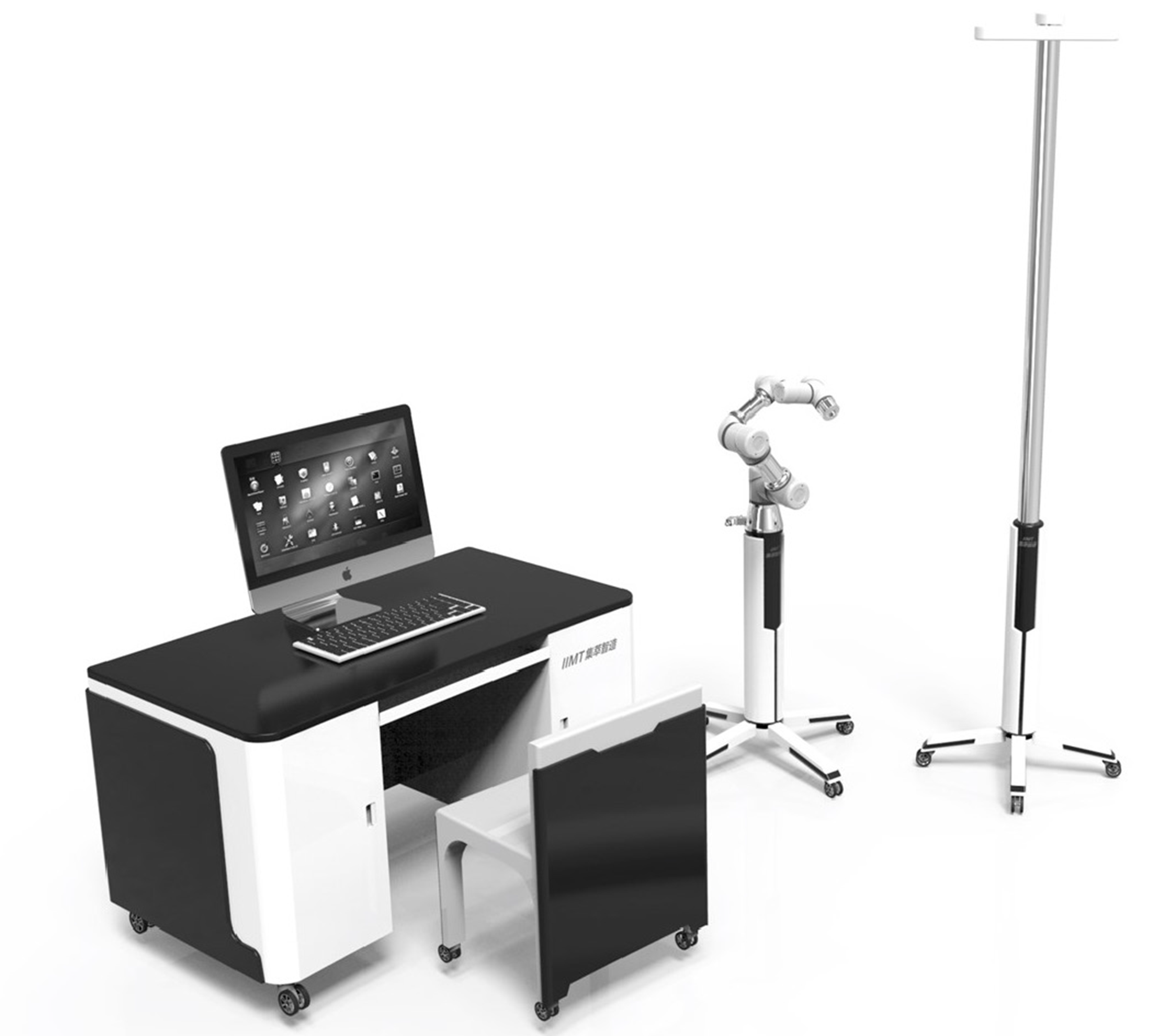
This binocular-stereo-vision-based navigation robot was independently designed by Nanjing Drum Tower Hospital, the affiliated hospital of Nanjing University Medical School, and the Institute of Intelligent Manufacturing Technology, Jiangsu Industrial Technology Research Institute. The navigation robot is composed of binocular stereo visual tracking equipment, an operation planning and controlling system and a six-axis robotic arm (UR3 robot, Universal Robots, Odense, Denmark). The binocular stereo visual tracking equipment is a binocular camera, which automatically identifies and locates the spatial position of the markers on the robot arm and operation object. The operation planning and controlling system consists of a program for image processing, spatial registration, cannulating trajectory design and movement control of the robot arm (Figure 1).
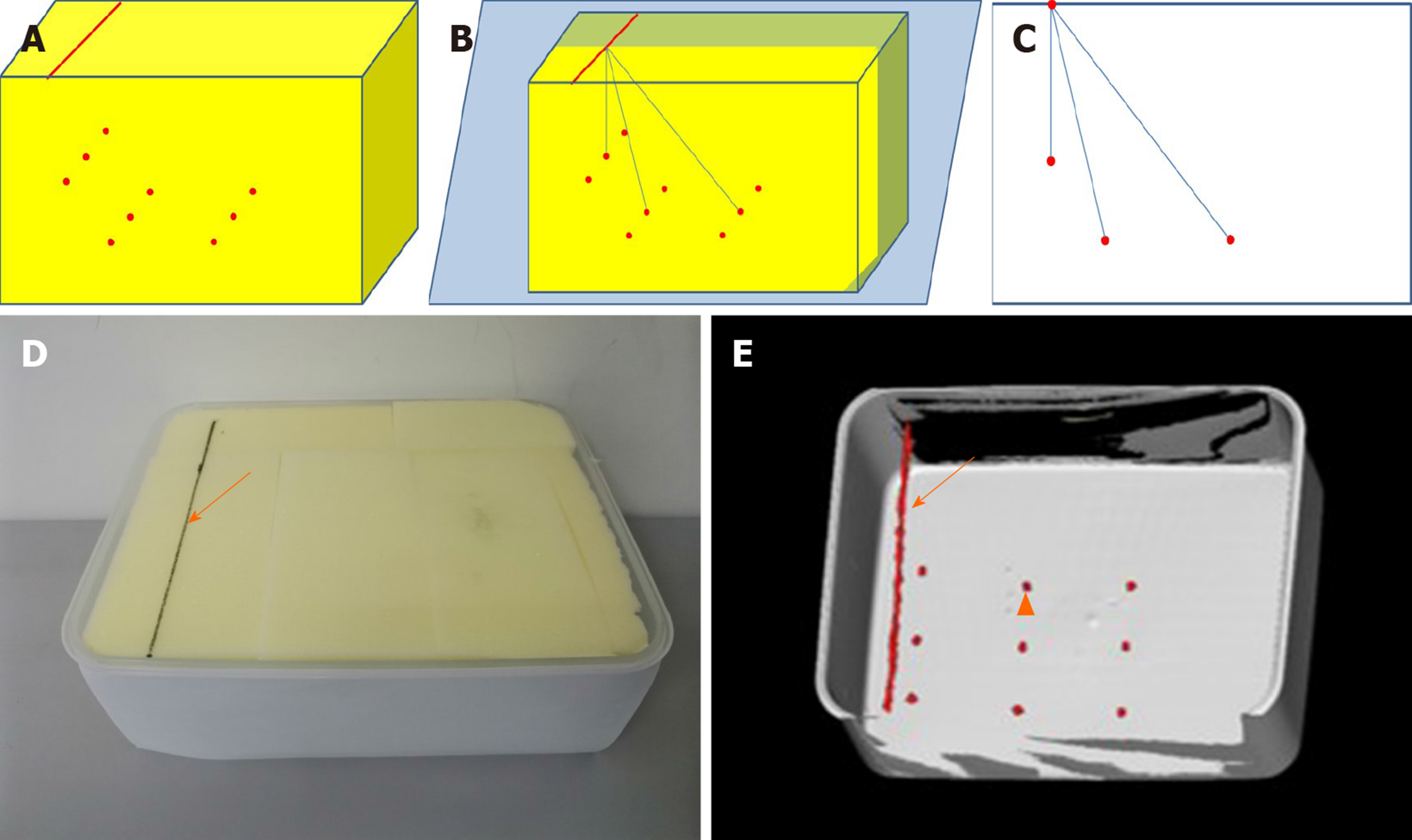
To investigate the accuracy of the navigation robot, one cuboid box model and three head phantoms were used. The box model was made of acrylic resin and filled with sponge. On the upper surface of the box, one insertion line was drawn with contrast agent parallel to the narrow edge. Nine lead spheres (three sets) with a 1 mm radius were inserted into different positions in the box. We used these artificial spheres targets to simulate respectively the short-distance, moderate-distance and long-distance operation goals. The absolute distances from the target to insertion line on the surface include 50 mm (three spheres), 100 mm (three spheres) and 150 mm (three spheres) (Figure 2).
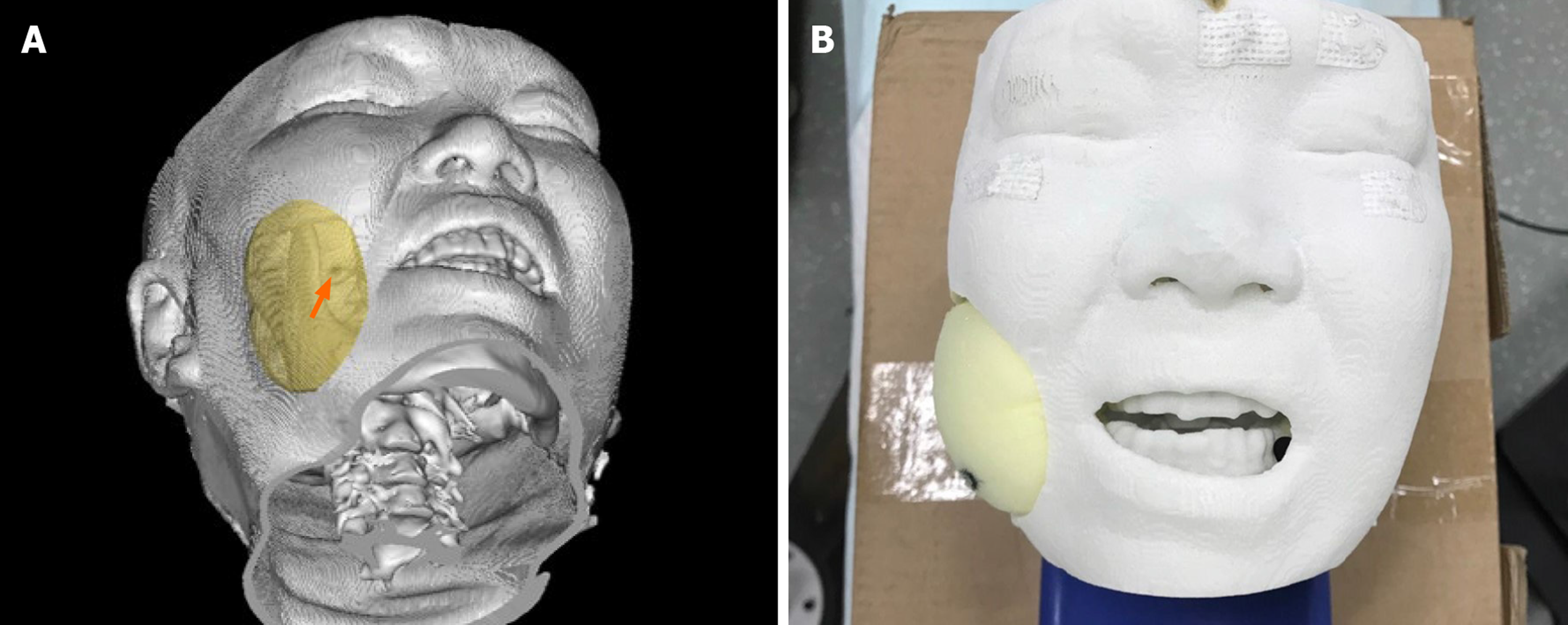
The head phantom (1:1 model of patient’s head) was also used for evaluating the clinical feasibility and stability of robot, by comparing with the free-hand operation. FO was set as the target to simulate the procedure of intervention therapy on the trigeminal ganglion. The head phantom was printed with nylon. To enhance the simulated effect, the skin of the cheek was replaced by a plasticized sponge, which fully packs the space from the face to FO (Figure 3). The long diameter and short diameter of FO were 4.9 mm and 3.4 mm on right side and 5.3 mm and 4.0 mm for left side.
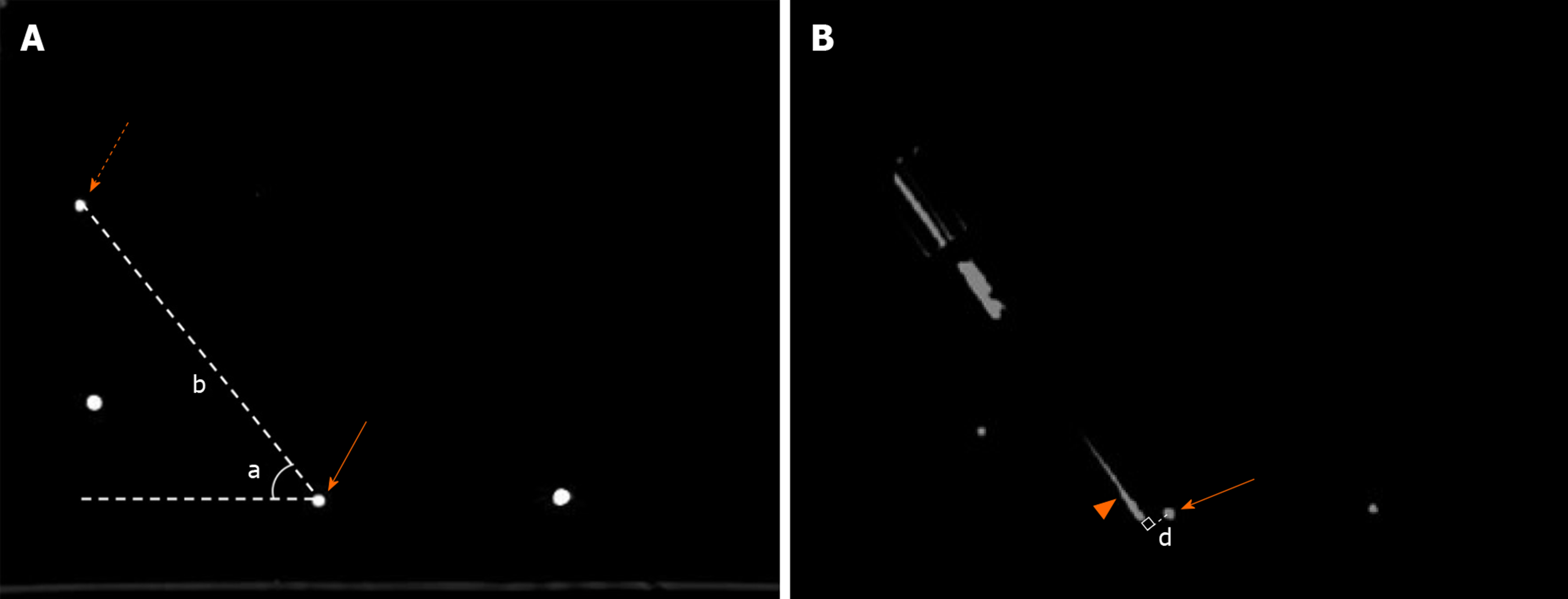
Box model was used in the first part of the experiment. On the box model, the puncture procedure was repeated three times for each goal. Therefore, a total of 27 insertion procedures were performed with the guidance of the navigation robot. When applying the robot-guided puncture, the box was placed on the bed panel with the insertion line perpendicular to the scanning coil. After trajectory planning, the insertion depth and insertion angle were measured. The insertion depth was the distance from entry point to target. The insertion angle was the angle of insertion trajectory and horizontal line. Needle was placed with the guide of robot. The Euclidian error was used to evaluate the accuracy of the navigational system. This parameter was the vertical dimension from the center of the target to the tip of real needle, which was measured on CT images (Figure 4). The Euclidian error for different targets were compared.
Head models were used for needle insertion targeting the FO in the second part of the experiment. The procedure was conducted by three attending doctors who have performed such an interventional operation at least 400 times. FO on each side of three head phantoms were operated twice (free hand and robotic guided). According to the navigation methods, free-hand group and robotic group were set in the study. In the free-hand group, FO placement was performed through the traditional Hartel approach manually[17]. In the robotic group, FO puncture was performed with the navigation of the robot. CT scanning was utilized for needle adjustment in both groups. The operation time and adjustment time were recorded. The one-time success rate of placement was also calculated and compared between two groups. Meanwhile, operation time and adjustment time of robot-guided puncture were also compared among three performers.
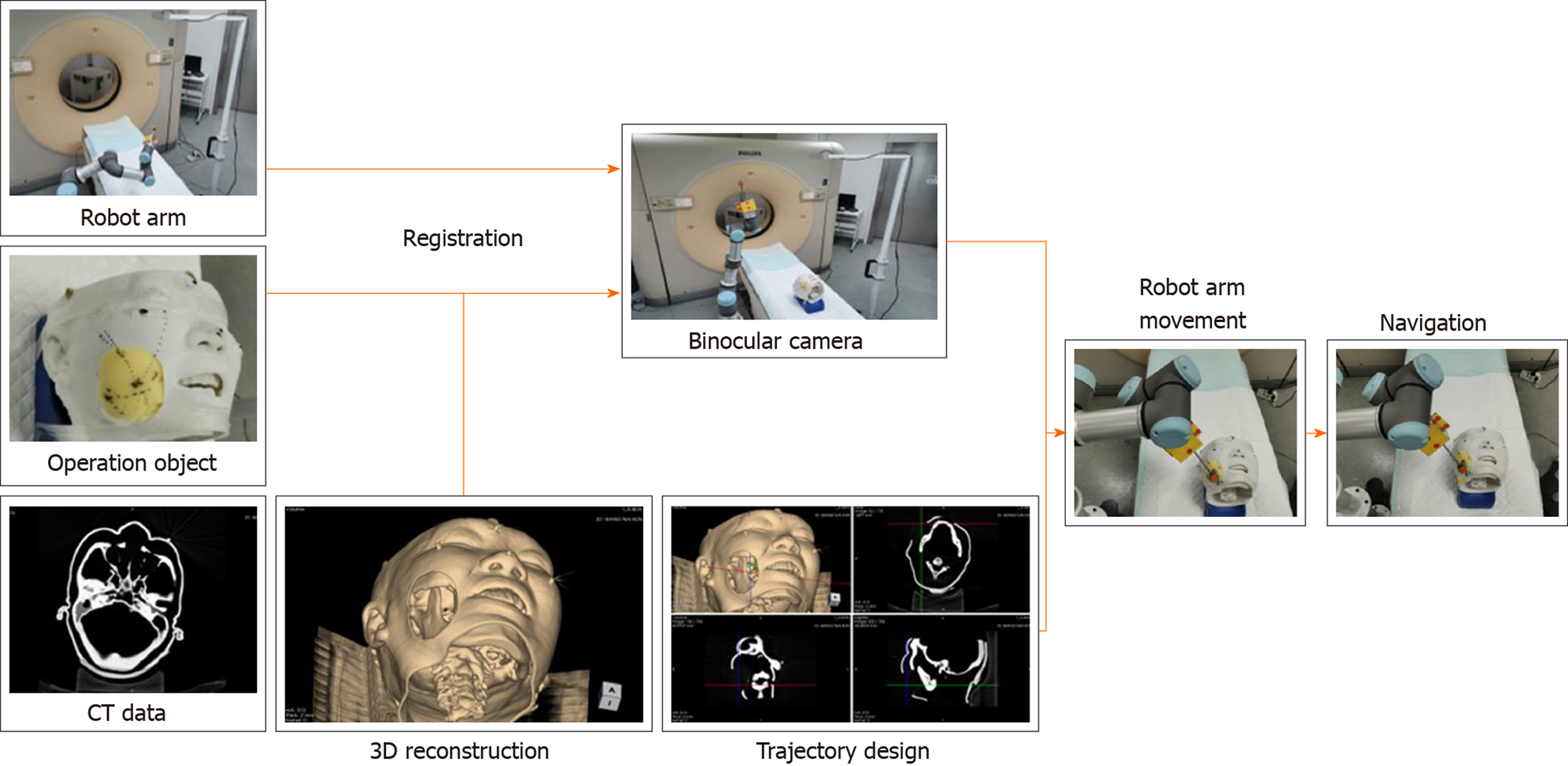
The basic work process of the binocular-stereo-vision-based navigation robot is illustrated in Figure 5. Before the interventional surgery placement, a CT scan of the operation object with four visual markers placed on the skin surface was conducted. The markers were black round labels with one lead sphere (a radius of 1 mm) in the center. The four markers were not placed in one plane. The scanning range of CT images covered the operation target and skin for percutaneous puncture. Then, the CT images were uploaded to the computer system. After three-dimensional reconstruction of the skin surface, the program acquired the spatial location of the markers for registration from cephalad to caudal. Concurrently, a puncture trajectory was designed with the target point and the entry point on the insertion line.
The robot arm and stereo camera were placed beside the operation table. A series of pictures were taken by a stereo camera for locating the markers on the operation object and the robot arm. In the coordinate system of the robot, the location of markers on the arm was obtained automatically after the posture was fixed. Then, the registration process was automatically finished by the system, and the robot arm started to move to the predetermined position for navigation. Through the guiding device at the terminal of the robot arm, the interventional tool was inserted along the planed approach to a certain depth.
The statistical analysis was conducted with SPSS Statistics Version 25 software (IBM Corp., Armonk, NY, United States). The normality distribution of the quantitative parameters was determined by the Shapiro-Wilk test. Quantitative data are presented as the mean ± SD or the median with 25th and 75th percentiles. The qualitative parameters are expressed as a number and percentage. The t-test, analysis of variance (least significant difference method) and Mann-Whitney U were used for the comparison of quantitative data between groups. Fisher's exact test was utilized for the comparison of qualitative data. The level of statistical significance was set to P < 0.05.
Puncture of three short-distance targets, three moderate-distance targets and three long-distance targets were all repeated three times. A total of 29 punctures were conducted. The puncture parameters of different targets are stated in Table 1. The difference of mean Euclidian error among three kinds of targets was statistically significant. The mean Euclidian error for long-distance target was significantly greater than those of short-distance target and moderate-distance target (P < 0.05). No significant differences were found between short and moderate distance targets (P > 0.05).
A total of 18 procedures were done in both groups. Between the two groups, no significant difference was found in operation time (P > 0.05), but the one-time success rate in robotic group was significantly higher than that in free-hand group (P < 0.05). Median adjustment time was also fewer in robotic group than in free-hand group (P < 0.05) (Table 2).
| Robotic group | Free-hand group | |
| Operation time in sec | 389 ± 85 | 422 ± 166 |
| Adjustment time | 0 (0, 1) | 2 (1, 2)a |
| One-time success rate | 72.2% (13/18) | 11.1% (2/18) |
In robotic group, the average operation times were 403 ± 87 s, 358 ± 71 s and 406 ± 100 s for three operators. Median adjustment times were 0 (0, 1), 0 (0, 0.5) and 0 (0, 1) for three performers. No significant differences in operation time and adjustment time were found among performers (P > 0.05).
The tracking method is the pivotal process in surgical navigation. This method is used to match the spatial position of target, instrument or robot arm, and this can influence the accuracy of the whole navigation system. The tracking systems applied in most commercial navigation equipment is the active optical tracking system[18-20]. The active optical tracking system is superior to other methods, such as electromagnetic tracking systems, but it is expensive and inconvenient for further development[19]. Our team has recently designed an optical tracking method based on binocular stereo vision with a high-resolution ratio and low cost. Black round labels were used as markers pasted on the skin of operation region and the robot arm. The three-dimensional location of label center can be recognized by the ZED binocular camera for registration and tracking[16]. The error of this tracking method was evaluated in our previous study. In this research, we further evaluated the position accuracy of the navigation robot system based on this tracking method.
The interventional therapies for chronic pain vary from one another, and the depths of different operation targets are also variant. To evaluate the position error of the robot as comprehensively as possible, we set three kinds of simulated targets in the box model with three levels of puncture depth. As the thinnest scanning thickness of our CT is 1 mm, the radius of sphere target was set as 1 mm to reduce the influence of scanning. When compared with other equipment[21-24], the results of our research are promising given the same accuracy of the navigation robot. For short- and medium-distance targets, the position errors were 1.7 ± 0.9 mm and 2.4 ± 1.0 mm, respectively. Although the target is still, this accuracy is acceptable and compatible for most interventional treatments. For long-distance targets, the position error was 4.4 ± 1.4 mm and relatively large for surgical navigation. The inaccuracies of this robot system may have come from several sources. The first one is the system error, and the error of the tracking system may account for the main part. The mechanical error of the robot arm is very small because the Universal Robot arm is used for industry processes, and the error is significantly less than 1 mm. The second factor may be the bending of the needle. With the existence of an inclined plane, the longer the needle is inserted, the more obvious the needle bends. Furthermore, the orientation of the inclined plane may also affect the accuracy[25].
Trigeminal neuralgia is a common chronic pain disease[26]. Most of the interventional therapies for this disease require placement of the FO, which is a difficult and challenging procedure[27]. We compared the difference of FO punctures performed by freehand and under guidance of the robot to evaluate the clinical potential. The result showed that robot assistance can significantly raise the success rate of punctures. Furthermore, the operation time was also shortened. The longest and shortest diameters of the FO inside hole were X and Y mm. The insertion depth was Z mm. Considering the placement distance, the diameter of the FO is relatively larger than the position error. Finally, five robot-assisted cases were not completed in one time. Timewise, this robot system is reasonable. The preparation time included preoperative scanning, image processing, trajectory design, registration and the robot arm moving. The whole preparation took approximately 5 min, and the registration time was approximately 1 min. Although no difference was found between the operation time of the two groups, the whole time was still slightly shorter with the robot. Our data showed no significant difference in operation time and adjustment time among the performers. This result indicated that our robot system is stable and not easily influenced by the operator.
Our study has several limitations. The first one was the still model. When operating on patients under local anesthesia, the inevitable dodge, body twist and breathing movements can influence the puncture. However, these factors were not simulated in our study. To reduce the influence of these factors in clinic, based on our experience, three measures can be taken: Fixation with headrest, sufficient local anesthesia before registration and appropriate sedation. Second, the needle used in our experiment has a cutting edge, which may induce a needle-tissue force. Pen-point needles should be used for the improved evaluation of position error. The test time was small. More tests with different angles at different depths should be conducted.
In conclusion, in the laboratory environment, the accuracy of the binocular-stereo-vision-based navigation robot is acceptable for interventional treatment with a target at a 100 mm depth or less. The robot navigation is stable, and accuracy of FO placement can be improved with the guidance of robot. In spite of the need for improved performance, we believe this navigation tool deserves clinical application and promotion.
Medical robot is a promising surgical tool, but no specific one has been designed for interventional treatment of chronic pain. We developed a computed tomography-image based navigation robot using new registration method with binocular vision. This kind of robot is appropriate for minimal invasive interventional procedures and easy to operate.
The feasibility, accuracy and stability of this new robot need to be tested before clinical application.
To assess quantitatively the feasibility, accuracy and stability of the binocular-stereo-vision-based navigation robot for minimally invasive interventional procedures.
A box model was designed for assessing the accuracy for targets at different distances. Nine (three sets) lead spheres were embedded in the model as puncture goals. The entry-to-target distances were set 50 mm (short-distance), 100 mm (medium-distance) and 150 mm (long-distance). Puncture procedure was repeated three times for each goal. The Euclidian error of each puncture was calculated and statistically analyzed. Three head phantoms were used to explore clinical feasibility and stability. Three independent operators conducted foramen ovale placement on head phantoms (both sides) by freehand or under the guidance of robot (18 punctures with each method). The operation time, adjustment time and one-time success rate were recorded, and the two guidance methods were compared.
On the box model, the mean puncture errors of navigation robot were 1.7 ± 0.9 mm for the short-distance target, 2.4 ± 1.0 mm for the moderate target and 4.4 ± 1.4 mm for the long-distance target. On the head phantom, no obvious differences in operation time and adjustment time were found among the three performers (P > 0.05). The median adjustment time was significantly less under the guidance of the robot than under free hand. The one-time success rate was significantly higher with the robot (P < 0.05). There was no obvious difference in operation time between the two methods (P > 0.05). The accuracy measurement was conducted on still models, and further clinical research on patients need to be conducted.
In laboratory environment, accuracy of binocular-stereo-vision-based navigation robot is acceptable for target at 100 mm depth or less. Compared with freehand, foramen ovale placement accuracy can be improved with robot guidance.
Based on our research, binocular-stereo-vision-based navigation robot is a promising system in the future. It deserves promotion after clinical verification, especially for interventional treatment of chronic pain.
Manuscript source: Unsolicited manuscript
Specialty type: Medicine, research and experimental
Country of origin: China
Peer-review report classification
Grade A (Excellent): 0
Grade B (Very good): B
Grade C (Good): 0
Grade D (Fair): 0
Grade E (Poor): 0
P-Reviewer: Cheng JG S-Editor: Zhang L L-Editor: Filipodia P-Editor: Liu JH
| 1. | Hylands-White N, Duarte RV, Raphael JH. An overview of treatment approaches for chronic pain management. Rheumatol Int. 2017;37:29-42. [RCA] [PubMed] [DOI] [Full Text] [Cited by in Crossref: 123] [Cited by in RCA: 250] [Article Influence: 25.0] [Reference Citation Analysis (0)] |
| 2. | Villard J, Ryang YM, Demetriades AK, Reinke A, Behr M, Preuss A, Meyer B, Ringel F. Radiation exposure to the surgeon and the patient during posterior lumbar spinal instrumentation: a prospective randomized comparison of navigated versus non-navigated freehand techniques. Spine (Phila Pa 1976). 2014;39:1004-1009. [RCA] [PubMed] [DOI] [Full Text] [Cited by in Crossref: 108] [Cited by in RCA: 124] [Article Influence: 10.3] [Reference Citation Analysis (0)] |
| 3. | Ye L, Wen C, Liu H. Ultrasound-guided versus low dose computed tomography scanning guidance for lumbar facet joint injections: same accuracy and efficiency. BMC Anesthesiol. 2018;18:160. [RCA] [PubMed] [DOI] [Full Text] [Full Text (PDF)] [Cited by in Crossref: 10] [Cited by in RCA: 12] [Article Influence: 1.5] [Reference Citation Analysis (0)] |
| 4. | Huang B, Yao M, Feng Z, Guo J, Zereshki A, Leong M, Qian X. CT-guided percutaneous infrazygomatic radiofrequency neurolysis through foramen rotundum to treat V2 trigeminal neuralgia. Pain Med. 2014;15:1418-1428. [RCA] [PubMed] [DOI] [Full Text] [Cited by in Crossref: 28] [Cited by in RCA: 36] [Article Influence: 3.0] [Reference Citation Analysis (0)] |
| 5. | Han Z, Yu K, Hu L, Li W, Yang H, Gan M, Guo N, Yang B, Liu H, Wang Y. A targeting method for robot-assisted percutaneous needle placement under fluoroscopy guidance. Comput Assist Surg (Abingdon). 2019;1-9. [RCA] [PubMed] [DOI] [Full Text] [Cited by in Crossref: 1] [Cited by in RCA: 1] [Article Influence: 0.1] [Reference Citation Analysis (0)] |
| 6. | Le XF, Shi Z, Wang QL, Xu YF, Zhao JW, Tian W. Rate and Risk Factors of Superior Facet Joint Violation during Cortical Bone Trajectory Screw Placement: A Comparison of Robot-Assisted Approach with a Conventional Technique. Orthop Surg. 2020;12:133-140. [RCA] [PubMed] [DOI] [Full Text] [Full Text (PDF)] [Cited by in Crossref: 12] [Cited by in RCA: 24] [Article Influence: 3.4] [Reference Citation Analysis (0)] |
| 7. | Wang R, Han Y, Lu L. Computer-Assisted Design Template Guided Percutaneous Radiofrequency Thermocoagulation through Foramen Rotundum for Treatment of Isolated V2 Trigeminal Neuralgia: A Retrospective Case-Control Study. Pain Res Manag. 2019;2019:9784020. [RCA] [PubMed] [DOI] [Full Text] [Full Text (PDF)] [Cited by in Crossref: 2] [Cited by in RCA: 7] [Article Influence: 1.0] [Reference Citation Analysis (0)] |
| 8. | Aydoseli A, Akcakaya MO, Aras Y, Sabanci PA, Unal TC, Sencer A, Hepgul K, Unal OF, Barlas O, Izgi N. Neuronavigation-assisted percutaneous balloon compression for the treatment of trigeminal neuralgia: The technique and short-term clinical results. Br J Neurosurg. 2015;29:552-558. [RCA] [PubMed] [DOI] [Full Text] [Cited by in Crossref: 21] [Cited by in RCA: 25] [Article Influence: 2.3] [Reference Citation Analysis (0)] |
| 9. | Khan A, Meyers JE, Siasios I, Pollina J. Next-Generation Robotic Spine Surgery: First Report on Feasibility, Safety, and Learning Curve. Oper Neurosurg (Hagerstown). 2019;17:61-69. [RCA] [PubMed] [DOI] [Full Text] [Cited by in Crossref: 64] [Cited by in RCA: 116] [Article Influence: 19.3] [Reference Citation Analysis (0)] |
| 10. | Keric N, Doenitz C, Haj A, Rachwal-Czyzewicz I, Renovanz M, Wesp DMA, Boor S, Conrad J, Brawanski A, Giese A, Kantelhardt SR. Evaluation of robot-guided minimally invasive implantation of 2067 pedicle screws. Neurosurg Focus. 2017;42:E11. [RCA] [PubMed] [DOI] [Full Text] [Cited by in Crossref: 78] [Cited by in RCA: 105] [Article Influence: 13.1] [Reference Citation Analysis (0)] |
| 11. | Hu X, Ohnmeiss DD, Lieberman IH. Robotic-assisted pedicle screw placement: lessons learned from the first 102 patients. Eur Spine J. 2013;22:661-666. [RCA] [PubMed] [DOI] [Full Text] [Cited by in Crossref: 154] [Cited by in RCA: 171] [Article Influence: 13.2] [Reference Citation Analysis (0)] |
| 12. | VanSickle D, Volk V, Freeman P, Henry J, Baldwin M, Fitzpatrick CK. Electrode Placement Accuracy in Robot-Assisted Asleep Deep Brain Stimulation. Ann Biomed Eng. 2019;47:1212-1222. [RCA] [PubMed] [DOI] [Full Text] [Cited by in Crossref: 18] [Cited by in RCA: 26] [Article Influence: 3.7] [Reference Citation Analysis (0)] |
| 13. | Ho AL, Pendharkar AV, Brewster R, Martinez DL, Jaffe RA, Xu LW, Miller KJ, Halpern CH. Frameless Robot-Assisted Deep Brain Stimulation Surgery: An Initial Experience. Oper Neurosurg (Hagerstown). 2019;17:424-431. [RCA] [PubMed] [DOI] [Full Text] [Cited by in Crossref: 26] [Cited by in RCA: 31] [Article Influence: 5.2] [Reference Citation Analysis (0)] |
| 14. | Wu XB, Wang JQ, Sun X, Han W. Guidance for the Treatment of Femoral Neck Fracture with Precise Minimally Invasive Internal Fixation Based on the Orthopaedic Surgery Robot Positioning System. Orthop Surg. 2019;11:335-340. [RCA] [PubMed] [DOI] [Full Text] [Full Text (PDF)] [Cited by in Crossref: 23] [Cited by in RCA: 43] [Article Influence: 6.1] [Reference Citation Analysis (0)] |
| 15. | Malham GM, Wells-Quinn T. What should my hospital buy next?-Guidelines for the acquisition and application of imaging, navigation, and robotics for spine surgery. J Spine Surg. 2019;5:155-165. [RCA] [PubMed] [DOI] [Full Text] [Cited by in Crossref: 45] [Cited by in RCA: 83] [Article Influence: 11.9] [Reference Citation Analysis (0)] |
| 16. | Jiang G, Luo M, Lu L, Bai K, Abdelaziz O, Chen S. Vision solution for an assisted puncture robotics system positioning. Appl Opt. 2018;57:8385-8393. [RCA] [PubMed] [DOI] [Full Text] [Cited by in Crossref: 6] [Cited by in RCA: 1] [Article Influence: 0.1] [Reference Citation Analysis (0)] |
| 17. | Fraioli MF, Cristino B, Moschettoni L, Cacciotti G, Fraioli C. Validity of percutaneous controlled radiofrequency thermocoagulation in the treatment of isolated third division trigeminal neuralgia. Surg Neurol. 2009;71:180-183. [RCA] [PubMed] [DOI] [Full Text] [Cited by in Crossref: 26] [Cited by in RCA: 27] [Article Influence: 1.5] [Reference Citation Analysis (0)] |
| 18. | Sato I, Nakamura R. Positioning error evaluation of GPU-based 3D ultrasound surgical navigation system for moving targets by using optical tracking system. Int J Comput Assist Radiol Surg. 2013;8:379-393. [RCA] [PubMed] [DOI] [Full Text] [Cited by in Crossref: 7] [Cited by in RCA: 7] [Article Influence: 0.5] [Reference Citation Analysis (0)] |
| 19. | Zhou Z, Wu B, Duan J, Zhang X, Zhang N, Liang Z. Optical surgical instrument tracking system based on the principle of stereo vision. J Biomed Opt. 2017;22:65005. [RCA] [PubMed] [DOI] [Full Text] [Cited by in Crossref: 29] [Cited by in RCA: 23] [Article Influence: 2.6] [Reference Citation Analysis (0)] |
| 20. | Maybody M, Stevenson C, Solomon SB. Overview of navigation systems in image-guided interventions. Tech Vasc Interv Radiol. 2013;16:136-143. [RCA] [PubMed] [DOI] [Full Text] [Cited by in Crossref: 33] [Cited by in RCA: 37] [Article Influence: 3.1] [Reference Citation Analysis (0)] |
| 21. | Ghelfi J, Moreau-Gaudry A, Hungr N, Fouard C, Véron B, Medici M, Chipon E, Cinquin P, Bricault I. Evaluation of the Needle Positioning Accuracy of a Light Puncture Robot Under MRI Guidance: Results of a Clinical Trial on Healthy Volunteers. Cardiovasc Intervent Radiol. 2018;41:1428-1435. [RCA] [PubMed] [DOI] [Full Text] [Cited by in Crossref: 15] [Cited by in RCA: 16] [Article Influence: 2.0] [Reference Citation Analysis (0)] |
| 22. | Ben-David E, Shochat M, Roth I, Nissenbaum I, Sosna J, Goldberg SN. Evaluation of a CT-Guided Robotic System for Precise Percutaneous Needle Insertion. J Vasc Interv Radiol. 2018;29:1440-1446. [RCA] [PubMed] [DOI] [Full Text] [Cited by in Crossref: 26] [Cited by in RCA: 53] [Article Influence: 6.6] [Reference Citation Analysis (0)] |
| 23. | Beyer LP, Michalik K, Niessen C, Platz Batista da Silva N, Wiesinger I, Stroszczynski C, Wiggermann P. Evaluation of a Robotic Assistance-System For Percutaneous Computed Tomography-Guided (CT-Guided) Facet Joint Injection: A Phantom Study. Med Sci Monit. 2016;22:3334-3339. [RCA] [PubMed] [DOI] [Full Text] [Full Text (PDF)] [Cited by in Crossref: 3] [Cited by in RCA: 6] [Article Influence: 0.6] [Reference Citation Analysis (0)] |
| 24. | Won HJ, Kim N, Kim GB, Seo JB, Kim H. Validation of a CT-guided intervention robot for biopsy and radiofrequency ablation: experimental study with an abdominal phantom. Diagn Interv Radiol. 2017;23:233-237. [RCA] [PubMed] [DOI] [Full Text] [Cited by in Crossref: 25] [Cited by in RCA: 22] [Article Influence: 2.8] [Reference Citation Analysis (0)] |
| 25. | van Gerwen DJ, Dankelman J, van den Dobbelsteen JJ. Needle-tissue interaction forces--a survey of experimental data. Med Eng Phys. 2012;34:665-680. [RCA] [PubMed] [DOI] [Full Text] [Cited by in Crossref: 167] [Cited by in RCA: 139] [Article Influence: 9.9] [Reference Citation Analysis (0)] |
| 26. | Mueller D, Obermann M, Yoon MS, Poitz F, Hansen N, Slomke MA, Dommes P, Gizewski E, Diener HC, Katsarava Z. Prevalence of trigeminal neuralgia and persistent idiopathic facial pain: a population-based study. Cephalalgia. 2011;31:1542-1548. [RCA] [PubMed] [DOI] [Full Text] [Cited by in Crossref: 153] [Cited by in RCA: 173] [Article Influence: 11.5] [Reference Citation Analysis (0)] |
| 27. | Cheng JS, Lim DA, Chang EF, Barbaro NM. A review of percutaneous treatments for trigeminal neuralgia. Neurosurgery. 2014;10 Suppl 1:25-33; discussion 33. [RCA] [PubMed] [DOI] [Full Text] [Cited by in Crossref: 31] [Cited by in RCA: 53] [Article Influence: 4.4] [Reference Citation Analysis (0)] |


