Published online Aug 16, 2023. doi: 10.12998/wjcc.v11.i23.5554
Peer-review started: April 10, 2023
First decision: June 12, 2023
Revised: June 25, 2023
Accepted: July 25, 2023
Article in press: July 25, 2023
Published online: August 16, 2023
Processing time: 128 Days and 5 Hours
Jackstone is a rare entity of calculi in urinary tracts and has the characteristic appearance resembling toy jacks. They are nearly always reported to occur in the urinary bladder, we first report a rare case of jackstone located in the obstructed renal calyx.
We report a 46-year-old man presenting with intermittent, painless gross hematuria and left flank pain. Urinary computed tomography revealed staghorn stones and secondary hydronephrosis. A jackstone with radiating branches was found in one of the dilated renal calyx. Percutaneous nephrolithotomy was performed and endoscopic images were recorded during the operation. Posto
Jackstones can occur in the renal collecting system besides the bladder. The unique appearance and imaging manifestations are the most important factors in the diagnosis of jackstones, and further exploration of the formation mechanism is required.
Core Tip: As a rare entity, jackstone with the characteristic appearance resembling toy jacks is usually found in the urinary bladder. This study discusses a rare case of a jackstone in a hydronephrotic renal calyx which had never been described before. Jackstones are commonly composed of calcium oxalate monohydrate or calcium oxalate dihydrate. The exact pathophysiology of jackstone development remains poorly understood. Outflow obstruction may be the most common cause. Thus, when removing the stones, the obstruction should also be evaluated and treated to avoid recurrence.
- Citation: Song HF, Liang L, Liu YB, Xiao B, Hu WG, Li JX. Jackstone in the renal calyx: A rare case report. World J Clin Cases 2023; 11(23): 5554-5558
- URL: https://www.wjgnet.com/2307-8960/full/v11/i23/5554.htm
- DOI: https://dx.doi.org/10.12998/wjcc.v11.i23.5554
Jackstones are named for their radiating appearance, which resembles toy jacks[1]. They have been reported in different animals[2] and humans and are usually found in the urinary bladder but rarely found in the kidneys[3,4]. The most common jackstones include those composed of calcium oxalate monohydrate and calcium oxalate dihydrate[5]. Here, we report a rare case of a jackstone composed of calcium oxalate monohydrate located in a hydronephrotic renal calyx.
A 46-year-old man presented with intermittent, painless gross hematuria and left flank pain after activity for one month.
About one month ago, the patient developed intermittent gross hematuria and distending pain of the left flank for unknown reasons, without frequency, urgency, and painful urination. He also had no fever, nausea, and vomiting.
The patient had a history of primary hypertension for one year, he took 80 mg valsartan orally per day. The blood pressure was controlled well. He also had bilateral saphenous varicose veins for 20 years and never treated. The patient was allergic to sulfonamides.
The patient’s personal and family history was not remarkable.
On physical examination, the vital signs were normal and there were no positive signs except percussive pain in the left renal region.
The routine urine analysis showed full field of red and white blood cells. The urine culture indicated Enterococcus faecalis (> 100000 CFU/mL). The patient’s serum creatine was slightly elevated (135 μmol/L). NMP22 and urine cytology were negative. No abnormality was found in other routine blood tests.
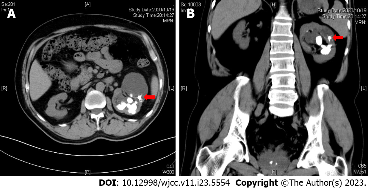
Urinary computed tomography (CT) revealed staghorn stones in the left kidney, filling the renal pelvis and several calices with secondary hydronephrosis. A stellate stone with characteristic radiating spicules (suspected to be a jackstone) was found in one of the dilated upper renal calyces, measuring 0.8 cm × 1.0 cm with a maximum density of 1240 Hounsfield units (HU) (Figure 1). There was no stone or obstruction found in the ureter. Prostate calculi were also found on CT image.
The final major diagnosis was kidney stone, other diagnoses included hydronephrosis, urinary infection, kidney injury, and prostate calculi.
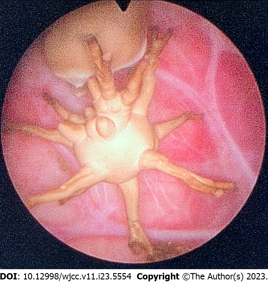
After antibiotics therapy (levofloxacin, 0.5 g, ivgtt) for three days, prone percutaneous nephrolithotomy was performed. Two standard channels were established to remove the stones. During the surgery, it was observed that the stones blocked the funnel of the upper calyx and a jackstone located in the dilated calyx (Figure 2). There was no obstruction at the ureteropelvic junction. All stones were completely removed using ultrasound lithotripsy and a double J stent and nephrostomy tubes were then placed.
The patient recovered smoothly with no complications occurred and was discharged 7 d after surgery. Nephrostomy tubes were removed during hospitalization. One month after, the left double-J stent was removed successfully. Postoperative infrared spectroscopy analysis demonstrated that the jackstone was composed of calcium oxalate monohydrate.
Jackstone is a type of urinary calculi with a distinctive appearance and are usually reported in the bladder. Despite its distinct shape, the clinical manifestations of jackstone are not unique or specific. Therefore, medical imaging examinations and visual inspection are necessary for diagnosis. Patients with jackstones often exhibit intermittent gross hematuria, obstructive lower urinary tract symptoms, and abdominal or flank pain, making these patients seek medical attention[3,6,7].
Jackstones located in the collecting system are extremely rare. In previous reports, jackstones were usually located in the renal pelvis[8,9]. Symeonidis et al[3] summarized 14 previously published cases of jackstones found in the urinary tract: 78.6% of patients had single jackstones; 2 cases had renal stones and the remaining 12 patients had bladder stones[3]. However, the exact pathophysiology of jackstone development remains poorly understood. Outflow obstruction may be the most common cause. Lim et al[7] reported two jackstone calculi in the renal pelvis in a 53-year-old man with ureteropelvic junction obstruction. They suggested that the capacious renal pelvis caused by obstruction enabled the formation of the jackstones[7]. In our case, the jackstone was located in an obstructed renal calyx, which has not been previously reported. This indicates that jackstone formation can be observed in any location where the obstruction is present in the urinary tract. However, in another case report by Goonewardena et al[8], a jackstone occurred in the renal pelvis without significant obstruction in the ureteropelvic junction demonstrated in the renogram curve. Therefore, the mechanism of jackstone's occurrence still remains uncertain and needs further clarification.
In our case, stone composition analysis using infrared spectroscopy revealed calcium oxalate monohydrate. According to previous reports, bladder jackstones were commonly composed of calcium oxalate monohydrate or calcium oxalate dihydrate. Grases et al[10] reported a jackstone in the renal pelvis composing of calcium oxalate monohydrate. Canela et al[5] used micro-CT and infrared spectroscopy to examine 98 jackstones, the largest case series of jackstones to date. They showed that jackstones had an X-ray transparent core within the outer projecting spines, with an outer shell that was always composed of calcium oxalate. Immunohistochemistry showed that the core was partially enriched with Tamm-Horsfall protein. They suggested that this protein-rich core might preferentially bind to more proteins in the urine, causing the spines to grow in a linear fashion and at a faster rate. Of note, in our case, the jackstone was fragmented intraoperatively, making it impossible to explore whether our jackstone could fit this pattern.
In conclusion, we report the case of a typical jackstone in a hydronephrotic renal calyx that has rarely been reported in the literature. The unique spike-like appearance and imaging manifestations resembling toy jacks were the most important factors for diagnosing jackstones. Although most studies attribute its occurrence to urinary tract obstruction, further investigation is still needed.
Provenance and peer review: Unsolicited article; Externally peer reviewed.
Peer-review model: Single blind
Specialty type: Urology and nephrology
Country/Territory of origin: China
Peer-review report’s scientific quality classification
Grade A (Excellent): 0
Grade B (Very good): B
Grade C (Good): 0
Grade D (Fair): D
Grade E (Poor): 0
P-Reviewer: Sureshkumar KK, United States; Vyshka G, Albania S-Editor: Yan JP L-Editor: A P-Editor: Yan JP
| 1. | Sweeney AP, Dyer RB. The "jackstone" appearance. Abdom Imaging. 2015;40:2906-2907. [RCA] [PubMed] [DOI] [Full Text] [Cited by in Crossref: 2] [Cited by in RCA: 1] [Article Influence: 0.1] [Reference Citation Analysis (0)] |
| 2. | Osborne CA, Jacob F, Lulich JP, Hansen MJ, Lekcharoensul C, Ulrich LK, Koehler LA, Bird KA, Swanson LL. Canine silica urolithiasis. Risk factors, detection, treatment, and prevention. Vet Clin North Am Small Anim Pract. 1999;29:213-230, xiii. [RCA] [PubMed] [DOI] [Full Text] [Cited by in Crossref: 12] [Cited by in RCA: 12] [Article Influence: 0.4] [Reference Citation Analysis (0)] |
| 3. | Symeonidis EN, Memmos D, Anastasiadis A, Mykoniatis I, Savvides E, Langas G, Baniotis P, Bouchalakis A, Tsiakaras S, Stefanidis P, Stratis M, Mutomba WF, Vakalopoulos I, Dimitriadis G. Jackstone: A Calculus "Toy" in the Bladder. A Case Report of Rare Entity and Comprehensive Review of the Literature. Acta Med Litu. 2022;29:149-156. [RCA] [PubMed] [DOI] [Full Text] [Full Text (PDF)] [Cited by in RCA: 3] [Reference Citation Analysis (0)] |
| 4. | Zhao J, Gu X. Jackstone Calculus in the Bladder. N Engl J Med. 2022;387:1123. [RCA] [PubMed] [DOI] [Full Text] [Cited by in RCA: 3] [Reference Citation Analysis (0)] |
| 5. | Canela VH, Dzien C, Bledsoe SB, Borofsky MS, Boris RS, Lingeman JE, El-Achkar TM, Williams JC Jr. Human jackstone arms show a protein-rich, X-ray lucent core, suggesting that proteins drive their rapid and linear growth. Urolithiasis. 2022;50:21-28. [RCA] [PubMed] [DOI] [Full Text] [Cited by in Crossref: 2] [Cited by in RCA: 4] [Article Influence: 1.0] [Reference Citation Analysis (0)] |
| 6. | Banerji JS. Jackstone Calculus. Mayo Clin Proc. 2019;94:2383-2384. [RCA] [PubMed] [DOI] [Full Text] [Cited by in RCA: 3] [Reference Citation Analysis (0)] |
| 7. | Lim KY, Sewell J, Harper M. Jackstones in the renal pelvis: A rare calculus. Urol Case Rep. 2022;42:101994. [RCA] [PubMed] [DOI] [Full Text] [Full Text (PDF)] [Cited by in RCA: 3] [Reference Citation Analysis (0)] |
| 8. | Goonewardena S, Jayarajah U, Kuruppu SN, Fernando MH. Jackstone in the Kidney: An Unusual Calculus. Case Rep Urol. 2021;2021:8816547. [RCA] [PubMed] [DOI] [Full Text] [Full Text (PDF)] [Cited by in Crossref: 3] [Cited by in RCA: 6] [Article Influence: 1.2] [Reference Citation Analysis (0)] |
| 9. | Mercurio M, Izzo F, Gatta GD, Salzano L, Lotrecchiano G, Saldutto P, Germinario C, Grifa C, Varricchio E, Carafa A, Di Meo MC, Langella A. May a comprehensive mineralogical study of a jackstone calculus and some other human bladder stones unveil health and environmental implications? Environ Geochem Health. 2022;44:3297-3320. [RCA] [PubMed] [DOI] [Full Text] [Cited by in Crossref: 1] [Cited by in RCA: 4] [Article Influence: 1.0] [Reference Citation Analysis (0)] |
| 10. | Grases F, Costa-Bauza A, Prieto RM, Saus C, Servera A, García-Miralles R, Benejam J. Rare calcium oxalate monohydrate calculus attached to the wall of the renal pelvis. Int J Urol. 2011;18:323-325. [RCA] [PubMed] [DOI] [Full Text] [Cited by in Crossref: 4] [Cited by in RCA: 5] [Article Influence: 0.3] [Reference Citation Analysis (0)] |


