Copyright
©The Author(s) 2021.
World J Clin Cases. Nov 26, 2021; 9(33): 10323-10327
Published online Nov 26, 2021. doi: 10.12998/wjcc.v9.i33.10323
Published online Nov 26, 2021. doi: 10.12998/wjcc.v9.i33.10323
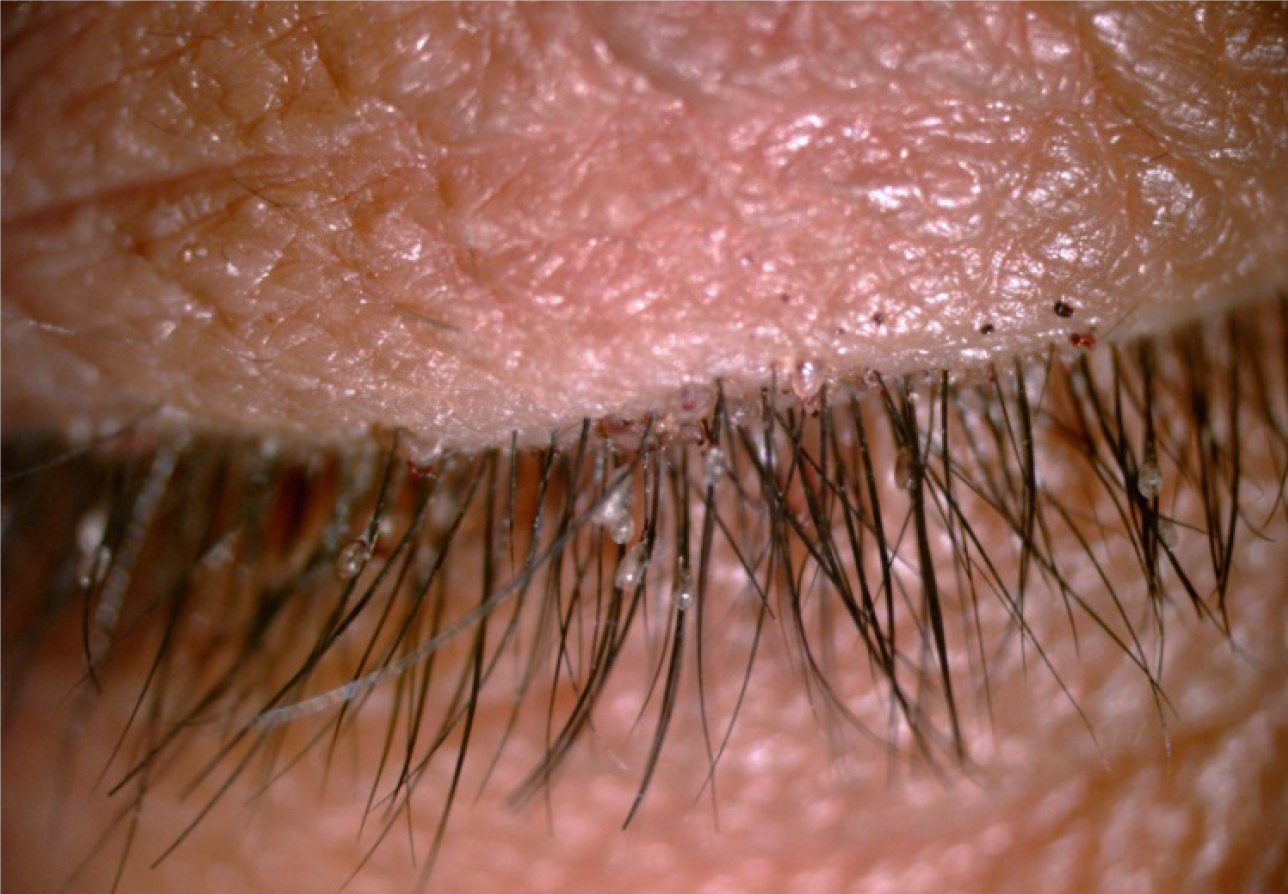
Figure 1 Slit-lamp examination of the patient’s right eyelashes and adjacent eyelids.
Some macula and empty shells are seen on the eyelashes.
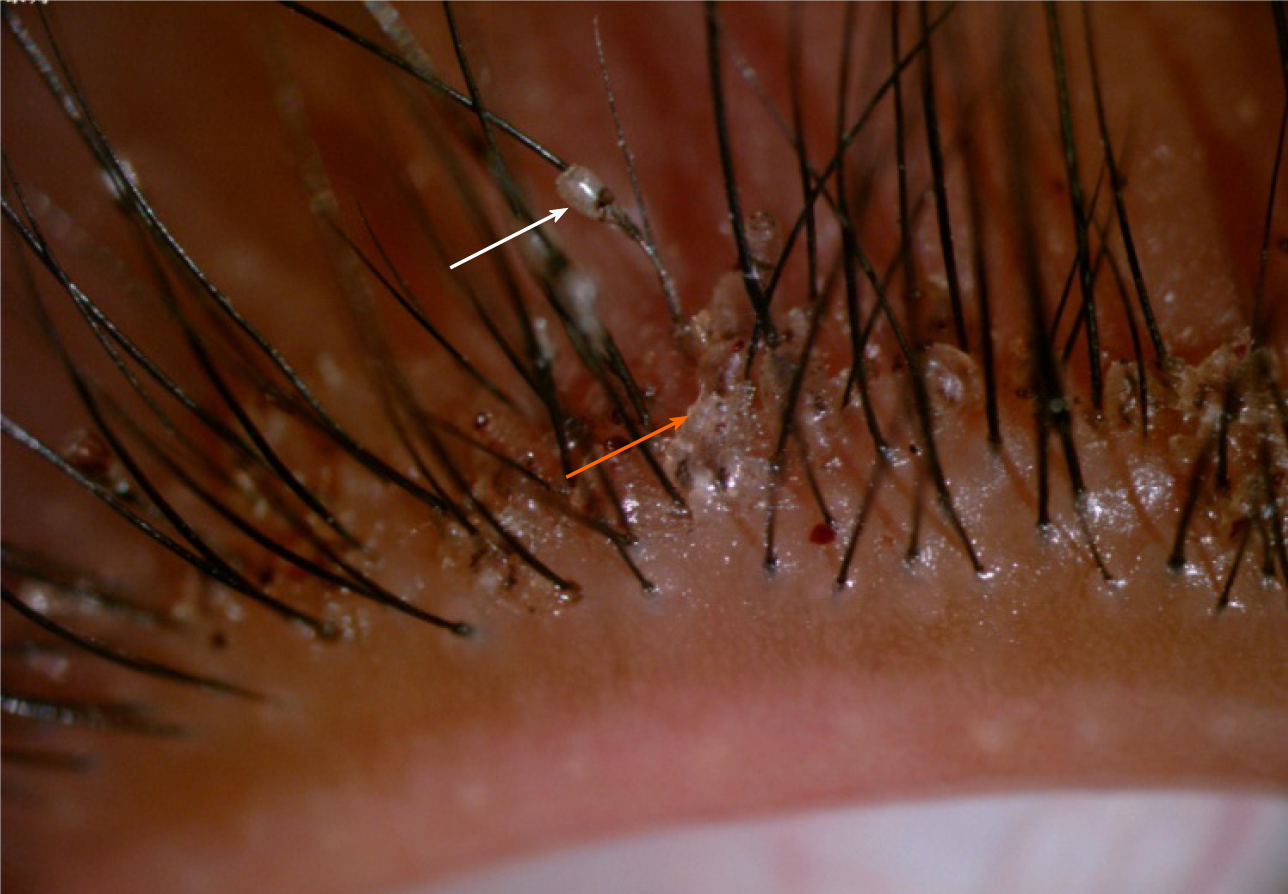
Figure 2 Photos of parasites and empty shells on the patient’s right eyelashes and adjacent eyelids.
Empty shells are denoted by white arrow; Parasites are denoted by orange arrow.
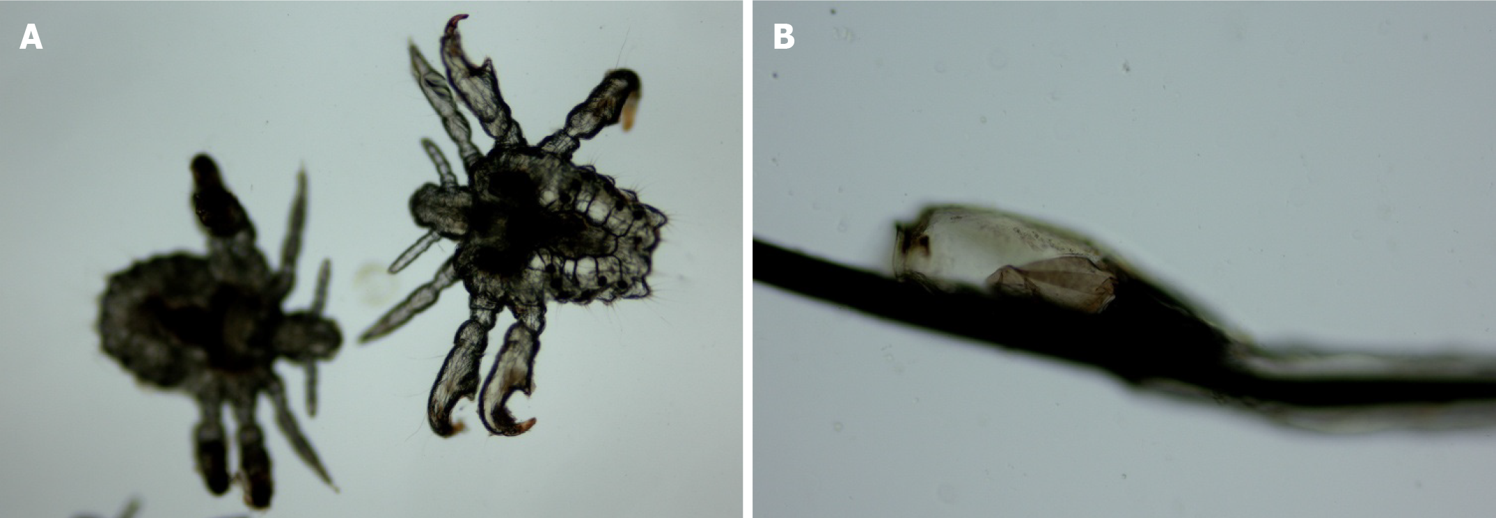
Figure 3 Photos of crab lice and nits taken from the patient’s right eyelashes and adjacent eyelids, as viewed under a light microscope.
Magnification 100 ×. A: Crab lice; B: Nits.
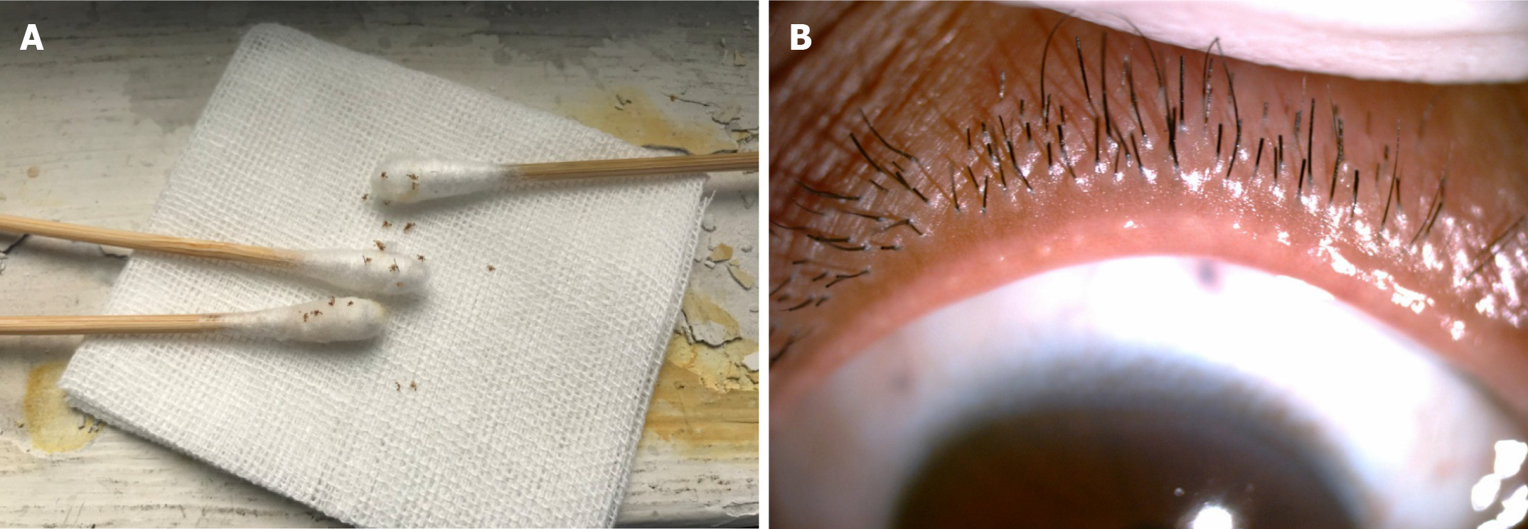
Figure 4 Some of crab lice taken from the patient and recovered eyelashes and eyelids.
A: Twenty crab lice removed on cotton swabs and gauze and 6 were subjected to examination under a light microscope; B: Cleared eyelashes (regrowth) and adjacent eyelids at 2-wk after treatment.
- Citation: Tang W, Li QQ. Crab lice infestation in unilateral eyelashes and adjacent eyelids: A case report. World J Clin Cases 2021; 9(33): 10323-10327
- URL: https://www.wjgnet.com/2307-8960/full/v9/i33/10323.htm
- DOI: https://dx.doi.org/10.12998/wjcc.v9.i33.10323


