©The Author(s) 2021.
World J Clin Cases. Oct 26, 2021; 9(30): 9144-9150
Published online Oct 26, 2021. doi: 10.12998/wjcc.v9.i30.9144
Published online Oct 26, 2021. doi: 10.12998/wjcc.v9.i30.9144
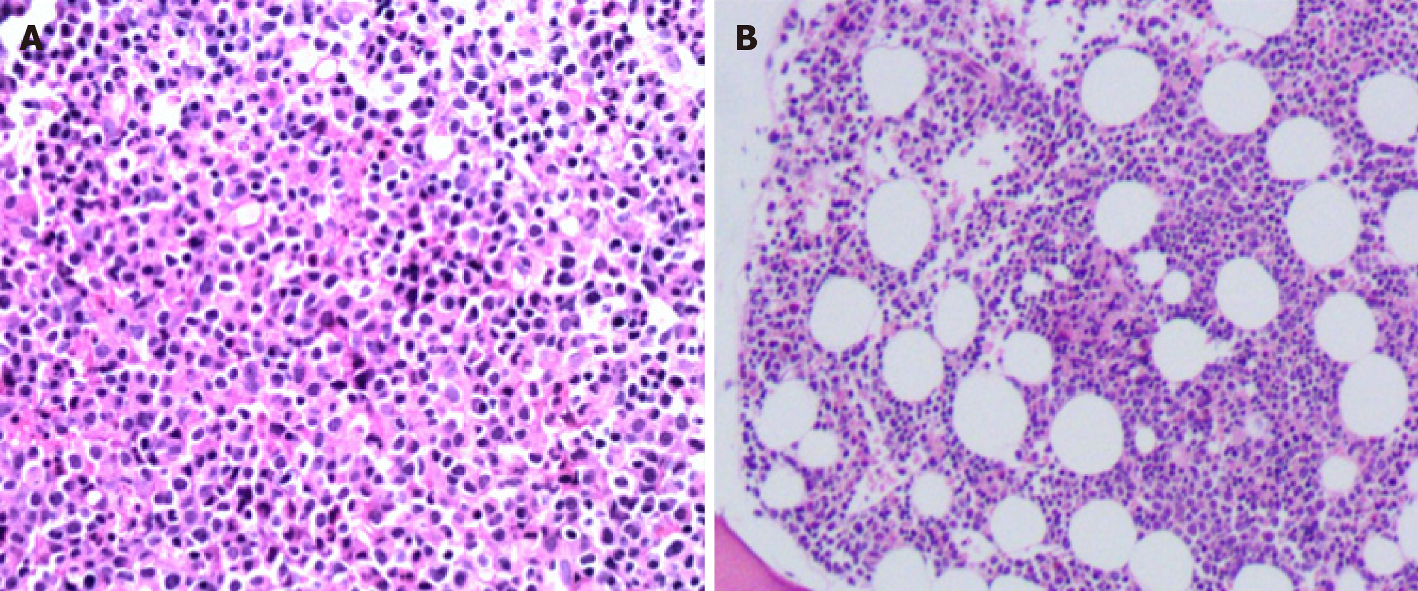
Figure 1 Pathological microscopic image with hematoxylin-eosin staining of lymph node biopsy in case once (A) and of marrow biopsy in case two (B).
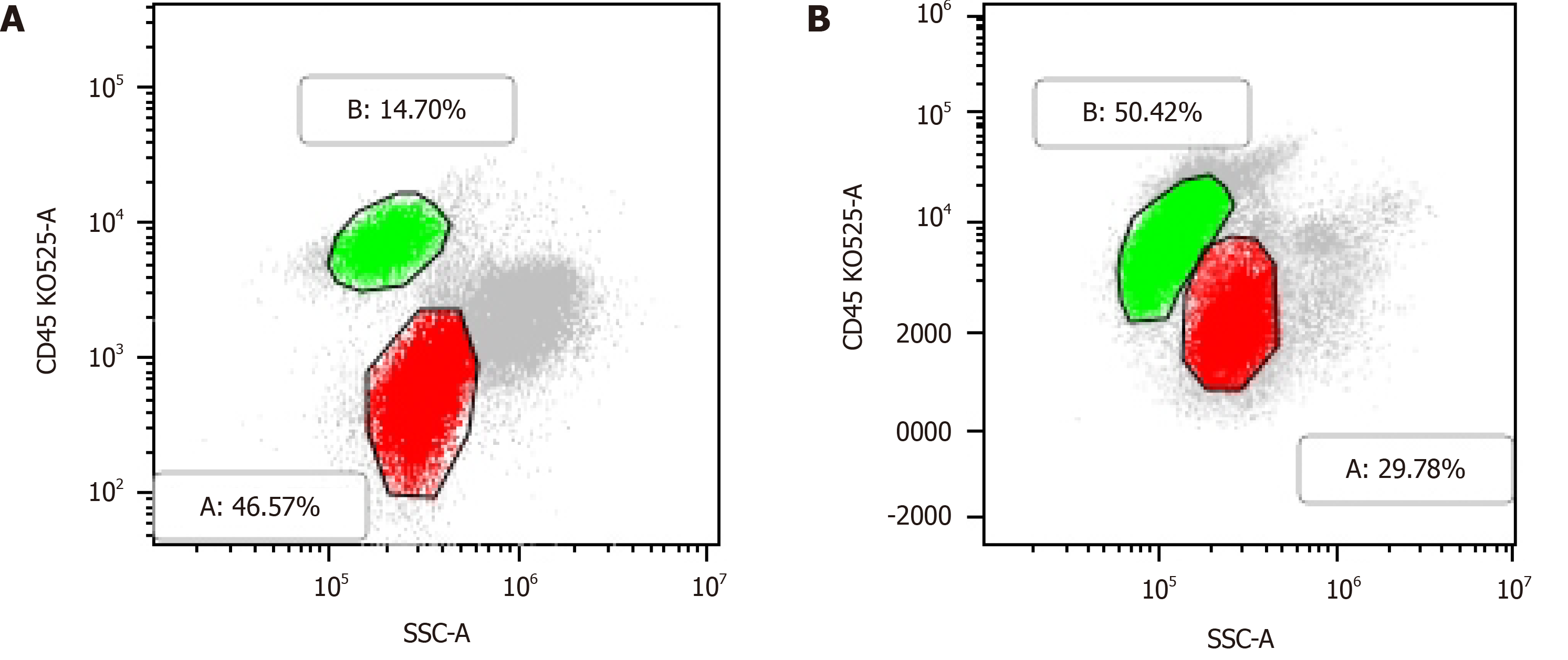
Figure 2 Flow cytometric analysis showed two separate populations of cells the red one positive for myeloblast markers (CD117, CD34, CD33, CD13, HLADR, CD38, MPO) and the green one for B lymphoid markers [CD19, CD5, CD22, CD20, CD23(weak), CD200, kappa light chain] in case one (A), and two separate populations of cells the red one positive for myeloblast markers (CD117, CD34, CD33, CD13, HLA-DR, CD38) and the green one for B lymphoid markers [CD19, CD5, CD22 (weak), CD20 (weak), CD23, CD200, Kappa light chain] in case two (B).
- Citation: Chen RR, Zhu LX, Wang LL, Li XY, Sun JN, Xie MX, Zhu JJ, Zhou D, Li JH, Huang X, Xie WZ, Ye XJ. Synchronous diagnosis and treatment of acute myeloid leukemia and chronic lymphocytic leukemia: Two case reports. World J Clin Cases 2021; 9(30): 9144-9150
- URL: https://www.wjgnet.com/2307-8960/full/v9/i30/9144.htm
- DOI: https://dx.doi.org/10.12998/wjcc.v9.i30.9144


