©The Author(s) 2021.
World J Clin Cases. Oct 16, 2021; 9(29): 8825-8830
Published online Oct 16, 2021. doi: 10.12998/wjcc.v9.i29.8825
Published online Oct 16, 2021. doi: 10.12998/wjcc.v9.i29.8825
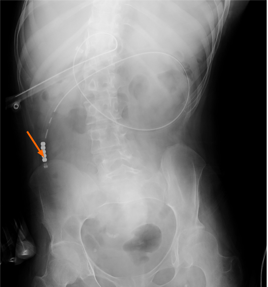
Figure 1 Radiograph confirming the proper placement of the percutaneous endoscopic gastrostomy with jejunal extension catheter's tip in the upper jejunum (arrow).
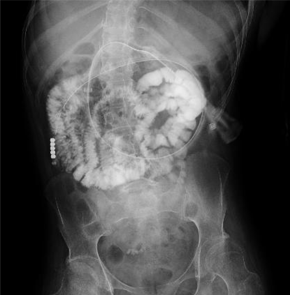
Figure 2 X-ray findings after the infusion of the contrast medium (gastrograffin) through the percutaneous endoscopic gastrostomy with jejunal extension catheter.
The contrast medium is visible from the upper jejunum to the lower part of the small intestine. However, it is not observed in the reflux to the duodenum.
- Citation: Ohmori H, Kodama H, Takemoto M, Yamasaki M, Matsumoto T, Kumode M, Miyachi T, Sumimoto R. Isolated neutropenia caused by copper deficiency due to jejunal feeding and excessive zinc intake: A case report. World J Clin Cases 2021; 9(29): 8825-8830
- URL: https://www.wjgnet.com/2307-8960/full/v9/i29/8825.htm
- DOI: https://dx.doi.org/10.12998/wjcc.v9.i29.8825


