©The Author(s) 2021.
World J Clin Cases. Oct 16, 2021; 9(29): 8812-8819
Published online Oct 16, 2021. doi: 10.12998/wjcc.v9.i29.8812
Published online Oct 16, 2021. doi: 10.12998/wjcc.v9.i29.8812
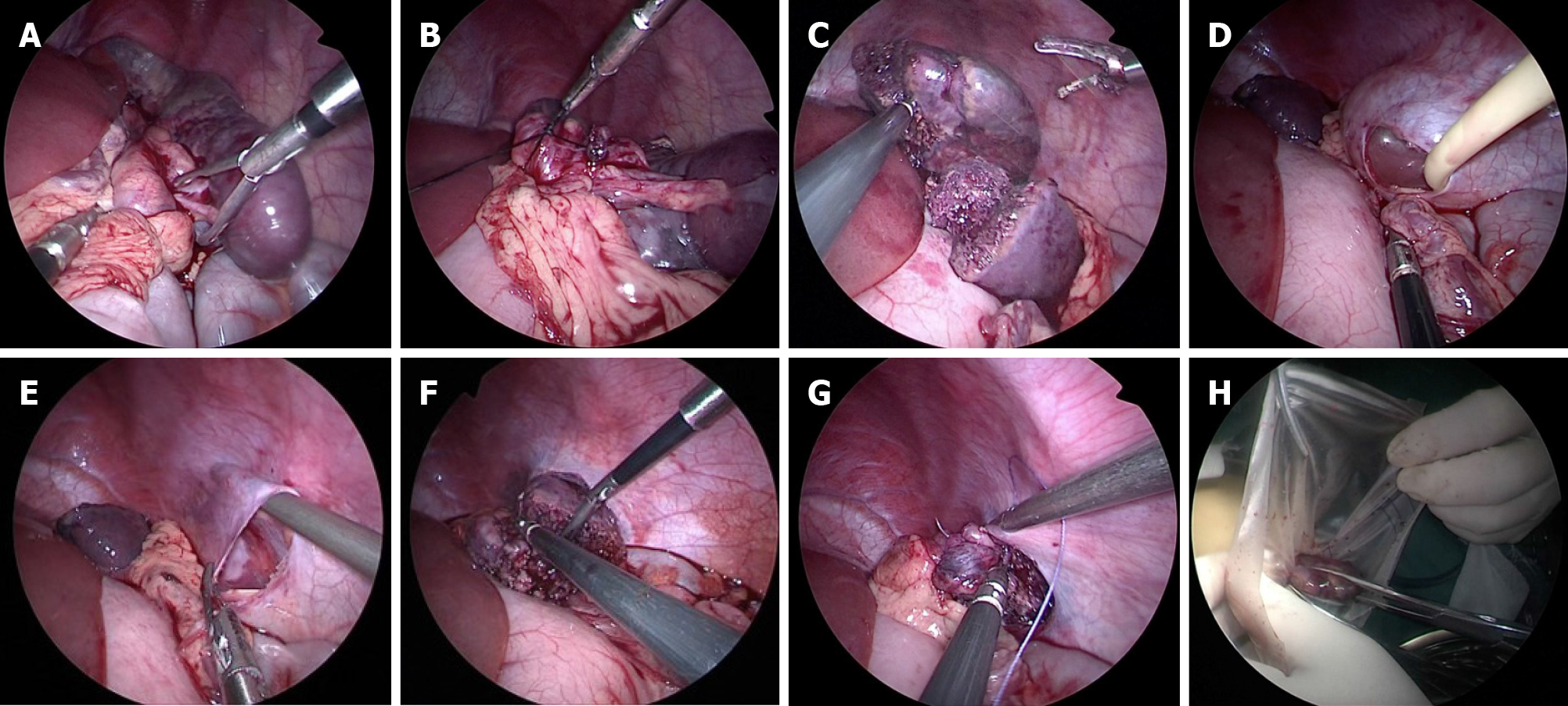
Figure 1 Laparoscopic exploration in case two.
A: Expose the spleen pedicle, clarify the direction of twisting, and reset the spleen; B: After silk thread ligation, the middle and upper pole vessels of the spleen are cut off, and the lower pole vessels of the spleen are preserved; C: Ultrasonic knife cuts off the spleen and retains the lower pole of the spleen; D: No. 14 Foley urinary catheter was introduced through Trocar to expand the balloon to form an extraperitoneal space; E: Formation of lateral retroperitoneal space; F: The remnant spleen was put into the lateral retroperitoneal space; G: Suture the peritoneal incision with 3-0 non-absorbable thread and fix the remaining spleen; H: The excised spleen tissue is bagged and pulled out from the umbilical incision and crushed.
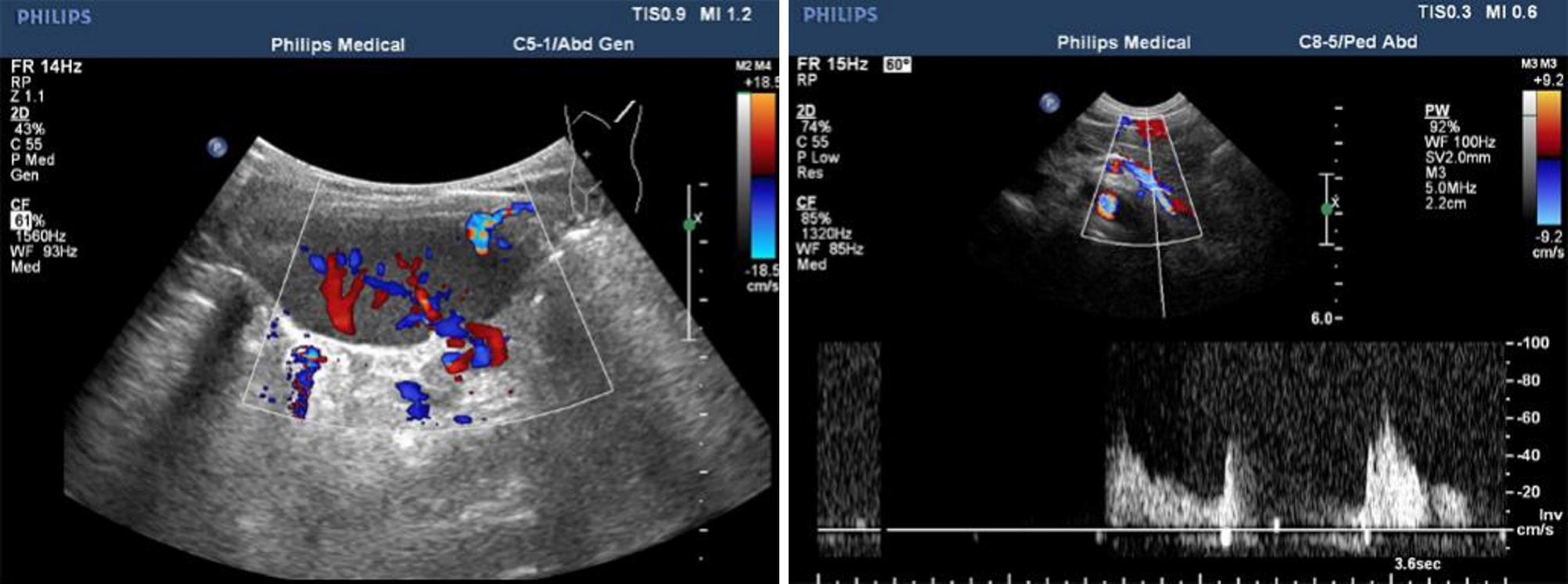
Figure 2 Ultrasound examination.
Eight months after operation, the size of residual spleen was 5.3 cm × 3.5 cm × 2 cm, the echo was homogeneous, and the blood supply was good.
- Citation: Sun C, Li SL. Successful treatment of floating splenic volvulus: Two case reports and a literature review . World J Clin Cases 2021; 9(29): 8812-8819
- URL: https://www.wjgnet.com/2307-8960/full/v9/i29/8812.htm
- DOI: https://dx.doi.org/10.12998/wjcc.v9.i29.8812


