Copyright
©The Author(s) 2021.
World J Clin Cases. Oct 16, 2021; 9(29): 8789-8796
Published online Oct 16, 2021. doi: 10.12998/wjcc.v9.i29.8789
Published online Oct 16, 2021. doi: 10.12998/wjcc.v9.i29.8789
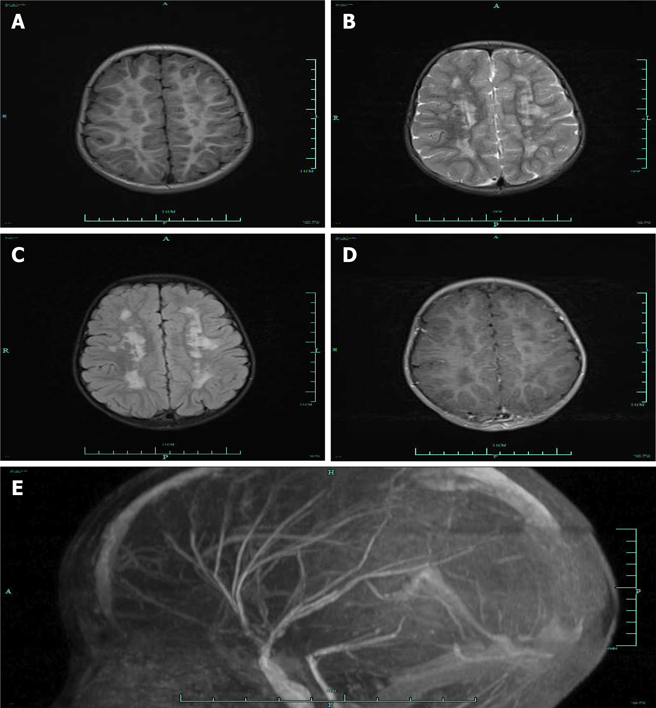
Figure 1 Cerebral magnetic resonance imaging (axial T1-weighted, T2-weighted, fluid attenuated inversion recovery images) for the patient with multi-systemic smooth muscle dysfunction syndrome multiple.
A-C: Cerebral Magnetic resonance imaging showed multiple aberrant signal shadows in bilateral paraventricular; D: There was no enhancement in contrast-enhanced scan; E: Lateral projection of magnetic resonance angiography indicated abnormally straight course of intracranial arteries.
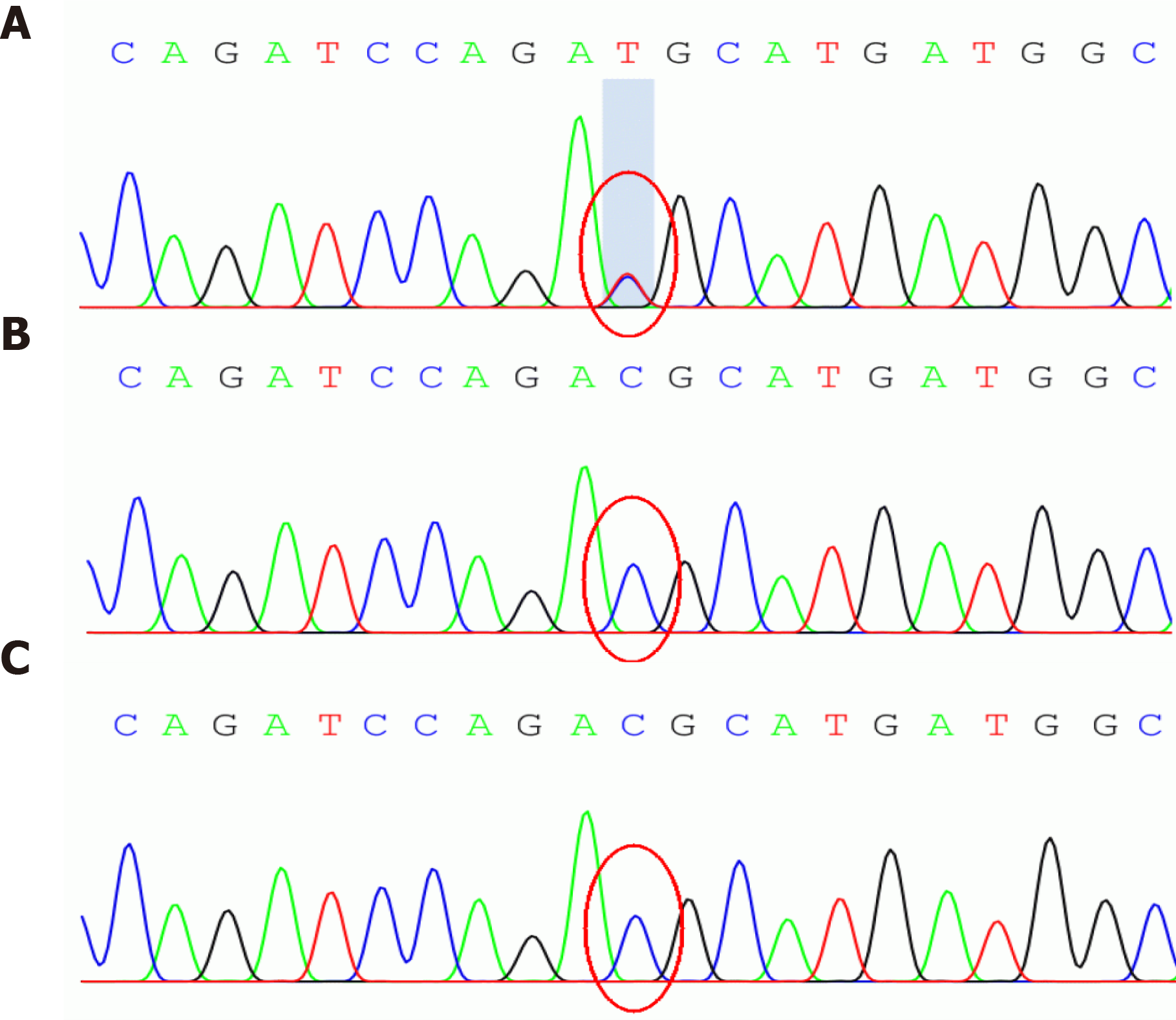
Figure 2 Sequencing analysis results for the patient and her parents.
The gene sequence map of the child showed the change of c.536G>A (the nucleotide cytosine mutation of coding region 536 became thymine). No mutation was identified in the sequencing data of her father and mother. A: The patient; B: Her father; C: Her mother; Green shadow indicated mutation sites.
- Citation: Yang WX, Zhang HH, Hu JN, Zhao L, Li YY, Shao XL. ACTA2 mutation is responsible for multisystemic smooth muscle dysfunction syndrome with seizures: A case report and review of literature. World J Clin Cases 2021; 9(29): 8789-8796
- URL: https://www.wjgnet.com/2307-8960/full/v9/i29/8789.htm
- DOI: https://dx.doi.org/10.12998/wjcc.v9.i29.8789


