Copyright
©The Author(s) 2021.
World J Clin Cases. Oct 6, 2021; 9(28): 8461-8469
Published online Oct 6, 2021. doi: 10.12998/wjcc.v9.i28.8461
Published online Oct 6, 2021. doi: 10.12998/wjcc.v9.i28.8461
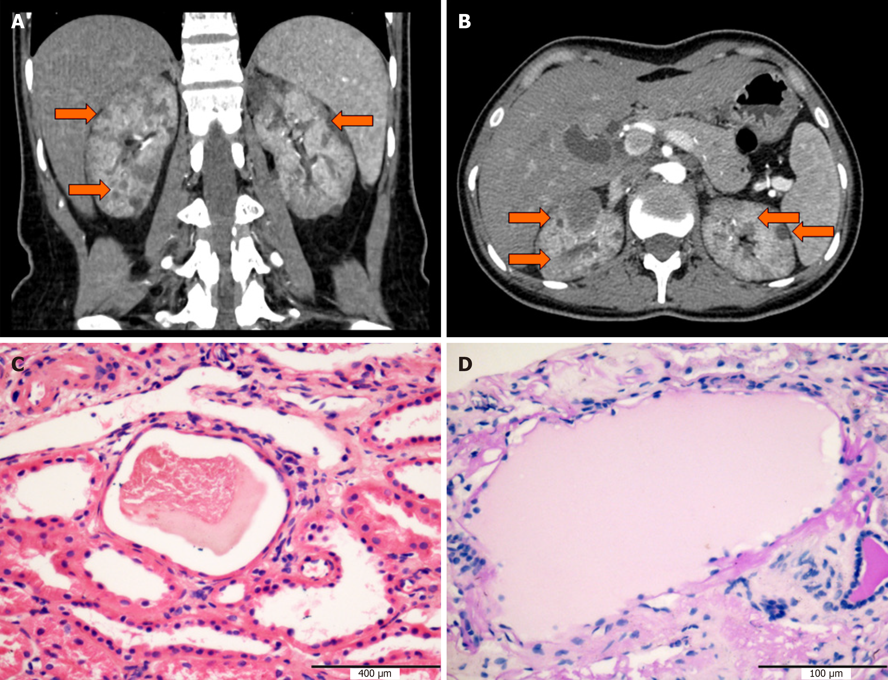
Figure 1 Computed tomography images and classic histological features of the kidney.
A and B: Abdominal computed tomography revealed multiple renal cysts in the proband (orange arrow), and cystic structure with serous-like substances was visible in tubular lumens; C: Section stained with hematoxylin-eosin; D: Section stained with Periodic acid-Schiff.
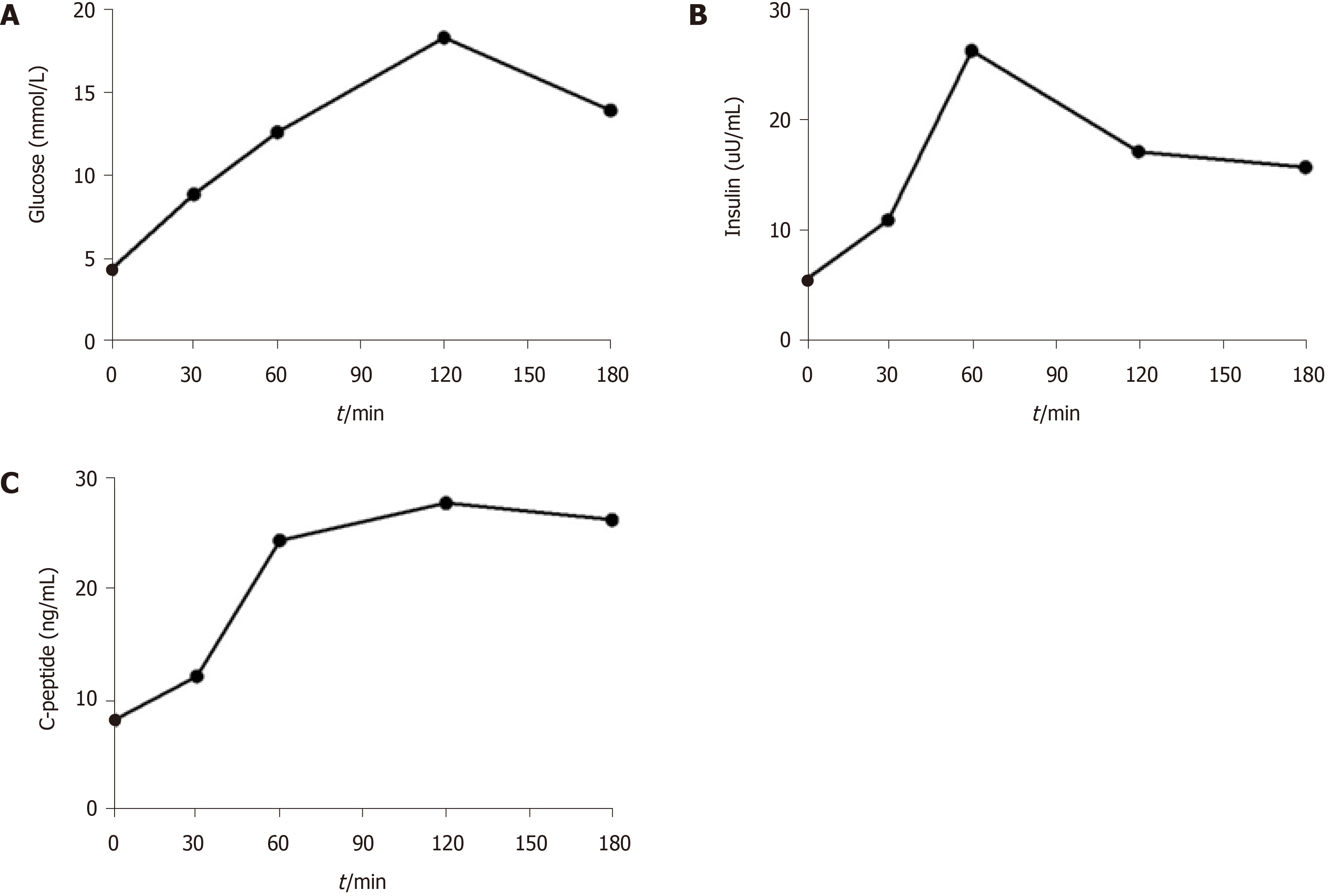
Figure 2 Oral glucose tolerance test.
A: Glucose; B: Insulin; C: C-peptide concentration during oral glucose tolerance test.
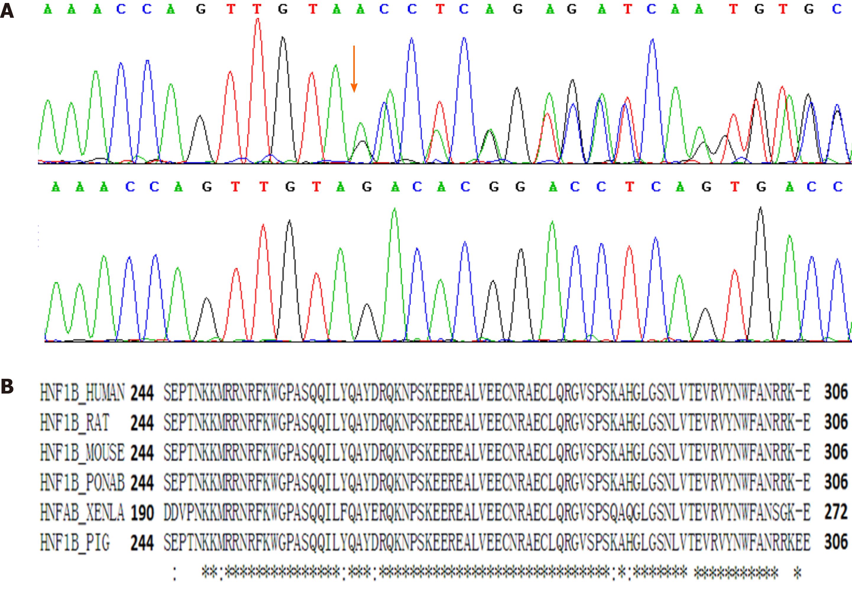
Figure 3 Mutation analysis of the renal cysts and diabetes patient.
A: Sanger sequencing of the renal cysts and diabetes patient (upper panel) and her parents (lower panel). Black arrow indicates the mutation position; B: The amino acid sequence of the DNA-binding domain of NHF1B is highly conserved among species.
- Citation: Xiao TL, Zhang J, Liu L, Zhang B. Hepatocyte nuclear factor 1B mutation in a Chinese family with renal cysts and diabetes syndrome: A case report. World J Clin Cases 2021; 9(28): 8461-8469
- URL: https://www.wjgnet.com/2307-8960/full/v9/i28/8461.htm
- DOI: https://dx.doi.org/10.12998/wjcc.v9.i28.8461


