©The Author(s) 2021.
World J Clin Cases. Aug 26, 2021; 9(24): 7009-7021
Published online Aug 26, 2021. doi: 10.12998/wjcc.v9.i24.7009
Published online Aug 26, 2021. doi: 10.12998/wjcc.v9.i24.7009
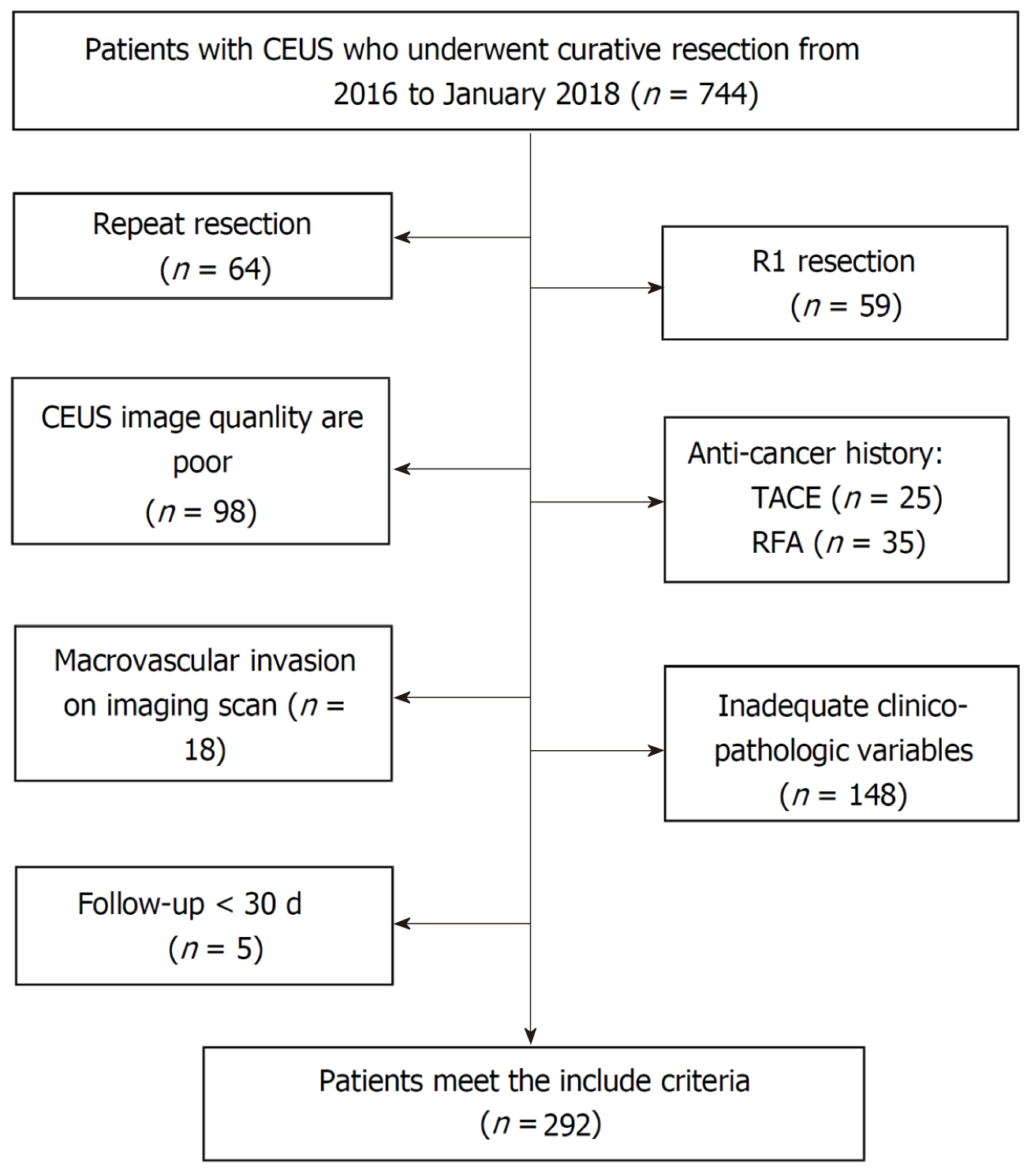
Figure 1 Flow program for patient inclusion.
CEUS: Contrasted-enhanced ultrasound.
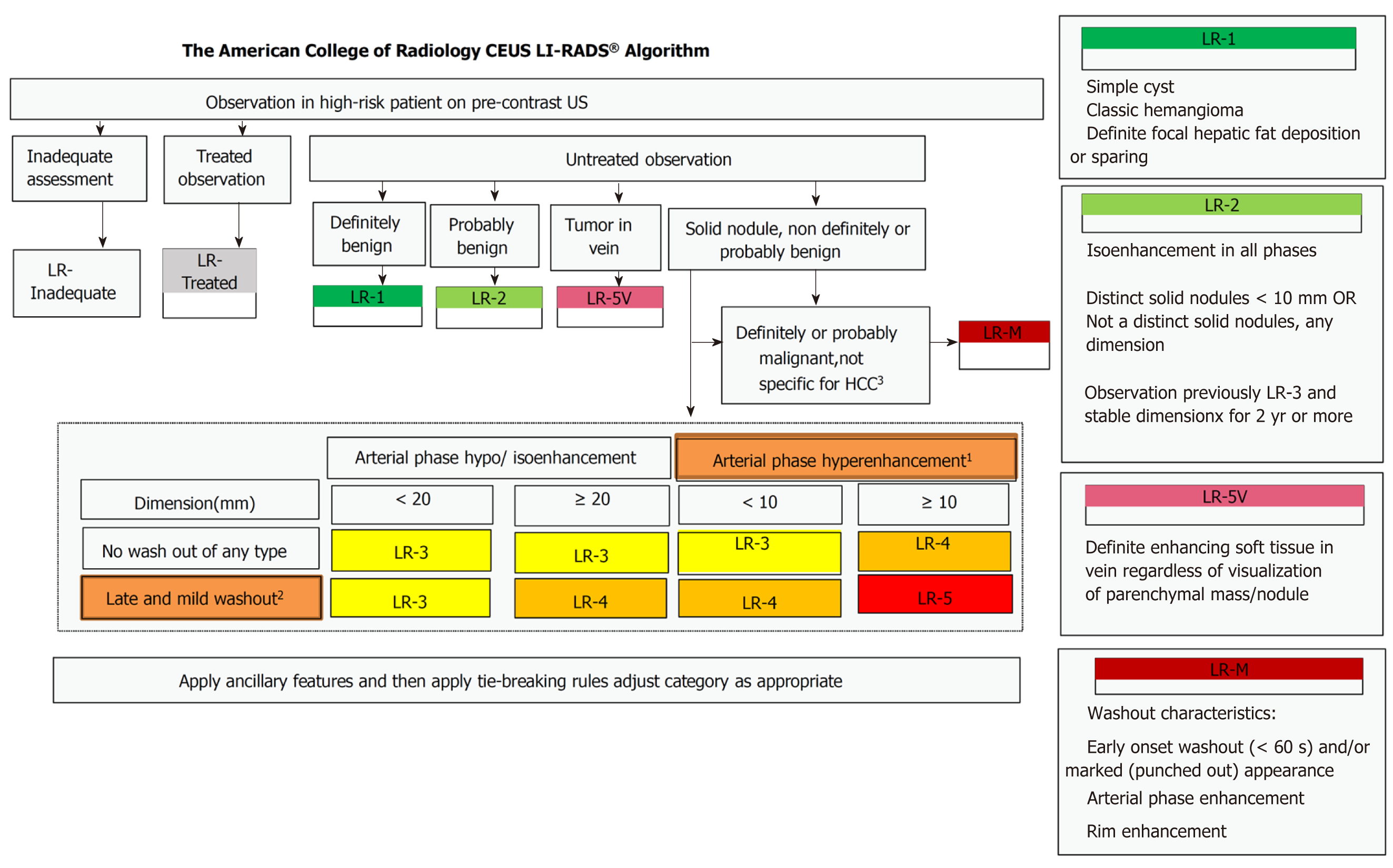
Figure 2 Official ACR algorithmic display for contrasted-enhanced ultrasound model and liver imaging reporting and data system.
1Arterial phase hyperenhancement: Whole or in part, no rim or peripheral discontinuous globular enhancement. 2Late in onset (≥ 60 s) and mild in degree: Whole or in part, with no part showing early or marked washout. 3Early onset washout (< 60 s) and/or marked (punched out) appearance and/or arterial phase rim enhancement. CEUS: Contrasted-enhanced ultrasound; LI-RADS: Liver imaging reporting and data system; US: Ultrasound; HCC: Hepatocellular carcinoma.
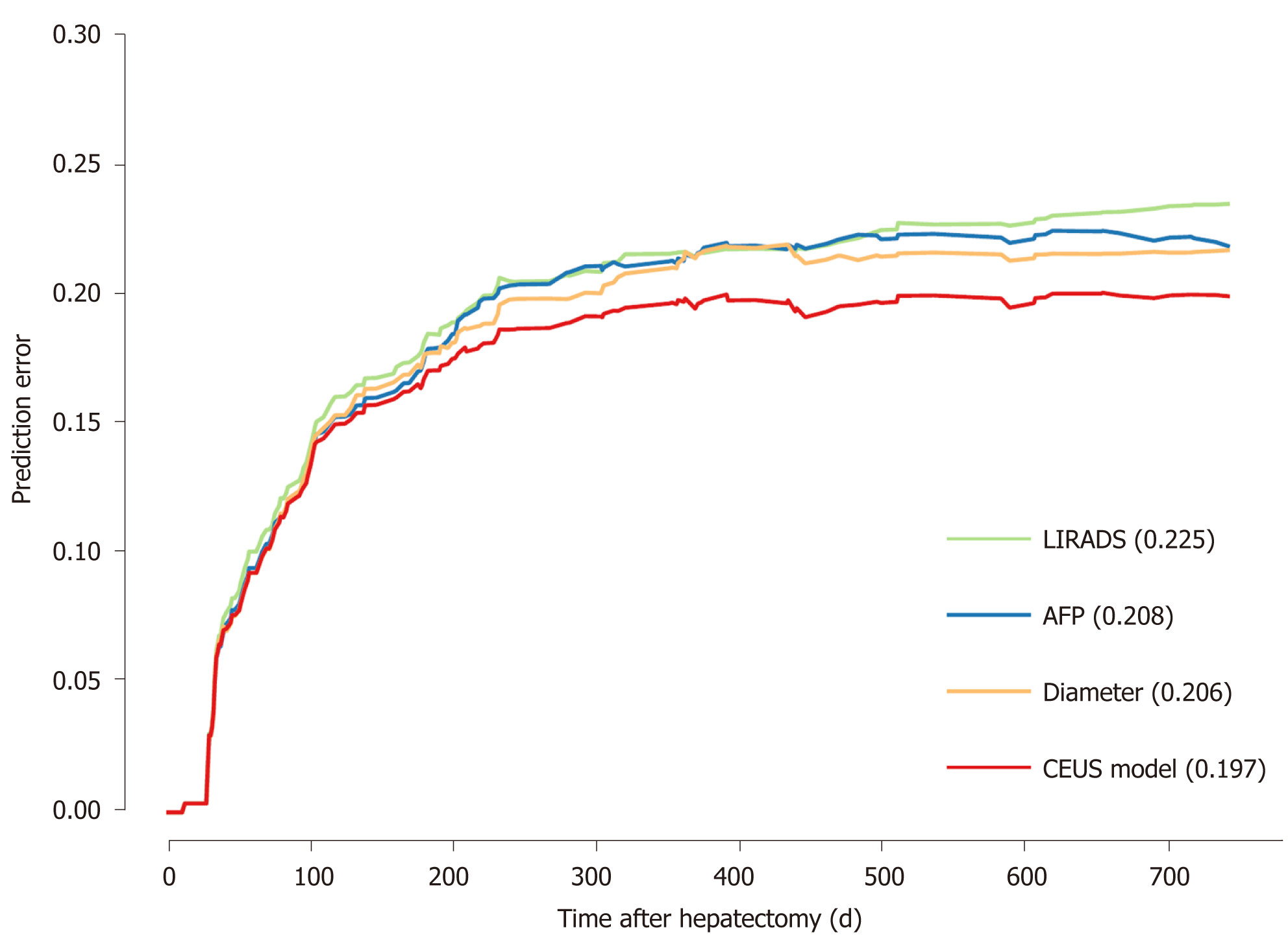
Figure 3 Prediction error of contrasted-enhanced ultrasound model and liver imaging reporting and data system, alpha-fetoprotein, and diameter.
AFP: Alpha-fetoprotein; LIRADS: Liver imaging reporting and data system; CEUS: Contrasted-enhanced ultrasound.
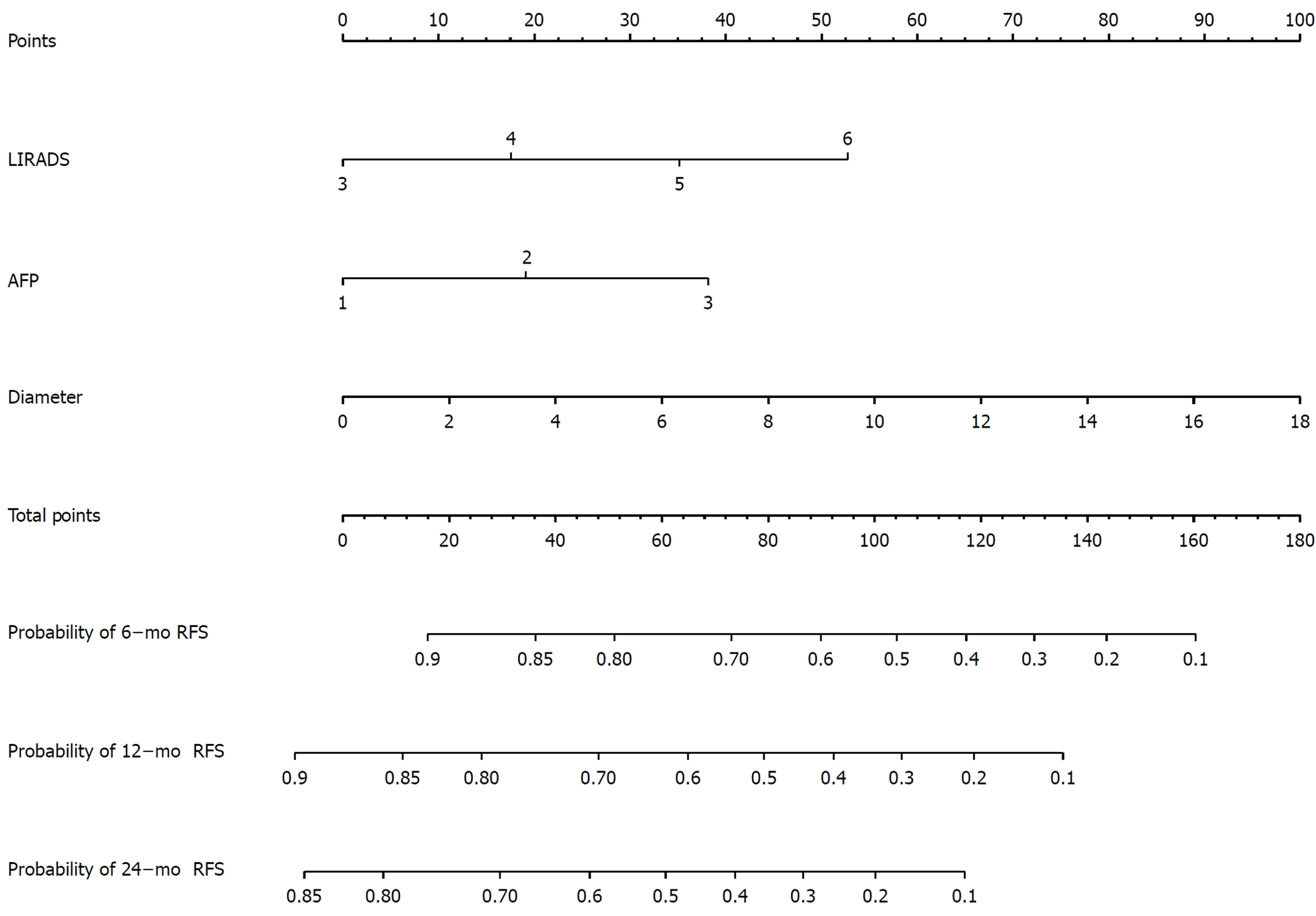
Figure 4 Nomogram structure based on liver imaging reporting and data system, alpha-fetoprotein, and tumor diameter.
LIRADS: Liver imaging reporting and data system; AFP: Alpha-fetoprotein; RFS: Relapse free survival.
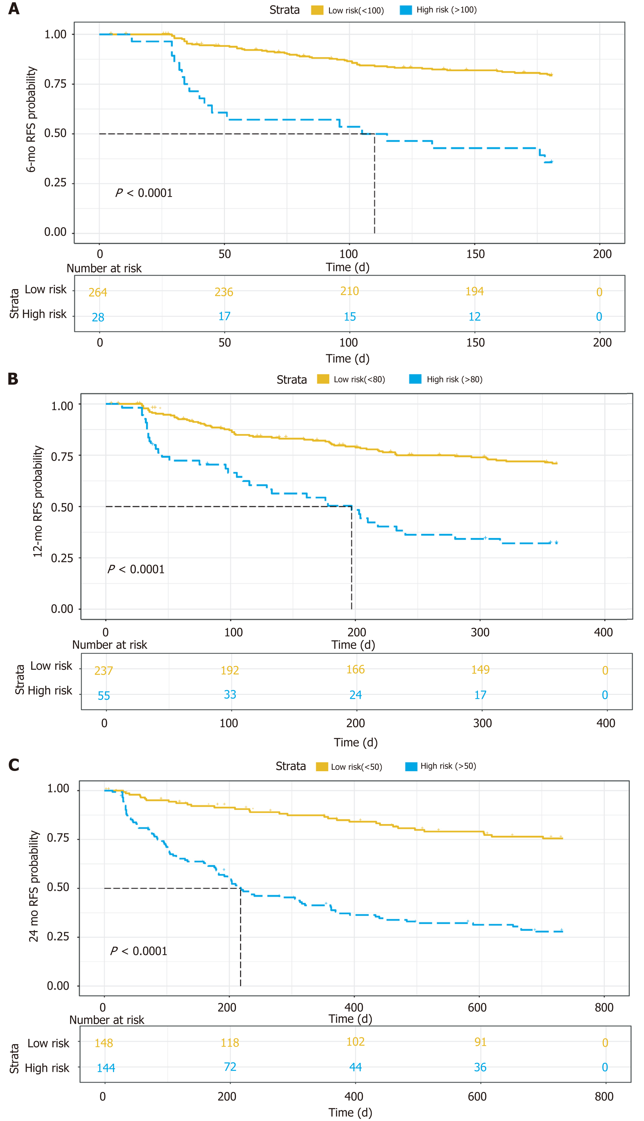
Figure 5 Kaplan–Meier curves for survival at 6 mo (A), 12 mo (B), and 24 mo (C).
RFS: Relapse free survival.
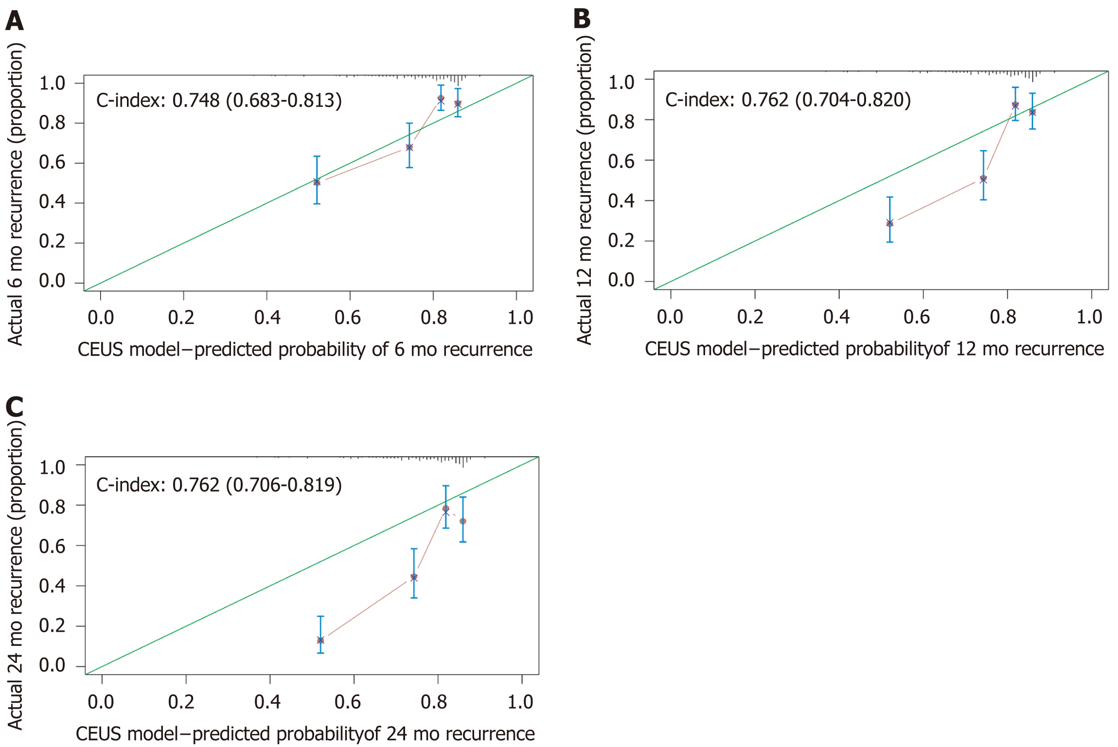
Figure 6 Calibrated curves for recurrence at 6 mo (A), 12 mo (B), and 24 mo (C).
CEUS: Contrasted-enhanced ultrasound.
- Citation: Tu HB, Chen LH, Huang YJ, Feng SY, Lin JL, Zeng YY. Novel model combining contrast-enhanced ultrasound with serology predicts hepatocellular carcinoma recurrence after hepatectomy. World J Clin Cases 2021; 9(24): 7009-7021
- URL: https://www.wjgnet.com/2307-8960/full/v9/i24/7009.htm
- DOI: https://dx.doi.org/10.12998/wjcc.v9.i24.7009


