©The Author(s) 2021.
World J Clin Cases. Jun 26, 2021; 9(18): 4748-4753
Published online Jun 26, 2021. doi: 10.12998/wjcc.v9.i18.4748
Published online Jun 26, 2021. doi: 10.12998/wjcc.v9.i18.4748
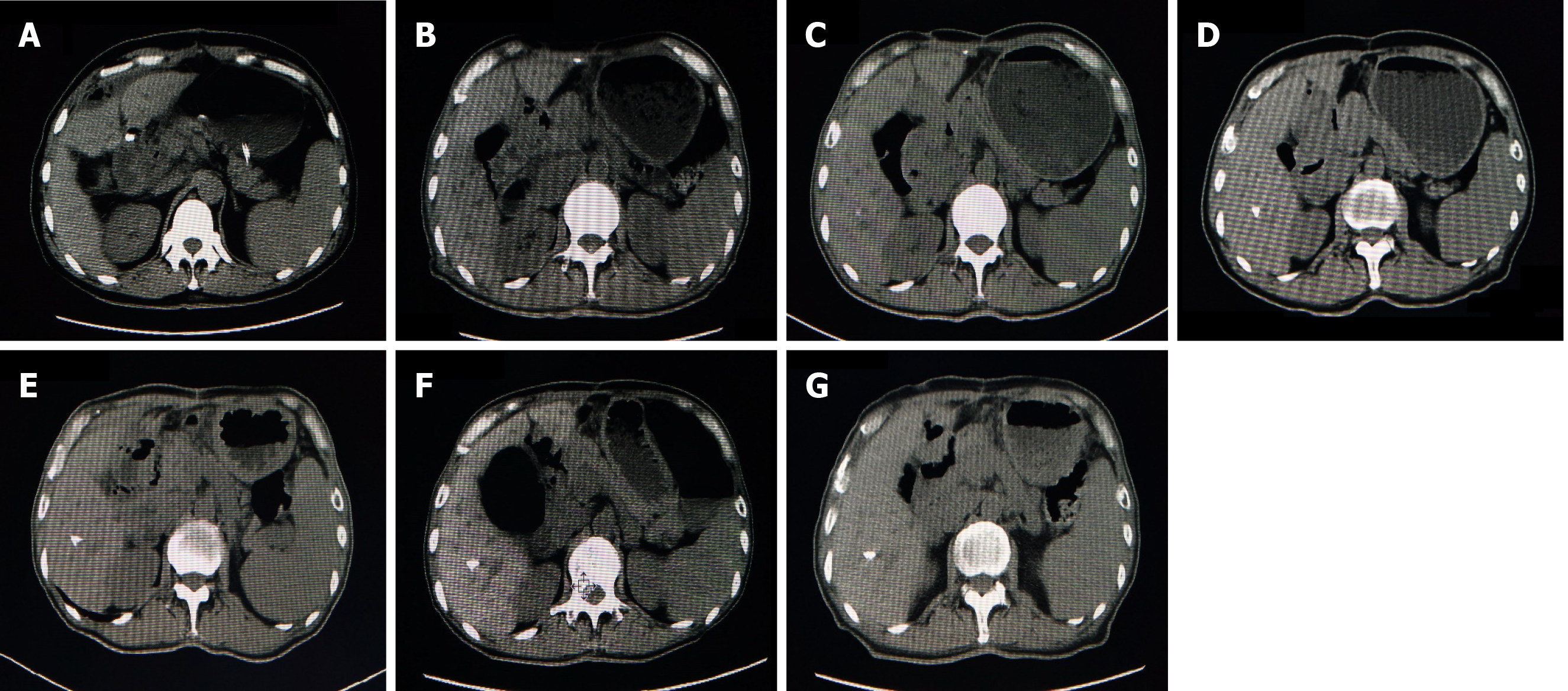
Figure 1 Computed tomography.
A: Computed tomography (CT) scan before the surgery; B: CT scan 1 year after the surgery; C: CT scan 5 years after the surgery; D: CT scan 2 years after the surgery; E: CT scan 3 years after the surgery; F: CT scan 4 years after the surgery; and G: CT scan 5 years after the surgery.
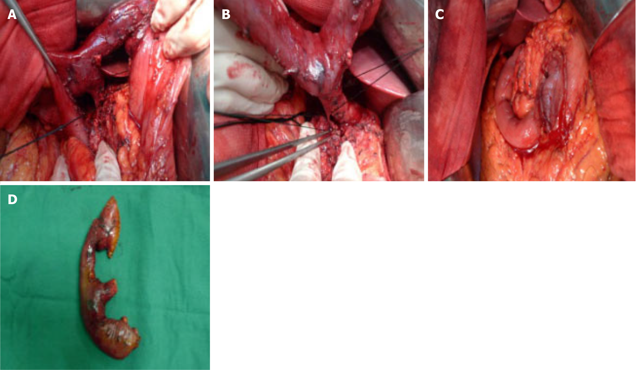
Figure 2 Intraoperative images.
A and B: Dissociation of the pancreatic duct and bile duct; C: Duodenectomy, lymphadenectomy around the pancreatic head, pancreatic-jejunal anastomosis, cholangio-jejunal anastomosis, gastro-jejunal anastomosis, and reconstruction in situ were completed; D: Surgical specimen.
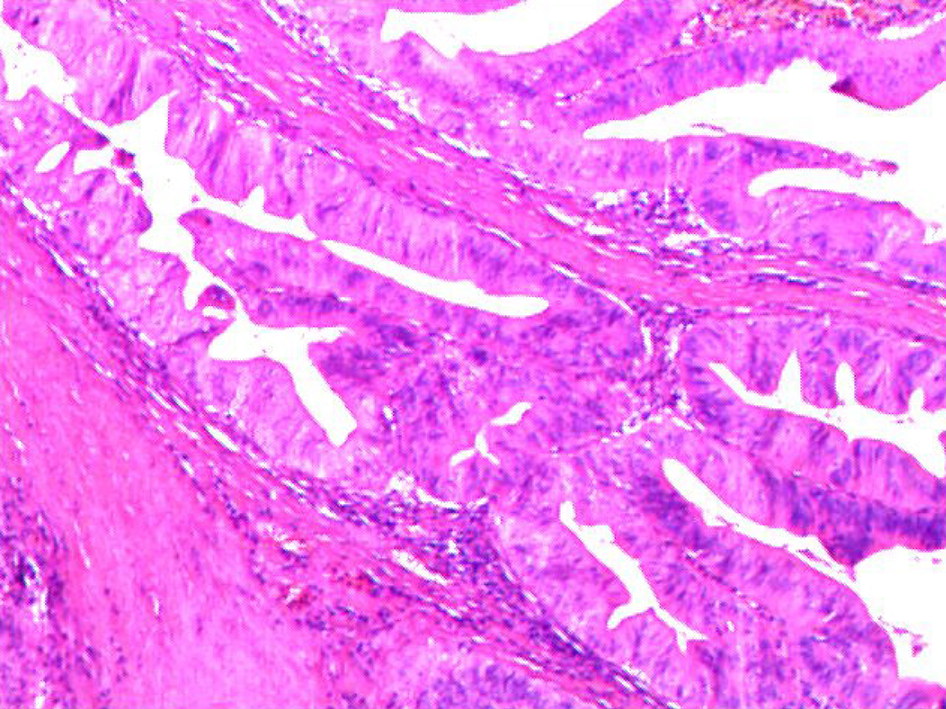
Figure 3 Postoperative pathology.
Duodenal papillary adenocarcinoma with full-thickness invasion of the intestinal wall (pT2N0M0; 200 ×).
- Citation: Wu B, Chen SY, Li Y, He Y, Wang XX, Yang XJ. Pancreas-preserving duodenectomy for treatment of a duodenal papillary tumor: A case report. World J Clin Cases 2021; 9(18): 4748-4753
- URL: https://www.wjgnet.com/2307-8960/full/v9/i18/4748.htm
- DOI: https://dx.doi.org/10.12998/wjcc.v9.i18.4748


