Copyright
©The Author(s) 2020.
World J Clin Cases. Nov 26, 2020; 8(22): 5611-5617
Published online Nov 26, 2020. doi: 10.12998/wjcc.v8.i22.5611
Published online Nov 26, 2020. doi: 10.12998/wjcc.v8.i22.5611
Figure 1 Three separated oval encapsulated masses with smooth surface.
The sectioned surface was grayish-white in color and cystic-solid lesion.
Figure 2 Pseudoglandular areas were lined by flat to cuboidal cells.
Figure 3 Immunohistochemistry stains showed strong S-100 protein positivity in the cells lining pseudoglandular cystic spaces as well as intervening cells.
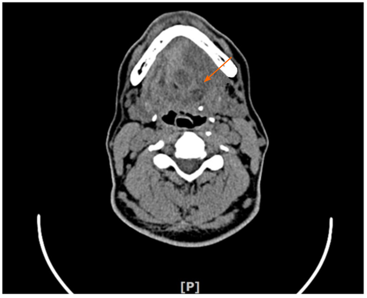
Figure 4 Computed tomography showed three low-density oval neoplasms under the tongue.
- Citation: Chen YL, He DQ, Yang HX, Dou Y. Multiple schwannomas with pseudoglandular element synchronously occurring under the tongue: A case report. World J Clin Cases 2020; 8(22): 5611-5617
- URL: https://www.wjgnet.com/2307-8960/full/v8/i22/5611.htm
- DOI: https://dx.doi.org/10.12998/wjcc.v8.i22.5611


