Copyright
©The Author(s) 2020.
World J Clin Cases. Oct 26, 2020; 8(20): 5057-5061
Published online Oct 26, 2020. doi: 10.12998/wjcc.v8.i20.5057
Published online Oct 26, 2020. doi: 10.12998/wjcc.v8.i20.5057
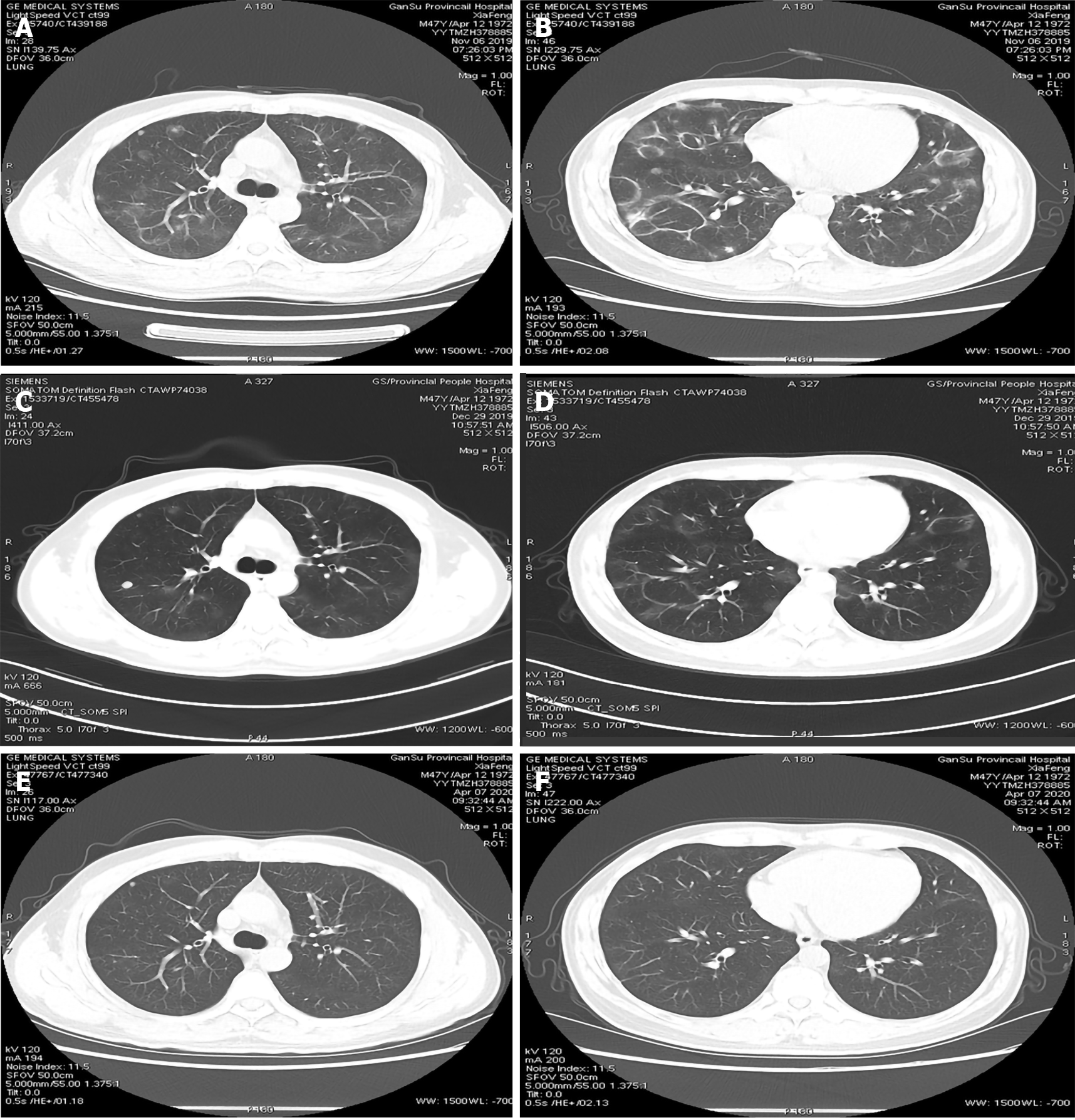
Figure 1 High-resolution computed tomography images.
A: High-resolution computed tomography (HRCT) showed multiple nodules and ground-glass shadows in both lungs; B: Circular high density shadows of various sizes were widely distributed in both lungs with lung markings inside them; C: HRCT showed that the original nodules and ground-glass shadows were significantly absorbed at 1 mo, but a new nodule was found in the right upper lobe; D: Circular high density shadows were significantly absorbed at 1 mo; E: HRCT showed that the original nodules and ground-glass shadows were significantly absorbed; F: HRCT showed that the original circular high density shadows were significantly absorbed.
- Citation: Wang HJ, Chen XJ, Fan LX, Qi QL, Chen QZ. Rare imaging findings of hypersensitivity pneumonitis: A case report. World J Clin Cases 2020; 8(20): 5057-5061
- URL: https://www.wjgnet.com/2307-8960/full/v8/i20/5057.htm
- DOI: https://dx.doi.org/10.12998/wjcc.v8.i20.5057


