Published online Oct 26, 2020. doi: 10.12998/wjcc.v8.i20.5062
Peer-review started: May 10, 2020
First decision: June 7, 2020
Revised: June 16, 2020
Accepted: September 2, 2020
Article in press: September 2, 2020
Published online: October 26, 2020
Processing time: 168 Days and 22 Hours
Fourth degree burns damage the full thickness of the skin and affect underlying tissues. Skin grafting after debridement is often used to cover the wounds of salvageable severe burns. A granulation wound can be formed by drilling the skull to the barrier layer to solve the problem of skull exposure. Low oxygen levels present at high altitudes aggravate ischemia and hypoxia which can negatively impact wound healing. The impaired healing in such cases can be ameliorated by hyperbaric oxygen therapy.
We describe a patient who presented with fourth degree burns to the left temporal and facial regions upon admission in December 2018. The periosteum of the skull and the deep fascia of the face were exposed. After the first stage of debridement and skin grafting, the temporal skin did not survive well. Granulation was induced by cranial drilling, and then a local flap was transferred to cover the wound. The left temporal and facial wounds were completely covered and the patient recovered well.
Skin grafting and flap transfer after early debridement to cover the wound and control infection were of great significance. In the later stages of the patient’s treatment, survival of the skin graft and skin flap was observed. The second stage repair was performed to achieve successful skin grafting by cranial granulation. Granulation was formed by drilling the skull, and then the wound was closed, which is suitable for cases with skull exposure and wounds with poor blood supply. We consider that hyperbaric oxygen treatment and improving tissue oxygen supply were beneficial in this patient.
Core Tip: We report the case of a female patient who suffered a fourth degree burn in the left temporal part of her face. This followed an episode of syncope which led to her collapse onto a fire basin. Skull periosteum and facial deep fascia were exposed resulting in complications with wound repair. As head and facial burn wounds were not suitable for early escharectomy, she was treated with systemic nutritional support, local wound exposure, debridement and skin grafting at a later stage. The patient was given a skin graft from the thigh, but some of the skin slices were observed to have survived poorly, resulting in further skull exposure. The skull was drilled to induce granulation, and the wound was closed with a local flap transfer. Due to the patient living in Xi’ning City which has an altitude as high as 2260 m above sea level, hyperbaric oxygen therapy was given repeatedly from initial admission until the local flap transfer operation to cover the exposed skull wounds. This was beneficial to the patient’s recovery and survival of the skin flap.
- Citation: Shen CM, Li Y, Liu Z, Qi YZ. Effective administration of cranial drilling therapy in the treatment of fourth degree temporal, facial and upper limb burns at high altitude: A case report. World J Clin Cases 2020; 8(20): 5062-5069
- URL: https://www.wjgnet.com/2307-8960/full/v8/i20/5062.htm
- DOI: https://dx.doi.org/10.12998/wjcc.v8.i20.5062
Fourth degree burns to the skin, also known as full thickness burns, and usually injure both layers of the skin[1] (dermis and epidermis) along with underlying tissues such as bone and muscle. The wound surface has no vesicle formation, appears waxy white or scorched yellow in color and develops skin necrosis following dehydration to form an eschar. It is necessary to ensure that the body’s natural wound repair processes in patients with severe burns are supported to restore good skin condition. Eschar formation and skin grafting are commonly used to cover the wounds of fourth degree burns after debridement. For fourth degree burns affecting a local small area, local flap transfer or free flap can be used to cover the wound.
Chronic wound healing is defined by wounds that cannot achieve anatomical and functional integrity through a normal, orderly and timely repair process[2]. The goal of closing the wound can be achieved clinically by controlling infection, removing necrotic tissue, negative pressure-based suction through vacuum sealing drainage, ligating veins, skin grafting and flaps. Common chronic refractory wounds include wounds caused by infection, pressure injury, diabetes mellitus, trauma, burns and arteriovenous ulcers. Accordingly, the treatment of chronic wounds is based on etiological treatment, such as complete wound debridement, decompression, control of patients' blood glucose and so on.
Cranial drilling is widely used in neurosurgery and it plays an important role in the drainage of chronic subdural hematomas, ventricle drainage and the treatment of deep brain lesions. Compared with traditional craniotomy, post-drilling drainage results in less trauma and has the key advantage of simpler operation. Moreover, it is associated with better improvement of neurological deficit than craniotomy, and the incidence of postoperative complications is also lower. The skull plate-barrier layer is rich in blood vessels. For patients with soft tissue defects of the scalp, the skull can be drilled to the barrier layer to form a granulation barrier. This facilitates a good base for the next step of skin grafting and the transfer of a skin flap to cover the wound surface.
Low atmospheric oxygen levels at high altitudes aggravates ischemia and hypoxia in chronic ulcer wounds. Hyperbaric oxygen therapy can improve the oxygen tension of wounds, increase the proliferation of fibroblasts, activate the function of white blood cells, control infection, promote the synthesis and maturation of collagen, promote the formation of blood vessels and accelerate wound healing.
The patient, a 45-year-old woman was admitted to hospital 2 h after she suffered flame burns to the face and left upper limb after syncope in December 2018. She referred to using shampoo that she had previously used numerous times at home, then fainting for unknown reasons. This resulted in her falling into a fire basin and she was taken to hospital by her family.
The patient had been in good health.
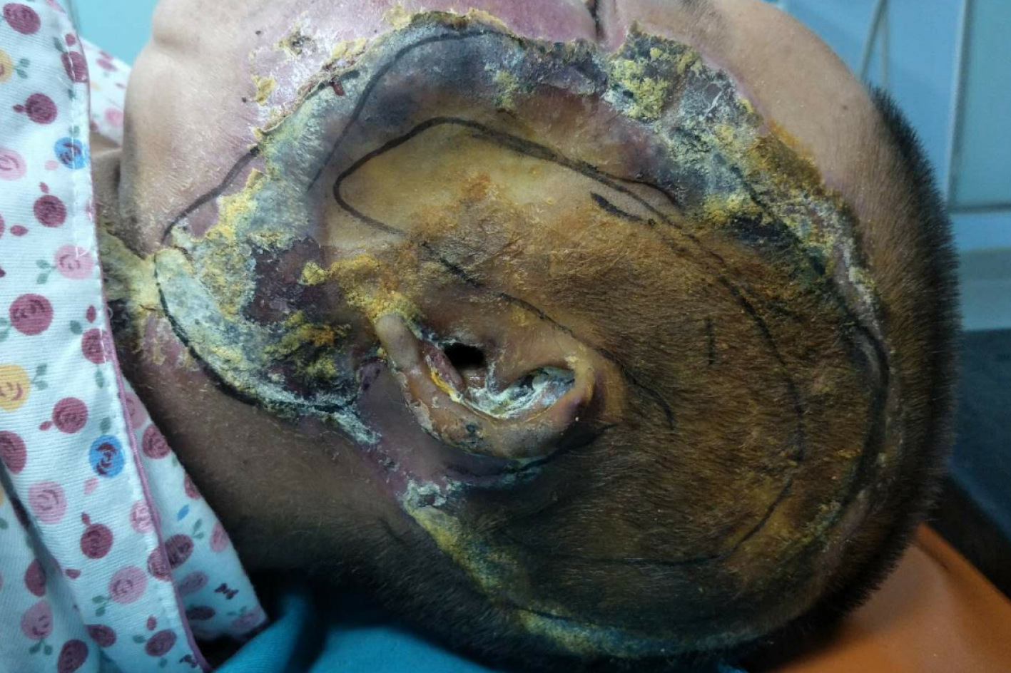
The patient was conscious, but mentally incapacitated with generally stable vital signs and no abnormalities detected in the heart and lungs. The patient's body temperature fluctuated within the normal range after admission. The total burn area of the patient was up to 4% total body surface area (TBSA). The left temporal burn wound was about 1% of the TBSA and had formed black eschar which felt hard to touch. The burn wound on the left part of face was about 1% of the TBSA, and the base of the wound was porcelain white. Branches of blood vessels could be seen in the wound and the patient lacked sensation in the area. The left eye was slightly swollen and unable to open. Although the patient could not see, she did have a sense of light. The left external ear was dry with necrotic skin. The burn wounds in the neck, shoulder and the left upper limb were 2% of the TBSA. The skin on these wounds was avulsed. There were several blisters of different sizes which felt tender. The blood supply in the extremities was normal and peripheral circulation was good. The oral and nasal mucosa were also normal (Figure 1).
Emergency computed tomography showed symmetrical speckled calcification of bilateral basal ganglia, swelling of the soft tissue in her left eyelid, left part of her face, left temporal forehead and ear. Radiography revealed a small amount of inflammation in bilateral maxillary sinuses.
Cranial magnetic resonance imaging scan + diffusion weighted imaging showed that there was no obvious abnormality on plain scan, but bilateral inferior turbinate hypertrophy and bilateral maxillary sinus submucosal cyst were observed. Vertebral artery ultrasound showed no obvious abnormality in bilateral vertebral arteries. An electrocardiogram showed that the axis of sinus arrhythmia was unbiased. Cardiac ultrasound showed that left ventricular diastolic function had decreased to grade I. Also, mitral valve and tricuspid regurgitation were observed. Ultrasound also showed a gallbladder polyp while a chest orthopedic film of the lung, heart, and diaphragm did not show obvious abnormalities.
Urgent routine blood examination was carried out after admission. The patient’s neutrophil count was 13.97 × 10⁹. Red blood cell count, hemoglobin and platelet count were normal. Blood glucose was 9.2 mmol/L. Potassium ion, sodium ion and chloride ion were all in the normal range. Blood bacterial culture was observed to be negative after admission. The patient was treated with oxygen inhalation, anti-inflammatory drugs, detumescence, rehydration, and debridement. Hyperbaric oxygen therapy was administered 10 times.
Fourth degree burns (head, face and neck); Cranial exposure.
Two weeks after admission, granulation of the wound was good. The patient received autologous skin transplantation on the left temporal area, the facial region and left upper limb under general anesthesia.
Three percent of the full thickness skin grafts were taken from the left thigh, the scab tissue on the left temporal area was removed during the operation to expose the temporal muscle. The superficial temporal artery was ligated with silk thread, and was embedded in the superficial fascia.
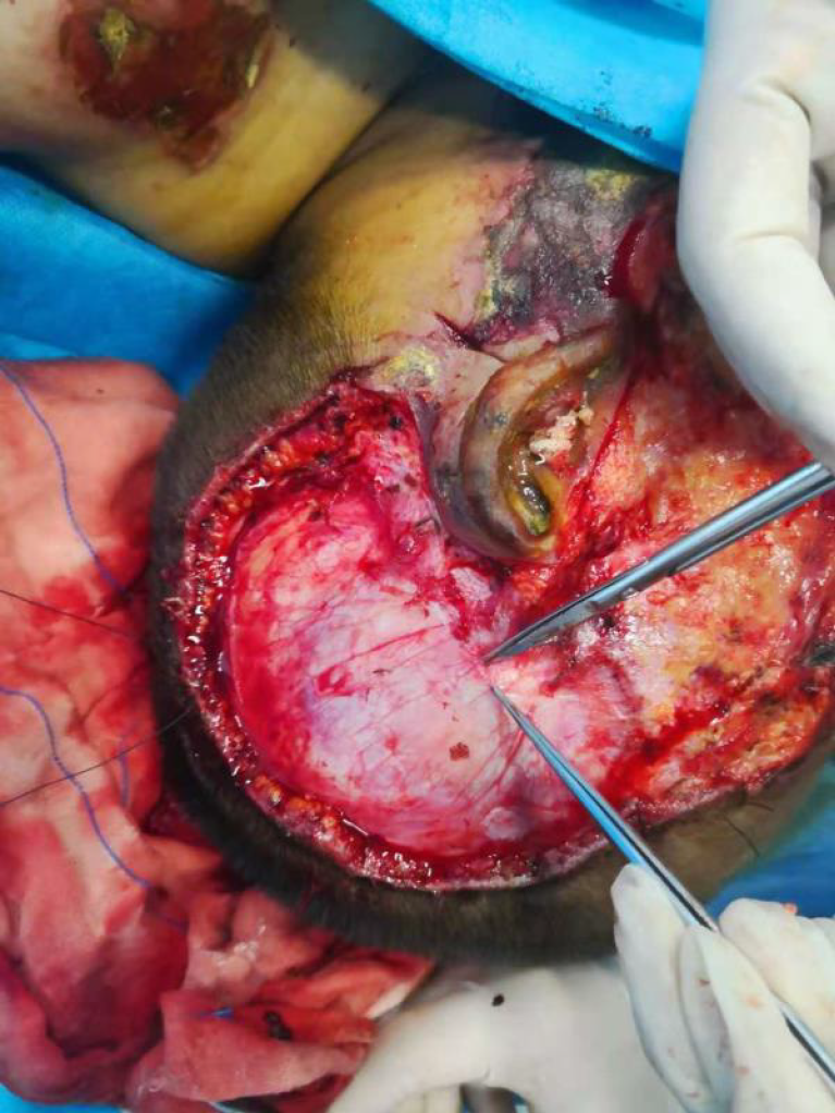
The scab tissue from the left facial area was transferred to the SMARS fascia (Figure 2). The reticular skin was placed in the left temporal area. The left facial wound was packed with silk threads. After removing the scab tissue on the left hand and left shoulder down to the base, the reticular skin patch was attached and bandaged under pressure. The patient was treated with anti-inflammatory drugs, hemostasis, detumescence and rehydration after the operation, and her recovery was stable. The patient was given routine oxygen treatment in a hyperbaric oxygen chamber which was repeated 10 times. On the 5th day after the operation, the packing suture was removed and the color of the skin was ruddy. Moreover, there was no blood accumulation and secretion under the skin as it was closely connected to the basement.
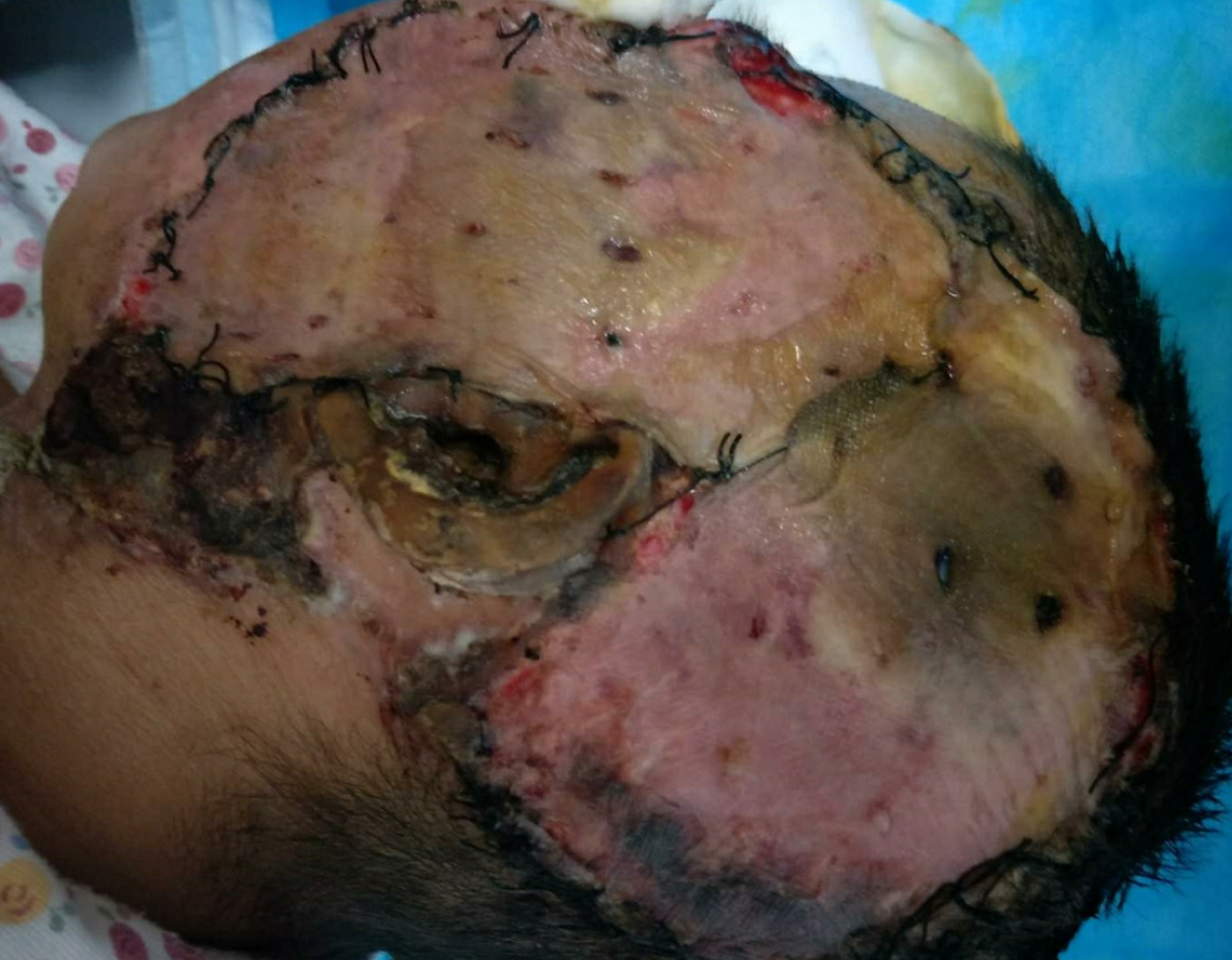
The patient underwent dressing changes every other day. However, on the 9th day after surgery, the left temporal skin flap was observed to have degenerated. Indeed, much of the skin had survived poorly (Figure 3). On the 15th day after surgery, an area of the left temporal skull measuring approximately 3 cm × 3 cm became exposed. However, the rest of the skin graft was closely connected to the basement and the color was normal. On the 34th day of admission, the patient underwent left skull drilling under general anesthesia. During the operation, the left exposed periosteum and the surrounding necrotic tissue were removed. A bone drill was used to drill the exposed part of the bone on the left side until a small number of bleeding points were observed. Oil yarn was placed in the wound where drilling had taken place and sterile dressing was used to cover it. Seven days after the drilling operation, a small amount of blood seepage could be seen in the drilled area. An aseptic dressing was used to cover the wound after iodophor disinfection. The patient was discharged from hospital with advice to have the dressing changed regularly at other hospitals.
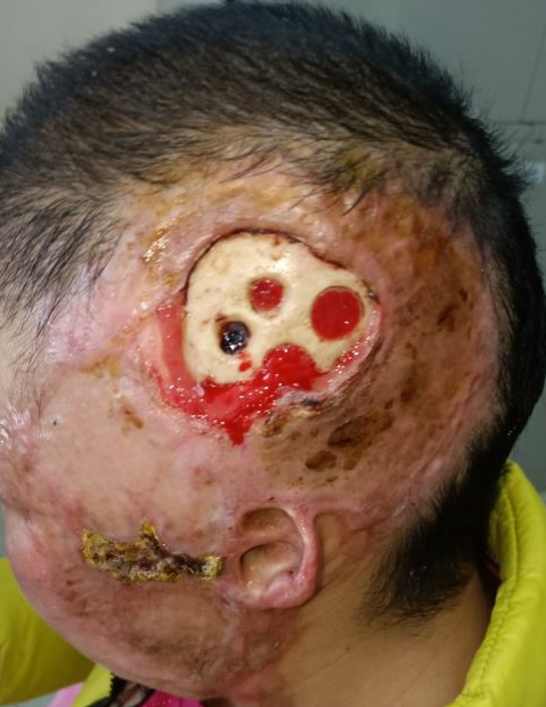
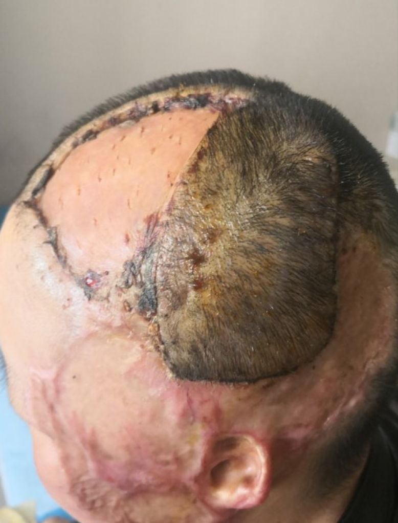
Two months later, after granulation was fully formed, an operation to transfer a skin flap to cover the skull was carried out under general anesthesia. Granulation was formed at the hole in the skull (Figure 4). Furthermore, bacterial cell cultures were negative in this patient. During the operation, the necrotic tissue around the exposed bone was removed. A rotating flap was designed above the parietal bone and the subcutaneous tissue of the skin was cut to the cap-shaped tendon film layer along the design incision. The skin flap was rotated to the exposed wound surface of the skull and stitched with the surrounding normal scalp. Then a graft measuring approximately 1% of the TBSA was excised from the left thigh to cover the flap area. The skin was packed and sutured with the surrounding normal tissue under pressure. A rubber drainage strip was placed under the flap to cover the head wound. The thigh donor area was covered with an aseptic dressing. The patient was treated with anti-inflammatory drugs, hemostasis, detumescence and rehydration after the operation and was given routine treatment in a hyperbaric oxygen chamber. Three days after the operation, the skin flap was rosy in color, no blood or secretions had accumulated under the flap, and the external dressing of the donor skin area was aseptic.
The packaged suture was removed 5 d after the operation, and it was found that the skin sheet had survived well with tight connection to the substrate. The patient recovered steadily. Seven days after the operation, she was discharged and instructed to remove the suture and have the dressing changed regularly. On the 14th day after surgery, the severe burn wounds in the head and face were completely healed (Figure 5).
The wound area on the face had formed scar tissue, and her hearing in the left ear was normal. When the patient was discharged from hospital, the head wound showed normal hair growth in the flap area, while the surgical repair of the dermis in this area was left to be fully considered at a later stage.
The patient described in this case report fainted due to an unknown reason and fell into a fire pot. Emergency head computed tomography and magnetic resonance imaging ruled out organic pathological changes in the nervous system as the etiology of syncope. An electrocardiogram and color doppler echocardiography were undertaken to rule out cardiogenic syncope. The patient’s blood glucose was 9.2 mmol/L. The patient had no history of diabetes, and her blood glucose returned to normal shortly afterwards. Thus, we considered that the patient suffered stress hyperglycemia. The elevated blood glucose level measured was transient and was considered to be due to stress. The patient’s family denied a family history of syncope and seizures. Although the patient’s electroencephalogram was normal, it could not rule out syncope caused by epileptic seizures. When the patient was admitted to the hospital, the color of her lip mucous membrane was normal. Carbon monoxide poisoning was suspected possibility due to the burning of coal in a confined space for heating in the patient’s home. The patient was given a mask for oxygen inhalation therapy immediately after admission and then treated with hyperbaric oxygen. Hyperbaric oxygen can accelerate the release of carboxyhemoglobin, promote carbon monoxide removal, and restore the oxygenation function of hemoglobin. Increasing the blood oxygen partial pressure is believed to rapidly correct any hypoxic ischemia[3]. Her oxygen saturation was then 96%.
The fourth degree burn on the head and face damaged the full thickness of the skin to the subcutaneous fascia. The structure of this area is loose, blood vessels and nerves are abundant, and edema after burn is serious. Due to reticulation of veins, they can communicate with the intracranial cavernous sinus, thereby enabling secondary intracranial infection to occur. Hence, head and facial burn wounds should be urgently addressed. Local coverage of the wound is encouraged together with enhancement of primary healing. The patient was treated with exposure therapy of the left facial and temporal burns on admission. When edema around the facial wound had resolved and the burned tissue began to dissolve and loosen, autologous skin transplantation to the left temporal wound, facial and left upper limb was conducted under general anesthesia. During the operation, eschar and granulation tissue were removed down to the normal substrate. In order to ensure the recovery of the patient's facial appearance, we excised thick skin from the patient's left thigh and transferred it to the wound. The edge of the skin was sutured with normal tissue. An aseptic dressing covered the surgical area which was bandaged under pressure.
After the first stage operation, survival of skin in the temporal area was poor. In particular, there was a small amount of necrotic tissue in the basal part of the temporal region where the skin was black and necrotic. An exposed surface of bone was also observed. For patients with skull exposure, free flaps may be considered to cover the wound. However, the free flap has high medical requirements. In addition, the cost of hospital stay is also high. We considered stretching the scalp to cover the wound with an expander, but in this case, the exposed bone was surrounded by primary surgical skin. A scar was formed, and the tissue was brittle. Therefore, it was not suitable to use a dilator to expand the scalp in this case[4]. Instead, local flap transfer was our first choice to cover the wound.
Therefore, craniotomy was performed at the second stage. After the formation of a window to the bone of the skull, the exudate of the barrier layer formed granulation tissue within 3 wk. The surface of the skull developed granulation particles, then formed slices, and finally formed a granulation barrier to cover the exposed skull. The time required for granulation is related to the size of the window and the depth of the hole. The larger the window and the narrower the hole, the faster granulation is formed. However, it may decrease the supportive function of the skull, which concomitantly reduces its protection of brain tissue. Conversely, the smaller the window and the wider the hole, the longer the granulation time. This makes the formation of a granulation barrier more difficult. The temporal skull is about 5 mm × 10 mm in size, and the outer plate of the skull is drilled into the barrier layer. The action should be gentle in order to prevent the drilling going too deep and damaging the brain tissue through the inner plate. During the operation, a bone drill was used to drill about 1 cm and the window was about 0.5 cm. Oil yarn was used to fill the drilling site, and the dressing was changed once every other day to observe the formation of granulation.
By the third stage operation, the wound had become chronic. The formation of chronic wounds is usually accompanied by tissue ischemia and hypoxia[5]. Moreover, bacteria tend to thrive in chronic wounds[6]. Necrotic tissues became more noticeable as the blood supply became poor. The donor flap designed to cover the exposed bone area matched the skin color, texture, hair and thickness of the recipient area, making the transfer convenient.
After severe burns, the loss of body fluid and the increase in vascular permeability lead to a sharp decrease in effective blood volume and insufficient tissue perfusion due to the destruction of the skin’s barrier function. The local blood vessels in this patient were damaged and the oxygen supply to her wounds was reduced[7,8]. The patient had lived at high altitude for a long time, and the special geographical environment in this area aggravated her wound ischemia and hypoxia. Rapid transient hypoxia can increase the speed of wound healing, while prolonged hypoxia will lead to delayed wound healing[9]. Hyperbaric oxygen therapy can increase the oxygen content in the tissue and promote vasodilation of the tissue to a certain extent, thus improving the oxygen tension in the wound and increasing the proliferation of fibroblasts. Hyperbaric oxygen can increase the growth of wound blood vessels during healing[10]. In addition, hyperbaric oxygen reduces the production of proinflammatory cytokines and inhibits the infiltration of inflammatory cells[9]. New research has shown that hyperbaric oxygen can relieve neuropathic pain caused by burns, which in turn can alleviate pain in burn patients[11]. It can also promote survival of skin flaps.
For severe burn patients at high altitudes, it is important to ensure their good nutritional status. Moreover, skin grafting and flap transfer after early debridement to cover the wound and control infection are of great significance. In the later stages of our patient’s treatment, the survival of her skin graft and skin flap was observed. The second stage repair was performed to achieve successful skin grafting following cranial granulation. Granulation was formed by drilling the skull, and then the wound was closed, which is suitable for cases with skull exposure and wounds with poor blood supply. We consider that hyperbaric oxygen treatment and improving tissue oxygen supply were beneficial in this patient. On the one hand, it was beneficial for the treatment of underlying disease, while on the other hand, it ensured the survival of her skin graft and skin flap.
The authors appreciate all the burn and plastic surgery staff for their efforts in treating the patient, and also thank the patient's family for their care.
Manuscript source: Unsolicited manuscript
Specialty type: Medicine, research and experimental
Country/Territory of origin: China
Peer-review report’s scientific quality classification
Grade A (Excellent): 0
Grade B (Very good): B
Grade C (Good): 0
Grade D (Fair): 0
Grade E (Poor): 0
P-Reviewer: Pawar A S-Editor: Zhang H L-Editor: Webster JR P-Editor: Wang LL
| 1. | Gao G, Li W, Chen X, Liu S, Yan D, Yao X, Han D, Dong H. Comparing the Curative Efficacy of Different Skin Grafting Methods for Third-Degree Burn Wounds. Med Sci Monit. 2017;23:2668-2673. [RCA] [PubMed] [DOI] [Full Text] [Full Text (PDF)] [Cited by in Crossref: 6] [Cited by in RCA: 8] [Article Influence: 0.9] [Reference Citation Analysis (0)] |
| 2. | Rodríguez-Quintero JH, Márquez-Gutierrez EA, Morales-Maza J. Application of a paste-type acellular dermal matrix for coverage of chronic ulcerative wounds. Arch Plast Surg. 2019;46:285-286. [RCA] [PubMed] [DOI] [Full Text] [Full Text (PDF)] [Cited by in Crossref: 1] [Cited by in RCA: 1] [Article Influence: 0.1] [Reference Citation Analysis (1)] |
| 3. | Spina V, Tomaiuolo F, Celli L, Bonfiglio L, Cecchetti L, Carboncini MC. A Case of Carbon Monoxide-Induced Delayed Neurological Sequelae Successfully Treated with Hyperbaric Oxygen Therapy, N-Acetylcysteine, and Glucocorticoids: Clinical and Neuroimaging Follow-Up. Case Rep Neurol Med. 2019;2019:9360542. [RCA] [PubMed] [DOI] [Full Text] [Full Text (PDF)] [Cited by in Crossref: 6] [Cited by in RCA: 11] [Article Influence: 1.6] [Reference Citation Analysis (0)] |
| 4. | Sokoya M, Inman J, Ducic Y. Scalp and Forehead Reconstruction. Semin Plast Surg. 2018;32:90-94. [RCA] [PubMed] [DOI] [Full Text] [Cited by in Crossref: 10] [Cited by in RCA: 23] [Article Influence: 2.9] [Reference Citation Analysis (0)] |
| 5. | Haesler E, Swanson T, Ousey K, Carville K. Clinical indicators of wound infection and biofilm: reaching international consensus. J Wound Care. 2019;28:s4-s12. [RCA] [PubMed] [DOI] [Full Text] [Cited by in Crossref: 26] [Cited by in RCA: 48] [Article Influence: 6.9] [Reference Citation Analysis (0)] |
| 6. | Knackstedt TJ. Reconstruction of a Large Scalp Defect Devoid of Periosteum. Dermatol Surg. 2018;44:1635-1638. [RCA] [PubMed] [DOI] [Full Text] [Cited by in Crossref: 5] [Cited by in RCA: 6] [Article Influence: 0.9] [Reference Citation Analysis (0)] |
| 7. | Xing D, Liu L, Marti GP, Zhang X, Reinblatt M, Milner SM, Harmon JW. Hypoxia and hypoxia-inducible factor in the burn wound. Wound Repair Regen. 2011;19:205-213. [RCA] [PubMed] [DOI] [Full Text] [Full Text (PDF)] [Cited by in Crossref: 49] [Cited by in RCA: 50] [Article Influence: 3.3] [Reference Citation Analysis (0)] |
| 8. | Schwacha MG, Nickel E, Daniel T. Burn injury-induced alterations in wound inflammation and healing are associated with suppressed hypoxia inducible factor-1alpha expression. Mol Med. 2008;14:628-633. [RCA] [PubMed] [DOI] [Full Text] [Cited by in Crossref: 29] [Cited by in RCA: 29] [Article Influence: 1.6] [Reference Citation Analysis (1)] |
| 9. | de Laat EH, van den Boogaard MH, Spauwen PH, van Kuppevelt DH, van Goor H, Schoonhoven L. Faster wound healing with topical negative pressure therapy in difficult-to-heal wounds: a prospective randomized controlled trial. Ann Plast Surg. 2011;67:626-631. [RCA] [PubMed] [DOI] [Full Text] [Cited by in Crossref: 29] [Cited by in RCA: 31] [Article Influence: 2.2] [Reference Citation Analysis (0)] |
| 10. | An H, Lee JT, Oh SE, Park KM, Hu KS, Kim S, Chung MK. Adjunctive hyperbaric oxygen therapy for irradiated rat calvarial defects. J Periodontal Implant Sci. 2019;49:2-13. [RCA] [PubMed] [DOI] [Full Text] [Full Text (PDF)] [Cited by in Crossref: 2] [Cited by in RCA: 4] [Article Influence: 0.6] [Reference Citation Analysis (1)] |
| 11. | Wu ZS, Lo JJ, Wu SH, Wang CZ, Chen RF, Lee SS, Chai CY, Huang SH. Early Hyperbaric Oxygen Treatment Attenuates Burn-Induced Neuroinflammation by Inhibiting the Galectin-3-Dependent Toll-Like Receptor-4 Pathway in a Rat Model. Int J Mol Sci. 2018;19. [RCA] [PubMed] [DOI] [Full Text] [Full Text (PDF)] [Cited by in Crossref: 13] [Cited by in RCA: 22] [Article Influence: 2.8] [Reference Citation Analysis (0)] |


