Copyright
©The Author(s) 2020.
World J Clin Cases. Oct 26, 2020; 8(20): 4844-4852
Published online Oct 26, 2020. doi: 10.12998/wjcc.v8.i20.4844
Published online Oct 26, 2020. doi: 10.12998/wjcc.v8.i20.4844
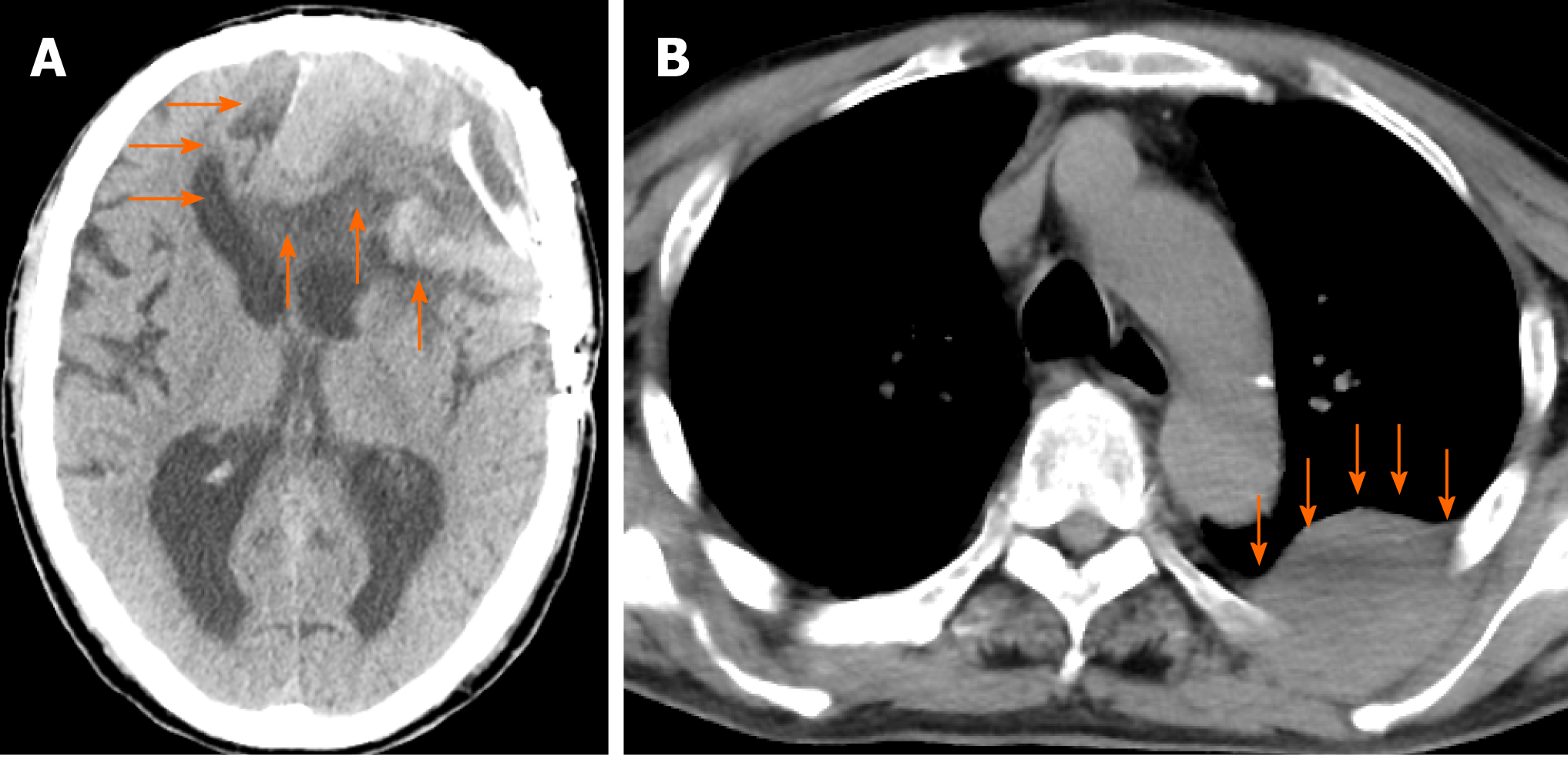
Figure 1 Computed tomography scan at admission.
A: A plain head computed tomography (CT) scan revealed the postoperative state and an 8 cm × 5.1 cm × 6.5 cm mixed-density mass at the left frontal lobe, accompanying a midline shift (orange arrow); B: A plain chest-abdomen CT scan revealed a 6 cm × 4.1 cm × 6.5 cm low-density mass in the chest wall at the superior segment of the left lung (orange arrow).
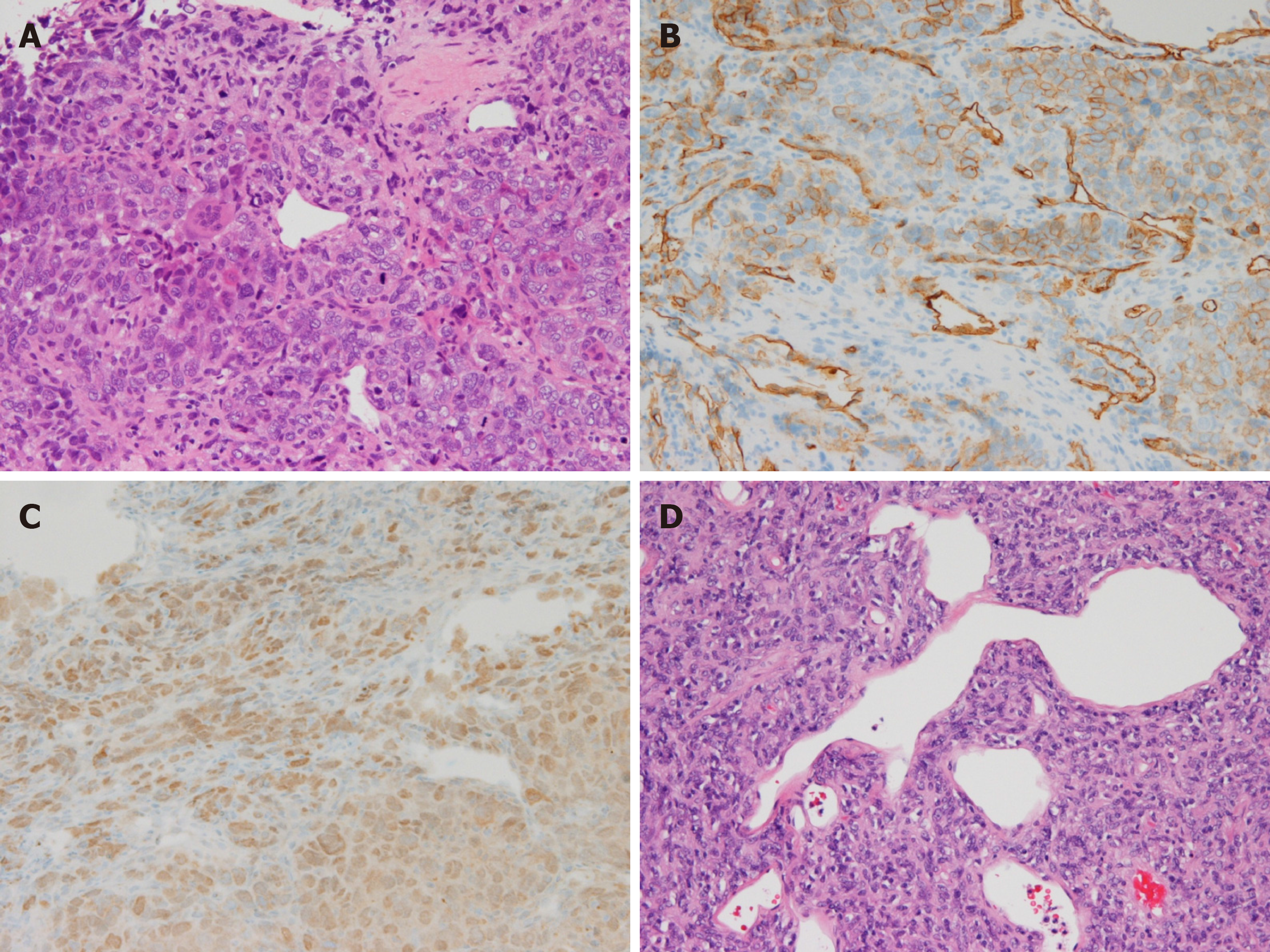
Figure 2 Histopathological image of the specimen.
A: Specimen of left chest wall mass. A solid proliferation of atypical polymorphic cells, including spindle-shaped cells, multinucleated giant cells, and blood vessels with a wall irregularity are dominant. Mitosis and pericytomatous patterns were also confirmed in part. Hematoxylin and eosin staining (magnified 1000 ×); B: Specimen of left chest wall mass. CD34-positive cells were confirmed through immunohistochemical staining (magnified 1000 ×); C: Specimen of left chest wall mass. STAT6-positive cells were confirmed through immunohistochemical staining (magnified 1000 ×); D: A back-ordered specimen of intracranial tumor. A solid proliferation of atypical polymorphic cells, including spindle-shaped cells, multinucleated giant cells, and blood vessels with a wall irregularity are dominant. Mitosis and pericytomatous patterns were also confirmed in part. Hematoxylin and eosin staining (magnified 400 ×).
- Citation: Usuda D, Yamada S, Izumida T, Sangen R, Higashikawa T, Nakagawa K, Iguchi M, Kasamaki Y. Intracranial malignant solitary fibrous tumor metastasized to the chest wall: A case report and review of literature. World J Clin Cases 2020; 8(20): 4844-4852
- URL: https://www.wjgnet.com/2307-8960/full/v8/i20/4844.htm
- DOI: https://dx.doi.org/10.12998/wjcc.v8.i20.4844


