©The Author(s) 2020.
World J Clin Cases. Jan 26, 2020; 8(2): 404-409
Published online Jan 26, 2020. doi: 10.12998/wjcc.v8.i2.404
Published online Jan 26, 2020. doi: 10.12998/wjcc.v8.i2.404
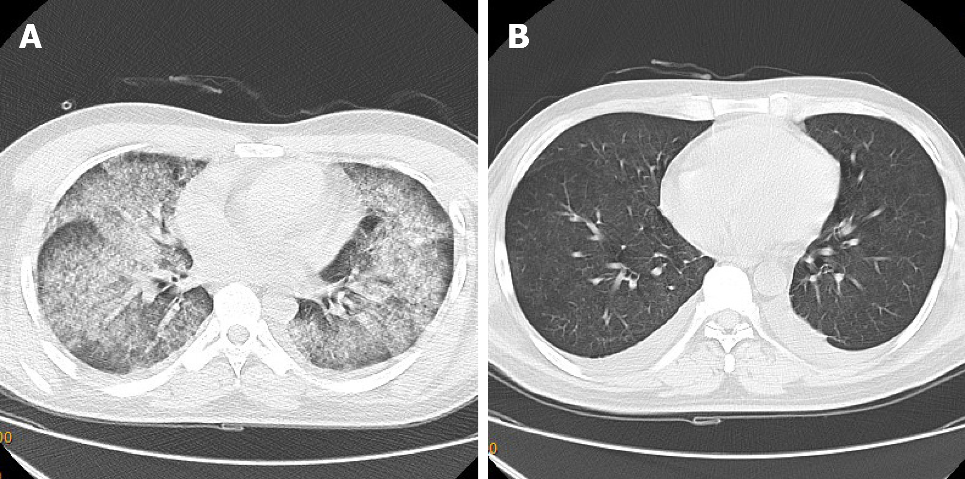
Figure 1 Chest computed tomography.
A and B: Chest computed tomography images showing bilateral diffuse exudation.
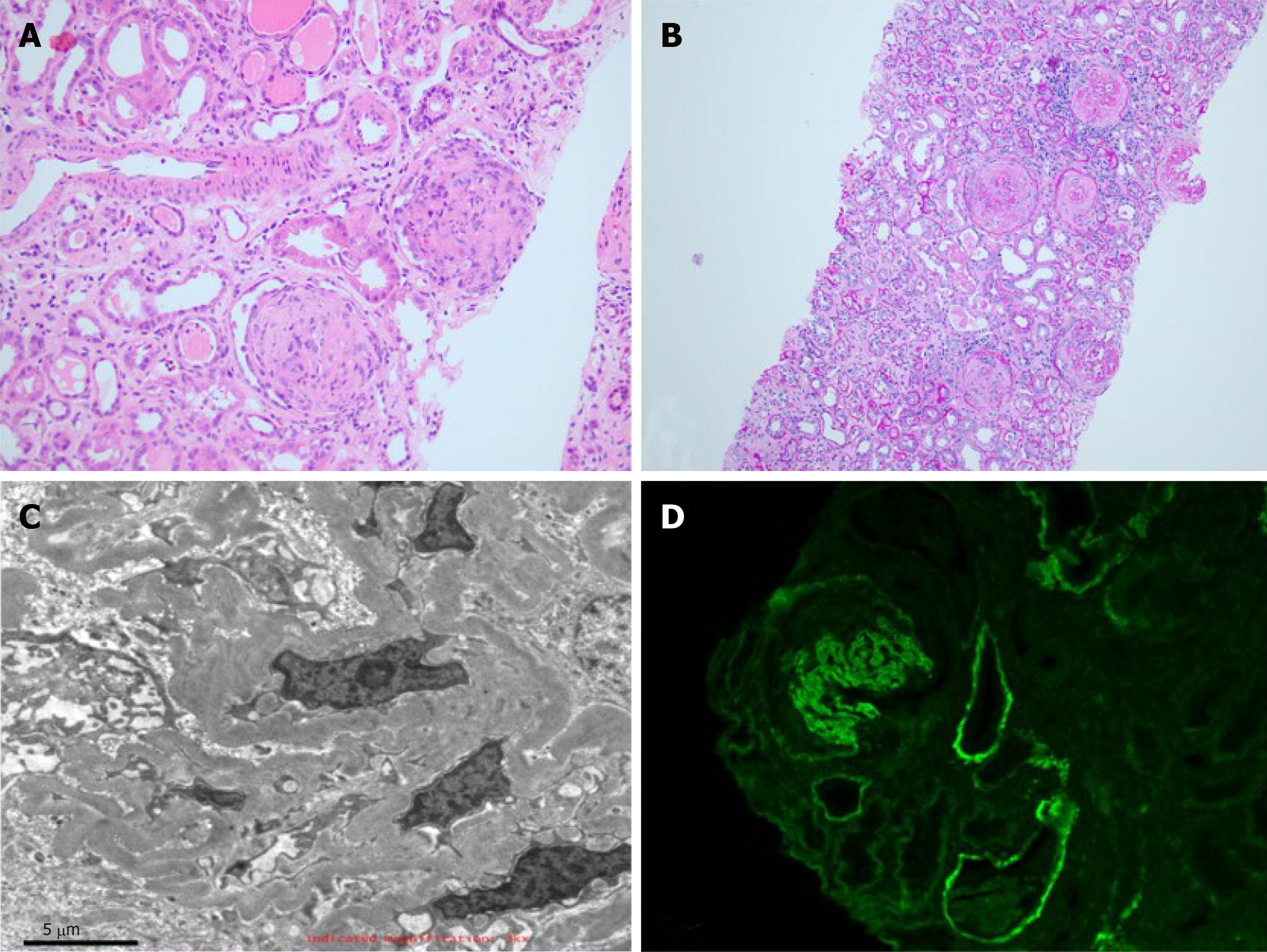
Figure 2 Electron microscopy and immunofluorescence.
A and B: Sections of the kidney taken at biopsy showing crescentic glomerulonephritis (hematoxylin and eosin staining; original magnification, ×40); C: Electron microscopy showed the occlusion of glomerular capillary loops and the infiltration of inflammatory cells in the interstitial region; D: Direct immunofluorescence showing linear IgG deposited along the glomerular basement membrane (original magnification, ×40).
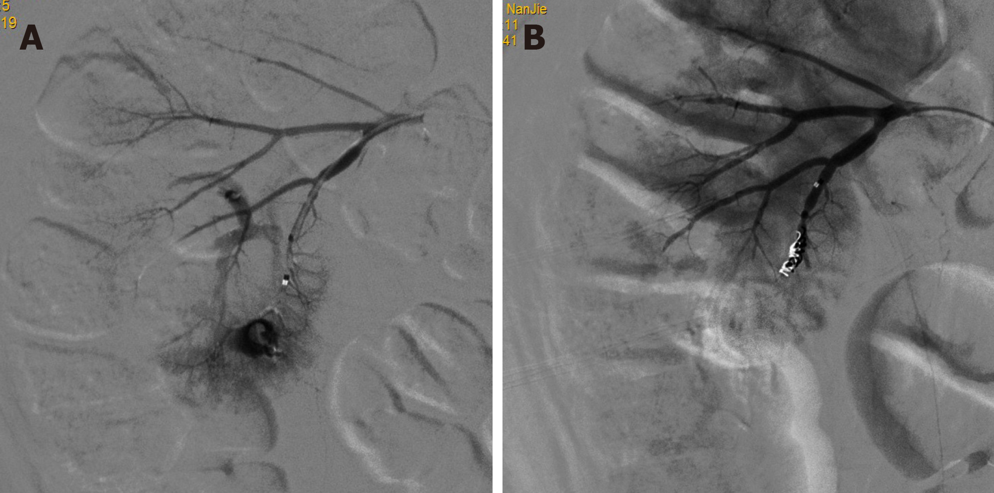
Figure 3 Radiography.
A and B: Radiographs showing hemorrhage of the renal artery and embolization.
Figure 4 Trends of hemoglobin, anti-glomerular basement membrane antibody, and C-reactive protein.
A: Hemoglobin; B: Anti-glomerular basement membrane antibody; C: C-reactive protein. GBM: Glomerular basement membrane.
- Citation: Li WL, Wang X, Zhang SY, Xu ZG, Zhang YW, Wei X, Li CD, Zeng P, Luan SD. Goodpasture syndrome and hemorrhage after renal biopsy: A case report. World J Clin Cases 2020; 8(2): 404-409
- URL: https://www.wjgnet.com/2307-8960/full/v8/i2/404.htm
- DOI: https://dx.doi.org/10.12998/wjcc.v8.i2.404


