Copyright
©The Author(s) 2020.
World J Clin Cases. Oct 6, 2020; 8(19): 4550-4557
Published online Oct 6, 2020. doi: 10.12998/wjcc.v8.i19.4550
Published online Oct 6, 2020. doi: 10.12998/wjcc.v8.i19.4550
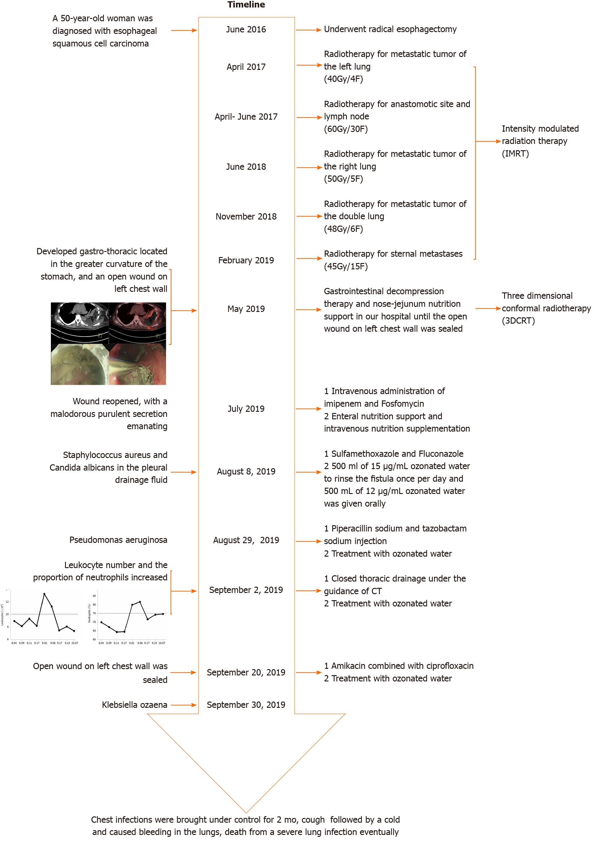
Figure 1 Timeline.
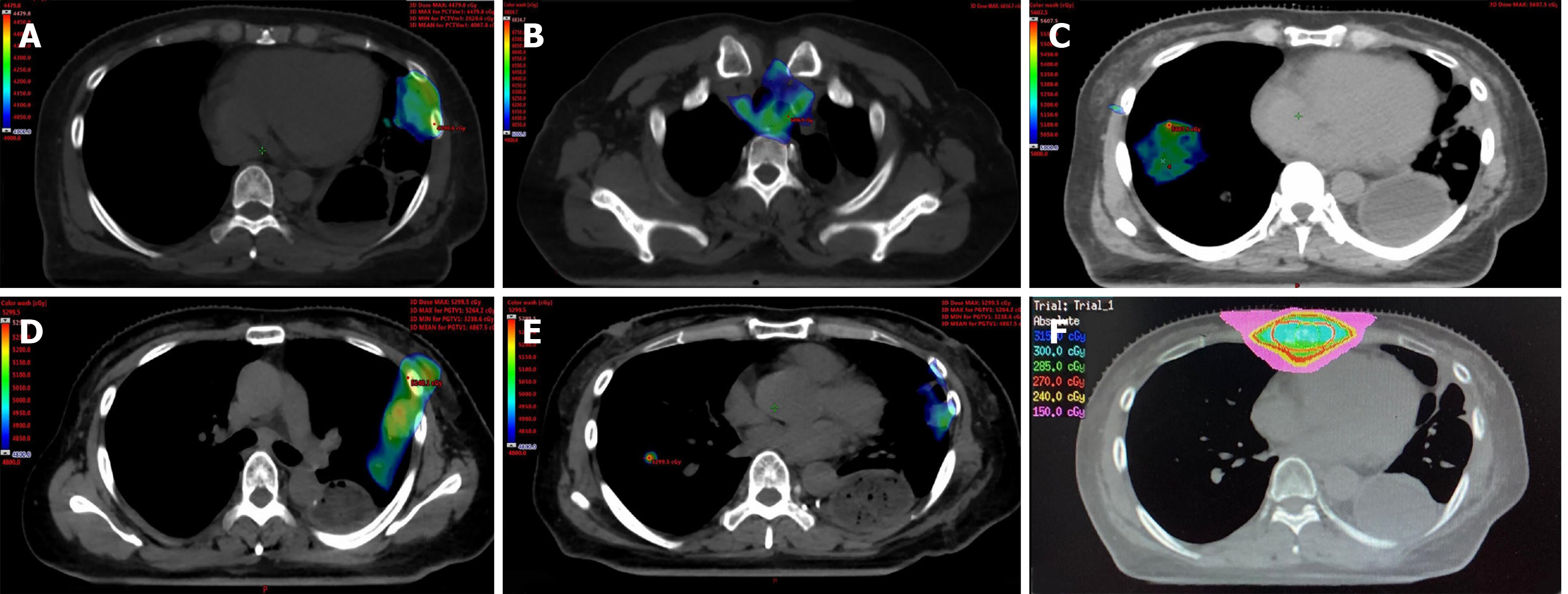
Figure 2 Cumulative isodose distribution.
A: Radiotherapy for metastatic tumor of the left lung (40 Gy/4 F) in April 2017; B: Radiotherapy for anastomotic site and lymph nodes (60 Gy/30 F) from April to June 2017; C: Radiotherapy for metastatic tumor of the right lung (50 Gy/5 F) in June 2018; D and E: Radiotherapy for metastatic tumors of both lungs (48 Gy/6 F) in November 2018; F: Radiotherapy for sternal metastases (45 Gy/15 F) in February 2019.
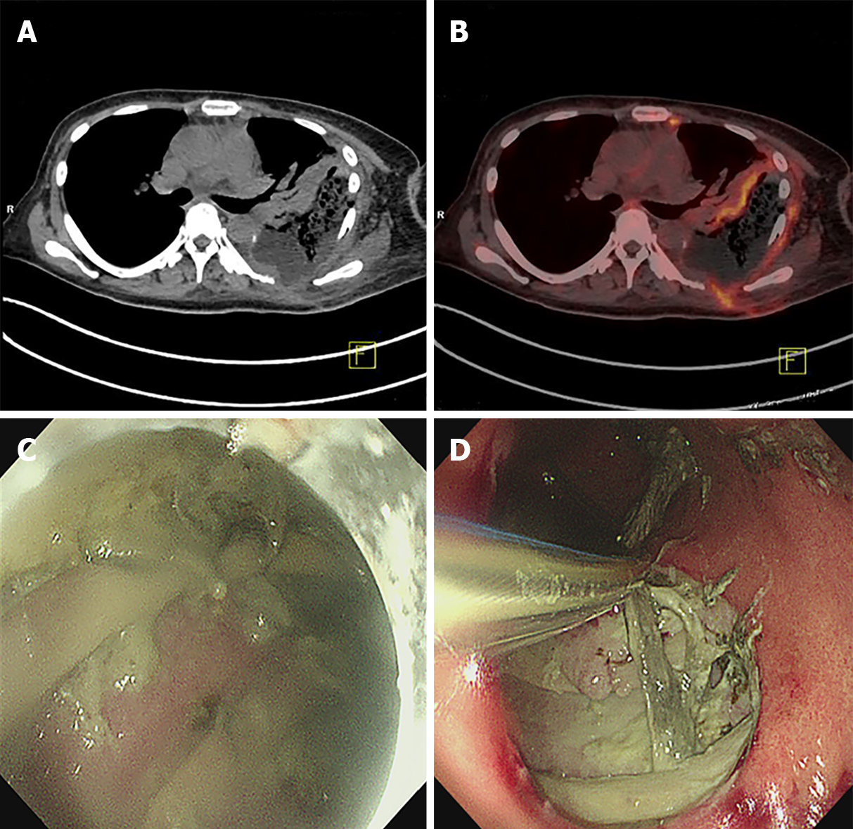
Figure 3 Positron emission tomography-computed tomography and endoscopy examination images of a 50-year-old woman with gastro-thoracic fistula after esophagectomy taken before treatment.
A and B: Computed tomography (CT) and fused CT images showing the formation of a gastro-thoracic fistula, with lesions involving the lateral chest wall; C and D: Endoscopic images showing the purulent gastric contents and local necrosis.
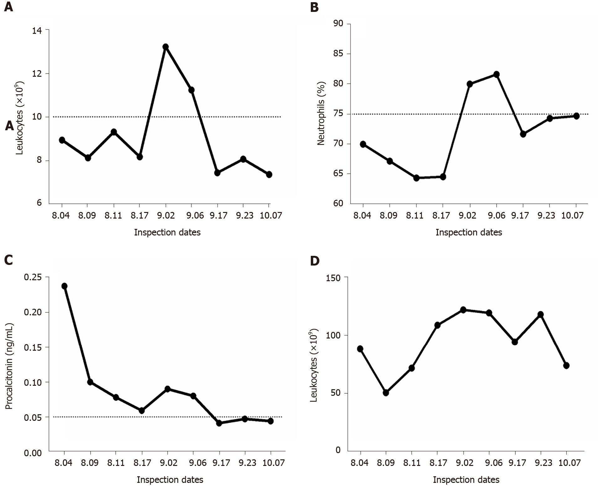
Figure 4 Infection indices of the patient during the course of treatment.
A and B: Leukocyte number and neutrophil proportion continued to rise in early September because of obstruction of the drainage tube and decreased gradually after the position of the drainage tube was changed; C: Procalcitonin was also declining, indicating that related symptoms, including severe bacterial infection and sepsis, were gradually improving; D: C-reactive protein levels fluctuated, related to the changes in the underlying disease of the patient, including obstructive pneumonia.
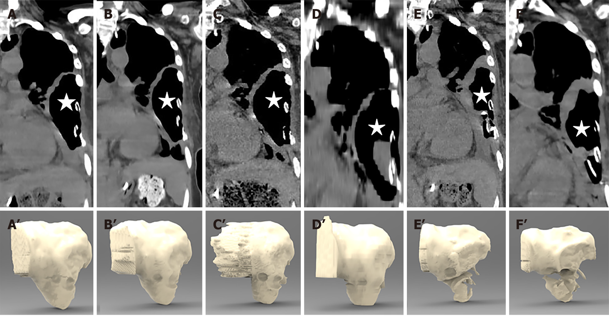
Figure 5 Computed tomography images and 3D stereograms of the gastro-thoracic fistula (asterisk) during the course of treatment.
A-F: Computed tomography examination of the patient on August 20, August 26, August 30, September 2, September 17, and October 8, 2019, respectively, showing that the wall of the thoracic abscess gradually thickened after 2 mo of adequate drainage, together with ozonated water rinse; A’-F’: Three-dimensional stereograms confirming that the volume of the fistula cavity was reduced markedly in late September.
- Citation: Wu DD, Hao KN, Chen XJ, Li XM, He XF. Application of ozonated water for treatment of gastro-thoracic fistula after comprehensive esophageal squamous cell carcinoma therapy: A case report. World J Clin Cases 2020; 8(19): 4550-4557
- URL: https://www.wjgnet.com/2307-8960/full/v8/i19/4550.htm
- DOI: https://dx.doi.org/10.12998/wjcc.v8.i19.4550


