Copyright
©The Author(s) 2020.
World J Clin Cases. Aug 26, 2020; 8(16): 3578-3582
Published online Aug 26, 2020. doi: 10.12998/wjcc.v8.i16.3578
Published online Aug 26, 2020. doi: 10.12998/wjcc.v8.i16.3578
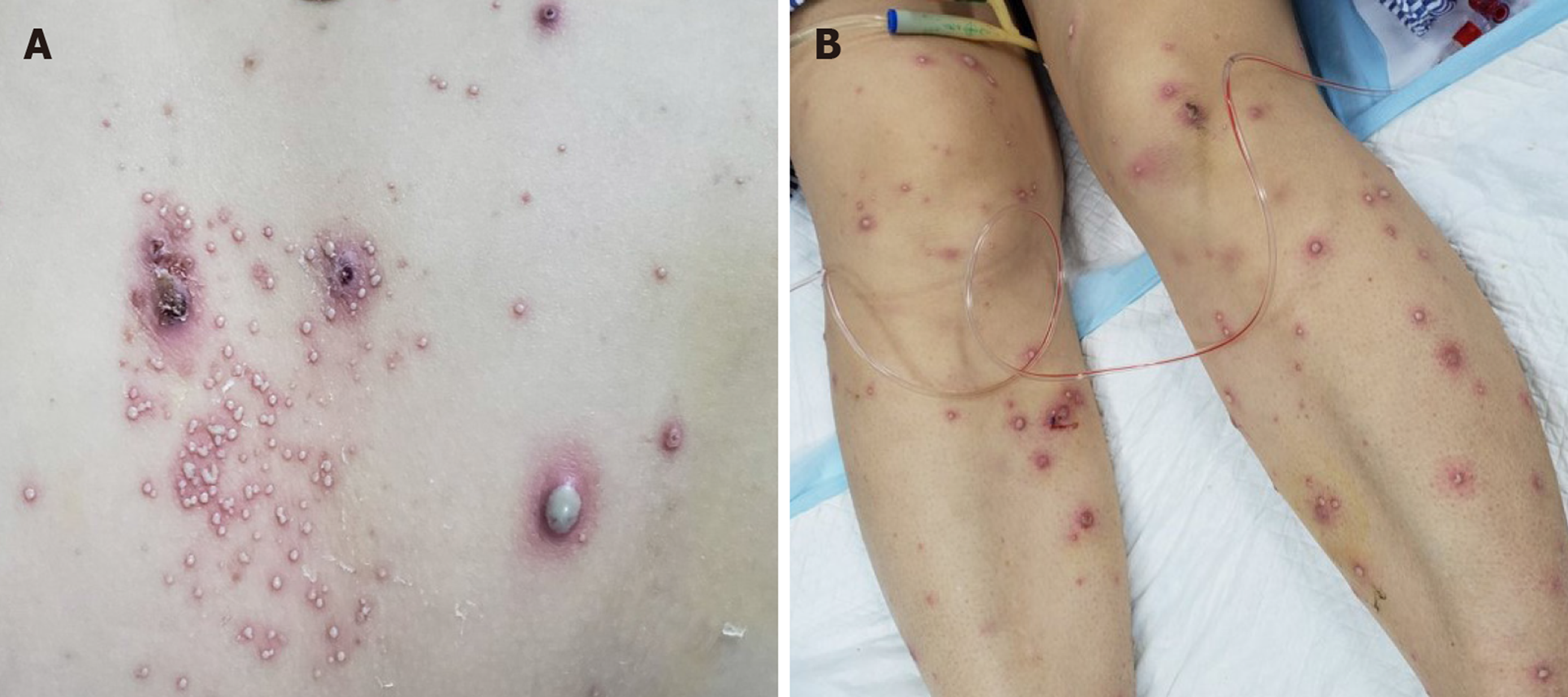
Figure 1 Diffuse pustular rash of the trunk and legs.
A: Diffuse, pustular rash of the trunk; B: Diffuse pustular rash of the legs.
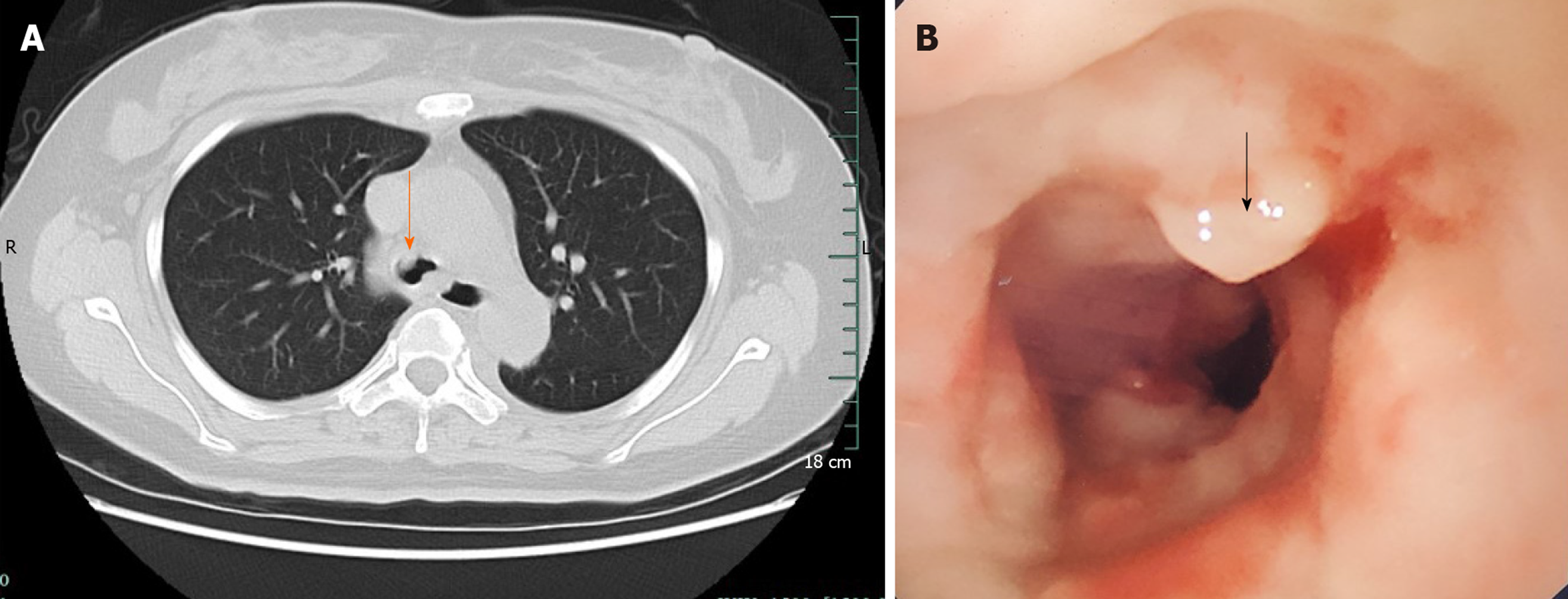
Figure 2 Lung computed tomography findings and the first tracheoscopy.
A: Lung computed tomography; B: Tracheoscopy. The main bronchus was occupied.
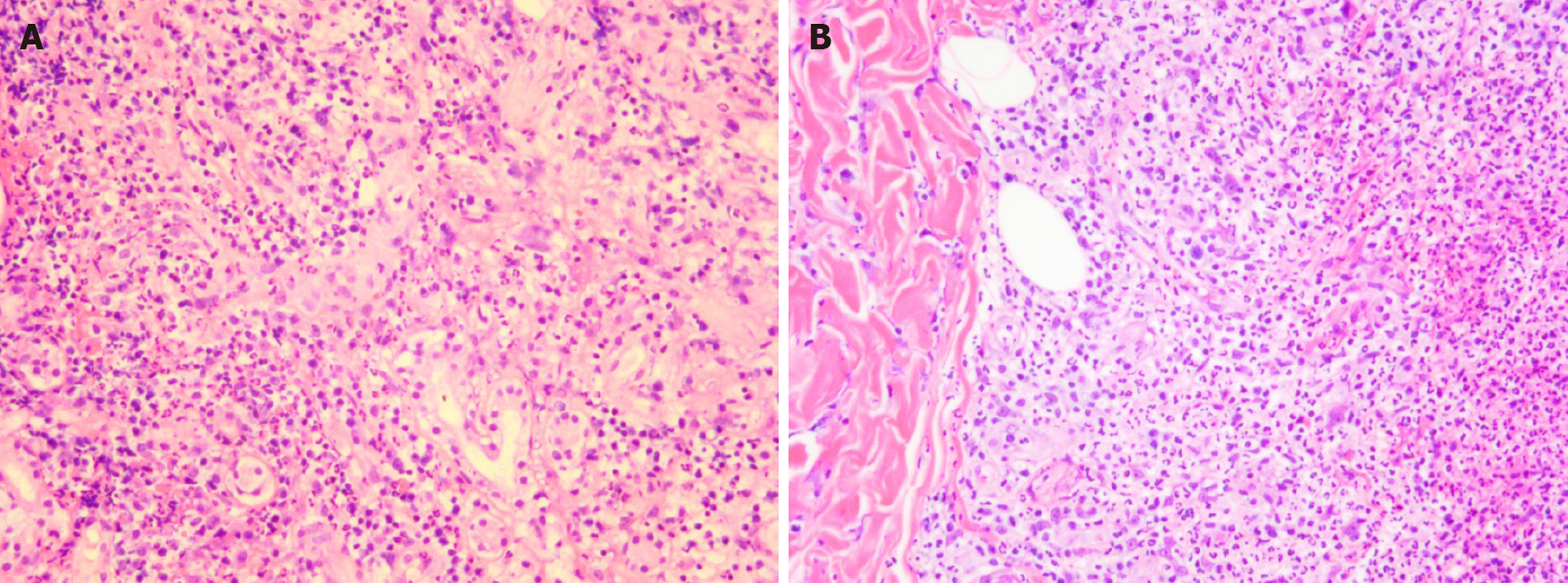
Figure 3 Skin and airway mucosa biopsy (magnification 100 ×).
A: Skin mucosa biopsy; B: Airway mucosa biopsy. Subepidermal bullous formation containing scattered neutrophils and eosinophils.
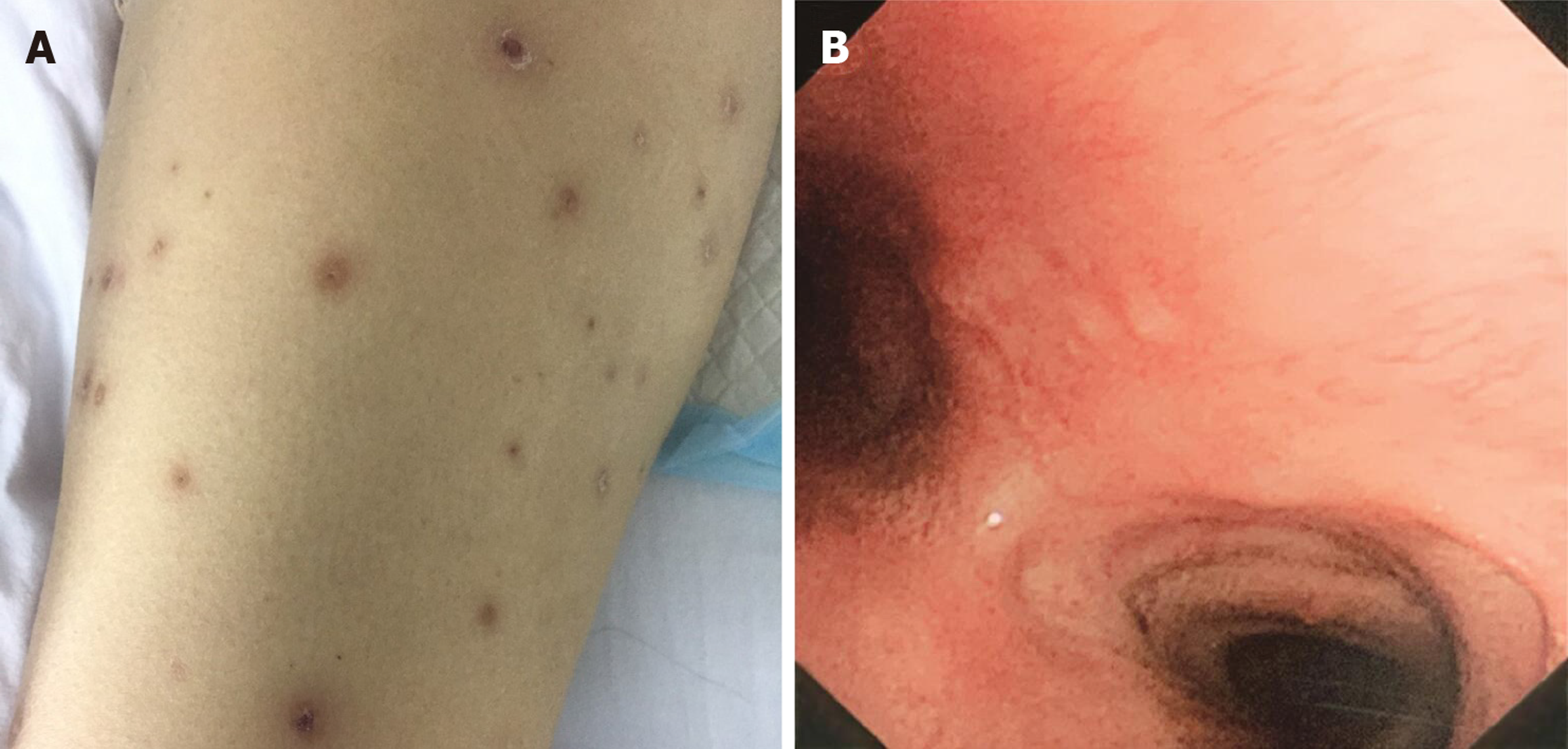
Figure 4 Leg rashes following transfer from the intensive care unit and final tracheoscopy.
A: Leg rashes following transfer from the intensive care unit; B: Final tracheoscopy. Main airway was unobstructed.
- Citation: Li LL, Lu YQ, Li T. Acute generalized exanthematous pustulosis with airway mucosa involvement: A case report. World J Clin Cases 2020; 8(16): 3578-3582
- URL: https://www.wjgnet.com/2307-8960/full/v8/i16/3578.htm
- DOI: https://dx.doi.org/10.12998/wjcc.v8.i16.3578


